
Sperm Formation Starts at Puberty
The male germ cells lie quietly in the wall of the seminiferous tubules right through childhood. Nothing is asked of them until puberty, and what changes then does not begin in the testis at all. It begins in the brain. At puberty the hypothalamus increases its secretion of gonadotrophin releasing hormone, usually written GnRH, and that single change sets the whole male reproductive machinery running. Hold the timing and the trigger together as one cause-and-effect pair rather than as two separate facts to be remembered. Sperm production does not start because the testis has grown large enough; it starts because a hypothalamic signal has risen.
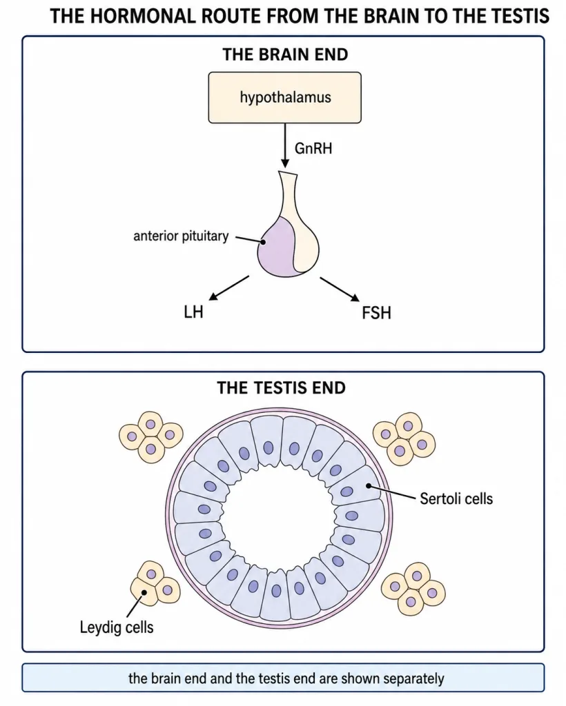
[NEET Important] GnRH itself never reaches the testis. Its target is the anterior lobe of the pituitary, and only the pituitary's own secretions travel on to the gonad. A great many mistakes come from shortening the route by one step and letting the hypothalamic hormone act on the testis directly.
The Route from the Brain to the Testis
The increased GnRH acts on the anterior pituitary and makes it secrete two gonadotrophins, luteinising hormone (LH) and follicle stimulating hormone (FSH). From here the route forks, and the fork is the part most worth learning properly, because the two hormones go to two different cells and produce two different results.
LH acts on the Leydig cells, also called the interstitial cells, which lie in the small spaces outside the tubules. Under this stimulus they synthesise and secrete androgens. FSH acts on the Sertoli cells, which sit within the wall of the tubule itself, and makes them secrete some of the factors that help the germ cells to be turned into sperms. So one gonadotrophin works through a hormone and the other works through a set of local factors. Taken end to end the route runs hypothalamus, then anterior pituitary, then the two cell populations of the testis, and only then the germ cells.
What the Androgens Do
The androgens have two quite separate collections of effects, and it pays to keep them in two mental columns.
Inside the reproductive apparatus, androgens stimulate the process of sperm formation, and they stimulate the growth, development and functions of the male accessory sex organs: the epididymis, the vas deferens, the seminal vesicles, the prostate and the urethra. These organs depend on a continuing androgen supply, which is why the LH arm of the axis matters for far more than sperm production alone.
Everywhere else in the body, androgens are responsible for the male secondary sexual characters. They stimulate muscular growth, the growth of facial and axillary hair, aggressiveness and a low pitch of the voice. They also act on the central nervous system and influence male sexual behaviour, or libido, and they have a stimulating effect on the rate of protein and carbohydrate metabolism.
The Two Somatic Populations of the Testis
Only some of the cells in and around a seminiferous tubule are germ cells. Two somatic populations serve them, and they are easy to confuse because their names are close in sound and their roles are close in place.
The Leydig or interstitial cells occupy the interstitial spaces between the tubules. They are the endocrine cells of the testis and their product, the androgens, leaves in the blood to act all over the body.
The Sertoli cells are the nurse cells. They lie inside the tubule wall among the developing germ cells, and they provide nutrition to those cells throughout their long development. During spermiogenesis the heads of the forming sperms become embedded in the Sertoli cells. A useful way to keep the pair straight is by position: the cell outside the tubule answers to LH and exports a hormone, while the cell inside the tubule answers to FSH and feeds the cells around it.
The Sperm and the Semen
A human sperm is a microscopic structure and a haploid one. Its body is marked off into four regions, and a plasma membrane envelops the whole spermatozoon rather than any one part of it. The neck is the region most often overlooked: it is short, it lies immediately behind the head, and it contains the centrioles.
Sperms alone are not what leaves the male tract. Semen is the name given to the complete discharge, and it has exactly two components: the sperms and the fluid called seminal plasma in which they are carried. Neither component on its own is semen.
[NEET Important] Learn the fertility figures as an exact pair. A human male ejaculates about 200 to 300 million sperms during a coitus. For that ejaculate to count as normal, at least 60 per cent of the sperms must have normal shape and size, and at least 40 per cent must show vigorous motility. Two numbers and two properties, and the larger number belongs to shape and size. Reversing the pair is the commonest error made with this piece of text.
Hold all of this as one connected chain rather than as a list. A rise in a hypothalamic hormone at puberty produces two pituitary gonadotrophins; those reach two different testicular cell types; and those two cells deliver a circulating hormone and a set of local factors respectively, so that the germ cells are supported and the rest of the male body is masculinised.
The Ovarian Follicle and Its Four Stages
Inside the ovary the female gamete never develops alone. It is always housed in a rounded sac of cells, and that sac passes through a fixed series of named stages while the cell inside it grows. Learning the series in order, and learning what is added at each step, is the most useful thing in this part of the chapter, because keeping the two apart is what most of this topic demands.
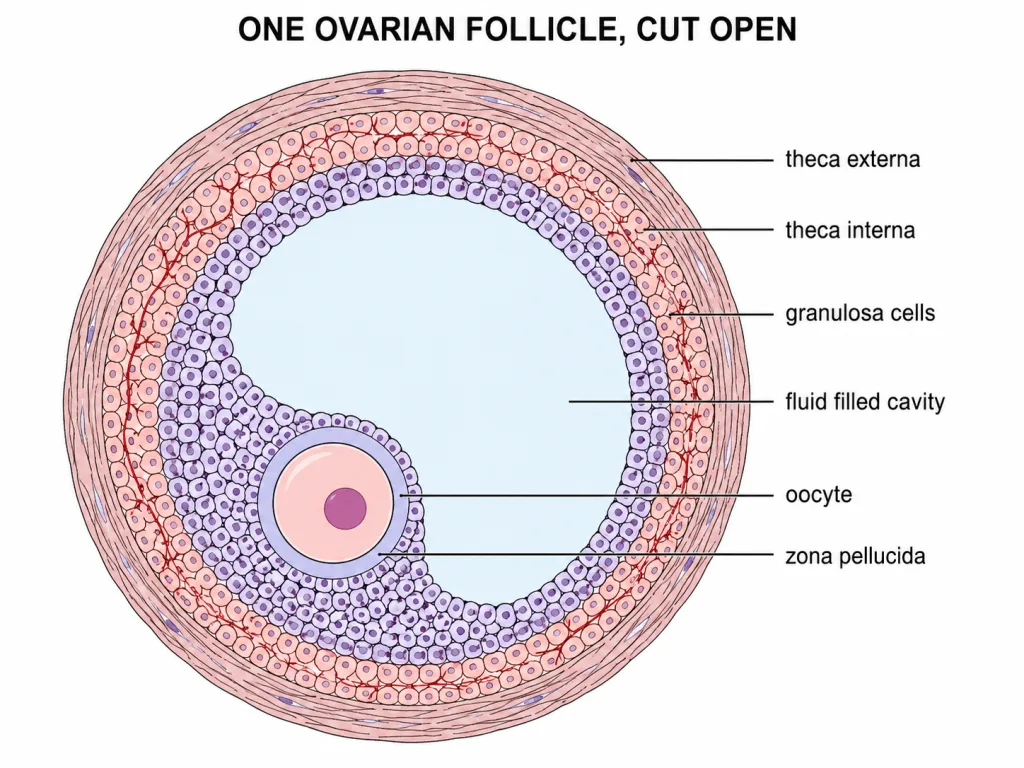
The earliest stage is the primary follicle. Here the growing gamete is surrounded by a layer of granulosa cells. As growth continues the sac becomes a secondary follicle: more layers of granulosa cells are laid down, and a fresh outer sheath called the theca appears around them. The theca is always external to the granulosa layers, never internal to them, and this is worth fixing in memory because the reverse is a common slip.
The next stage, the tertiary follicle, is marked by two changes. First, a fluid-filled cavity called the antrum opens up within the mass of granulosa cells. Second, the theca resolves into two distinct divisions, an inner theca interna and an outer theca externa. The antrum is the feature that separates a tertiary follicle from a secondary follicle at a glance, with no counting of cell layers. Finally the tertiary follicle changes into the mature Graafian follicle.
[NEET Important] Do not confuse the follicle with the cell inside it. The follicle series runs primary, secondary, tertiary, Graafian. The cell inside is the oocyte, and it has its own separate story. Mixing the two sets of names is the commonest error on this topic.
The Oocyte and the Unequal Division
As the follicle develops, the oocyte becomes surrounded by a new membrane of its own, the zona pellucida. This lies directly on the surface of the oocyte, inside the granulosa layers, so working outwards from the centre the arrangement runs oocyte, zona pellucida, granulosa cells, theca.
While the follicle is completing its change into the mature Graafian follicle, the primary oocyte inside it completes its first meiotic division. That division is remarkable because it is grossly unequal. One product, the secondary oocyte, is large and retains almost the whole of the nutrient-rich cytoplasm of the parent cell. The other, the first polar body, is tiny and carries little more than a nucleus. Both are haploid, so the reduction in chromosome number has happened in the ordinary way; it is only the cytoplasm that is shared out so unevenly. That hoarded cytoplasm is what will later support the earliest stages of development, and it is the reason the arrangement exists at all.
Ovulation, Menarche and Menopause
In human females a single ovum is released from one of the two ovaries near the middle of each menstrual cycle. Two points matter here: the number is one, not one from each ovary, and the timing is mid-cycle rather than at either end.
The menstrual cycle is a reproductive cycle found in female primates - monkeys, apes and human beings. One cycle is completed in about 28 or 29 days. Non-primate mammals such as cows, sheep, rats, deers, dogs and tigers do not show this cycle at all; they show an oestrus cycle instead. So the sorting rule is a taxonomic one, and it cannot be worked out from the size or the habitat of the animal.
The first menstruation begins at puberty and is called menarche. At the other end of reproductive life the cycles cease at around fifty years of age, and that is termed menopause.
[NEET Important] Lack of menstruation may be an indication of pregnancy, but it may also be caused by stress, poor health and other underlying causes. The hedge is part of the fact. Treating an absent flow as certain proof of pregnancy is factually wrong.
The Opening Phases of the Cycle
The cycle begins with the menstrual phase. Menstrual flow occurs in this phase and lasts for three to five days. The flow results from the breakdown of the endometrial lining of the uterus and of its blood vessels, which forms a liquid that comes out through the vagina. Note that three to five days is the length of the flow, not the length of the cycle; the two figures are frequently swapped.
The menstrual phase is followed by the follicular phase, also called the proliferative phase, and its defining feature is that two organs change at once. In the ovary, the primary follicles grow to become a fully mature Graafian follicle. At the same time, in the uterus, the endometrium regenerates through proliferation. A description that mentions only the ovarian half of this is incomplete.
These paired changes are induced by changes in the levels of pituitary and ovarian hormones. The secretion of the gonadotrophins, LH and FSH, increases gradually during the follicular phase. FSH stimulates follicular development, while the growing follicle itself secretes estrogens, and it is the estrogens that bring about the proliferative changes in the uterine lining. Keep the two sources distinct: the gonadotrophins come from the pituitary, the estrogens from the ovary itself. Sorting a hormone by its source, and not merely recognising its name, is the habit this part of the chapter rewards.
The Journey to the Site of Fusion
During copulation semen is released by the penis into the vagina, and this act is called insemination. The motile sperms then swim rapidly, pass through the cervix, enter the uterus and finally reach the junction of the isthmus and the ampulla of the fallopian tube. This ampullary-isthmic junction is where fusion takes place, and it is the only place in the tract where it normally does.
Getting there is not enough. Fusion can occur only if the ovum and the sperms are transported to that junction at the same time, and this is why not all acts of copulation lead to fertilisation and pregnancy.
[NEET Important] Learn the simultaneity condition, not just the site.
Fusion Itself
Fertilisation is the process of fusion of a sperm with an ovum. A sperm first comes in contact with the zona pellucida layer of the ovum. Three separate things then follow.
First, entry. The secretions of the acrosome help the sperm to enter through the zona pellucida and through the plasma membrane of the ovum. The work of getting in is chemical, done by enzymes released from the front of the head, not by the beating of the tail.
Second, exclusion. Contact induces changes in the membrane of the ovum that block the entry of additional sperms, ensuring that only one sperm can fertilise an ovum.
Third, completion. Those same secretions induce completion of the meiotic division of the secondary oocyte, which results in a second polar body and a haploid ovum, or ootid. Soon after, the haploid nucleus of the sperm and that of the ovum fuse to form a diploid zygote: n plus n gives 2n.
Which Gamete Decides the Sex
All ova carry one X chromosome besides 22 autosomes. Of the sperms, 50 per cent carry X and the other 50 per cent carry Y. After fusion the zygote therefore carries either XX or XY, depending on whether the sperm carrying X or the sperm carrying Y fertilised the ovum. It is thus the genetic makeup of the sperm that determines the sex of the embryo.
[NEET Important] Do not convert this into a genetics problem. The examinable point is the 50:50 split of the sperms and the paternal determination that follows from it.
Cleavage, Morula and Blastocyst
Mitotic division starts as the zygote moves through the isthmus of the oviduct towards the uterus, and this process is called cleavage. It yields 2, 4, 8 and 16 daughter cells, which are called blastomeres. The embryo holding 8 to 16 blastomeres is a morula, a compact solid ball with no cavity in it.
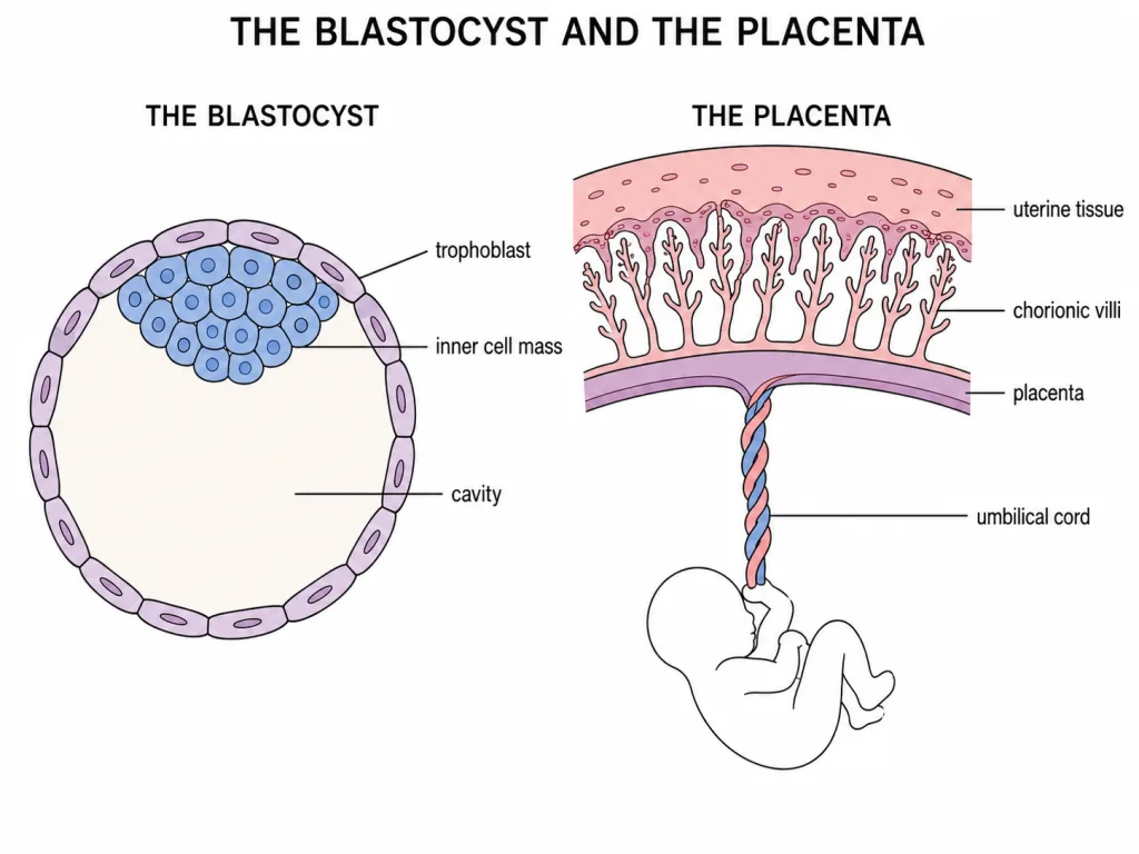
The morula continues to divide and is transformed into a blastocyst as it moves further into the uterus. Its blastomeres are now arranged into two populations: an outer layer called the trophoblast, and an inner group of cells attached to it called the inner cell mass. Their fates differ and are commonly swapped. The trophoblast gets attached to the endometrium; the inner cell mass gets differentiated as the embryo.
Implantation and the Placenta
After the trophoblast has attached, the uterine cells divide rapidly and cover the blastocyst, so that the blastocyst comes to lie embedded in the endometrium. This is implantation, and it leads to pregnancy. The active covering work is done by the mother's tissue.
After implantation, finger-like projections called chorionic villi appear on the trophoblast. These are surrounded by the uterine tissue and by maternal blood. The chorionic villi and the uterine tissue become interdigitated with each other and jointly form a structural and functional unit between the developing embryo and the maternal body, called the placenta. Jointness is the point. It facilitates supply of oxygen and nutrients to the embryo and removal of carbon dioxide and excretory material, and it is connected to the embryo by an umbilical cord.
The placenta also acts as an endocrine tissue and produces hCG, hPL, estrogens and progestogens. In the later phase of pregnancy a hormone called relaxin is also secreted, and its source is the ovary, not the placenta. hCG, hPL and relaxin are produced in women only during pregnancy. Separately, the levels of estrogens, progestogens, cortisol, prolactin and thyroxine are increased several-fold in maternal blood; these support foetal growth, the metabolic changes in the mother and the maintenance of pregnancy. The average duration of pregnancy is about nine months and is called the gestation period.
Parturition and Lactation
Parturition is the delivery of the foetus and is induced by a complex neuroendocrine mechanism. Signals originate from the fully developed foetus and from the placenta and induce mild uterine contractions, the foetal ejection reflex. This triggers release of oxytocin from the maternal pituitary; oxytocin acts on the uterine muscle to cause stronger contractions, which in turn stimulate further secretion of oxytocin. The loop feeds itself and the baby is expelled from the uterus.
The mammary glands undergo differentiation during pregnancy and start producing milk towards the end of pregnancy, by the process called lactation. Milk produced during the initial few days of lactation is called colostrum, and it contains several antibodies that are absolutely essential for developing resistance in the new-born. Breast-feeding during the initial period of infant growth is recommended by doctors for bringing up a healthy baby.