
The Male Reproductive System at a Glance
The human male reproductive system is located in the pelvis region of the body. It is not a single organ but a set of parts working together: a pair of testes, the accessory ducts that lead away from them, the accessory glands that add fluid along the way, and the external genitalia. Learning the system as these four groups makes every later detail easier to place, because each named structure belongs to exactly one group.
[NEET Important] The gonads of the male are a pair. A description that builds the system around a single gonad is wrong, however accurate the rest of the sentence may be.
The Scrotum and the Testis
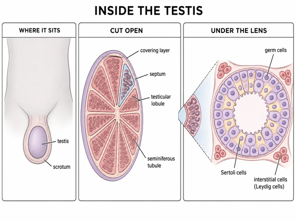
The testes are not held inside the abdominal cavity. They are carried in the scrotum, a pouch of skin that hangs outside the body wall. This position is not an accident of design. The scrotum keeps the temperature of the testes about 2 to 2.5 degrees C lower than the normal internal body temperature, and that cooler setting is necessary for the production of sperms. Both the direction of the difference and its size are stated facts about the scrotum, and neither can be guessed from the other.
Each testis is an oval body. In the adult it is about 4 to 5 cm in length and about 2 to 3 cm in width, and each one occupies its own scrotal sac. Note that the larger measurement belongs to the long axis; a testis is longer than it is broad.
Inside the Testis: Lobules, Tubules and Cells
A testis is not uniform inside. It is divided into about 250 compartments called testicular lobules. Each lobule is small, and it contains one to three highly coiled seminiferous tubules. These two figures travel together and are worth memorising as a pair: about 250 lobules in one testis, and one to three tubules in every lobule.
The inner side of each seminiferous tubule is lined by two quite different populations of cells. The first is the male germ cell line, the spermatogonia. The second is the Sertoli cells, and their stated job is to provide nutrition to the germ cells lying against them. A Sertoli cell is therefore a supporting and feeding partner rather than a hormone producing cell.
The regions outside the seminiferous tubules are called the interstitial spaces. They are not empty. They contain small blood vessels and the interstitial cells, which are also known as the Leydig cells. These cells synthesise and secrete the testicular hormones, the androgens.
[NEET Important] Two cell names, two addresses. Sertoli cells lie inside the tubule with the germ cells; Leydig cells lie between the tubules in the interstitial space. Fixing the address settles a large part of this topic.
The Ducts of the Male Tract
The vasa efferentia leave the testis and open into the epididymis. The epididymis itself is not a free structure floating nearby; it lies along the posterior surface of each testis, applied closely to the back of the organ. Because the two names vasa efferentia and vas deferens look alike on the page, it helps to attach each one to a place: the vasa efferentia belong to the testis it leaves, while the vas deferens belongs to the journey that follows.
The epididymis leads on to the vas deferens. This tube does not simply run downward. It ascends into the abdomen and there loops over the urinary bladder before turning towards the midline.
The urethra completes the outward path. It originates from the urinary bladder and extends through the penis to its external opening, which is called the urethral meatus. A single urethra serves both the urinary and the reproductive roles in the male, and its origin therefore lies at the bladder and not at any structure sitting on the gonad.
The Penis and the Accessory Glands
The penis is the male external genitalia. It is made of a special tissue that helps in erection, and this facilitates insemination. Unlike the testes, it is a single median organ and not a paired one. Its enlarged end is called the glans penis, and a loose fold of skin known as the foreskin covers that end.
Three kinds of accessory gland complete the system, and their numbers are easy marks. There is a pair of seminal vesicles, a single prostate, and a pair of bulbourethral glands. Two of the three kinds are paired and one is unpaired. Their secretions together constitute the seminal plasma, which is rich in fructose, calcium and certain enzymes. The secretion of the bulbourethral glands also helps in the lubrication of the penis.
The male system rewards a habit rather than a list: for every structure, ask what it is made of, where it sits, and what it contributes. Anatomy learned as addresses stays put far longer than anatomy learned as vocabulary.
The Plan of the Female Reproductive System
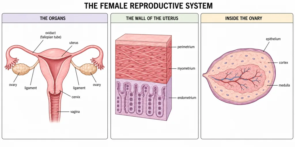
The female reproductive system lies in the pelvic region of the body. It is built from a pair of ovaries, a pair of oviducts, a uterus, a cervix, a vagina and the external genitalia. To this list one more organ must be added, and it is the one students most often leave out: the mammary glands. They are not merely glands of the skin. Structurally and functionally they are integrated with the reproductive system, and NCERT counts them as part of it.
[NEET Important] Two contrasts run right through this system and are worth fixing at the start. First, what is paired and what is single: the ovaries are paired, the oviducts are paired and the mammary glands are paired, but the uterus is one organ. Second, what is a producing organ and what is a conducting one: only the ovary makes anything, while the oviduct, the uterus and the vagina conduct.
The Ovary and Its Internal Zones
The ovaries are the primary female sex organs. Each is about 2 to 4 cm in length and lies one on either side of the lower abdomen. Each is anchored by ligaments running to the pelvic wall and to the uterus, so it is held in position rather than lying loose. Its role is a double one, and the doubleness is the point: the ovary produces the female gamete, the ovum, and it also secretes several steroid ovarian hormones.
Microscopic anatomy of the ovary is named in the syllabus, so it must be learnt properly. Each ovary is covered on its surface by a thin epithelium. That covering encloses the ovarian stroma, which is the substance of the organ. The stroma is not uniform. It is divided into a peripheral cortex and an inner medulla. The order to memorise is cortex outside, medulla inside. Reversing the two is the commonest error made about ovarian structure.
From the Ovary to the Uterus
Each oviduct, also called the fallopian tube, is about 10 to 12 cm long. It extends from the periphery of each ovary to the uterus, so the tube has one end near the site where the gamete is set free and the other end at the organ that will receive it. The tube does not touch the ovary directly. Its funnel-shaped end, the infundibulum, carries a fringe of finger-like projections called fimbriae, and their job is to help in the collection of the ovum after ovulation.
The uterus is a single organ, also known as the womb. It is shaped like an inverted pear and is supported by ligaments attached to the pelvic wall. At its lower end it opens into the vagina through a narrow opening called the cervix. Treat the cervix as a landmark: it tells you where one organ ends and the next begins, and nothing more.
[NEET Important] The wall of the uterus has three layers, and each has both a position and a property. The perimetrium is the thin external membrane. The myometrium is the middle, thick layer of smooth muscle, and it exhibits strong contractions during delivery of the baby. The endometrium is the inner glandular layer that lines the uterine cavity and undergoes cyclic changes during the menstrual cycle. Learn them as an ordered set from outside inwards, because questions built on these three usually give you the property and ask for the position, or the reverse.
The External Genitalia
The mons pubis is a cushion of fatty tissue covered by skin and hair. The labia majora are fleshy folds of tissue which extend down from the mons pubis and surround the vaginal opening. The labia minora are a pair of fleshy folds of tissue lying under the labia majora. So the sequence outwards to inwards is mons pubis, then majora, then minora, and the majora-outside, minora-under relation is what most items turn on.
The hymen is a membrane that partially covers the opening of the vagina. It is often torn during the first coitus, but it can also be broken by a sudden fall or jolt, by vigorous physical activity or by an inserted object, and in some women it persists even after coitus. For that reason the presence or absence of the hymen is not a reliable indicator of virginity or of sexual experience. The clitoris is a tiny finger-like structure that lies at the upper junction of the two labia minora, above the urethral opening.
The Mammary Glands
A pair of mammary glands is present, each containing glandular tissue along with a variable amount of fat. The glandular tissue of each breast is divided into 15 to 20 mammary lobes, and each lobe contains clusters of cells called alveoli. The cells of the alveoli secrete milk, which is stored in the cavities, or lumens, of the alveoli themselves. From there the milk moves along a fixed chain. The alveoli open into mammary tubules; the tubules of each lobe join to form a mammary duct; several mammary ducts join to form a wider mammary ampulla; and the ampulla is connected to a lactiferous duct through which the milk is sucked out.