
Nucleic Acids and Nucleotides
DNA (deoxyribonucleic acid) and RNA (ribonucleic acid) are nucleic acids - long polymers of nucleotides. DNA is the genetic material in most organisms; some viruses use RNA.
The building blocks
A nucleotide has three parts:
- A nitrogenous base,
- A pentose sugar - deoxyribose in DNA, ribose in RNA, and
- A phosphate group.
The nitrogenous bases are of two kinds:
- Purines (double-ring): Adenine (A) and Guanine (G).
- Pyrimidines (single-ring): Cytosine (C), Thymine (T) (only in DNA) and Uracil (U) (only in RNA, replacing thymine).
A base + sugar (without phosphate) is a nucleoside; adding the phosphate makes a nucleotide.
The polynucleotide chain
Nucleotides are joined by phosphodiester bonds between the phosphate of one and the 3'-OH of the sugar of the next, giving a sugar-phosphate backbone. A strand has a free 5'-phosphate end and a free 3'-OH end, so it has a defined polarity (5' to 3').
One-liners: nucleotide = base + sugar + phosphate; nucleoside = base + sugar (no phosphate); purines = A, G; pyrimidines = C, T (DNA), U (RNA); strands joined by phosphodiester bonds; each strand has 5' and 3' ends.
The DNA Double Helix (Watson and Crick, 1953)
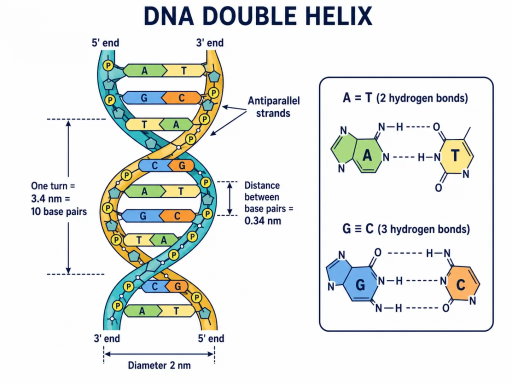
James Watson and Francis Crick proposed the double-helix model of DNA, building on the X-ray diffraction data of Maurice Wilkins and Rosalind Franklin. Its main features:
- Two polynucleotide strands coiled into a right-handed double helix, with the sugar-phosphate backbones outside and the bases pairing in the middle.
- The two strands are antiparallel - one runs 5' to 3' and the other 3' to 5'.
- Complementary base pairing by hydrogen bonds: adenine pairs with thymine (A=T) by two hydrogen bonds, and guanine pairs with cytosine (G≡C) by three hydrogen bonds. A purine always pairs with a pyrimidine, keeping the helix a uniform width.
Chargaff's rules and dimensions
- Chargaff's rules: in any double-stranded DNA the amount of A equals T and G equals C, so purines = pyrimidines (A + G = T + C).
- Dimensions: the helix is about 2 nm in diameter; one complete turn is 3.4 nm (34 angstrom) long and contains 10 base pairs, so adjacent base pairs are 0.34 nm apart.
One-liners: Watson-Crick 1953, X-ray by Wilkins & Franklin; strands antiparallel, right-handed; A=T (2 H-bonds), G≡C (3 H-bonds); Chargaff: A=T, G=C; one turn = 3.4 nm = 10 bp; 0.34 nm per bp; diameter 2 nm.
Packaging of DNA
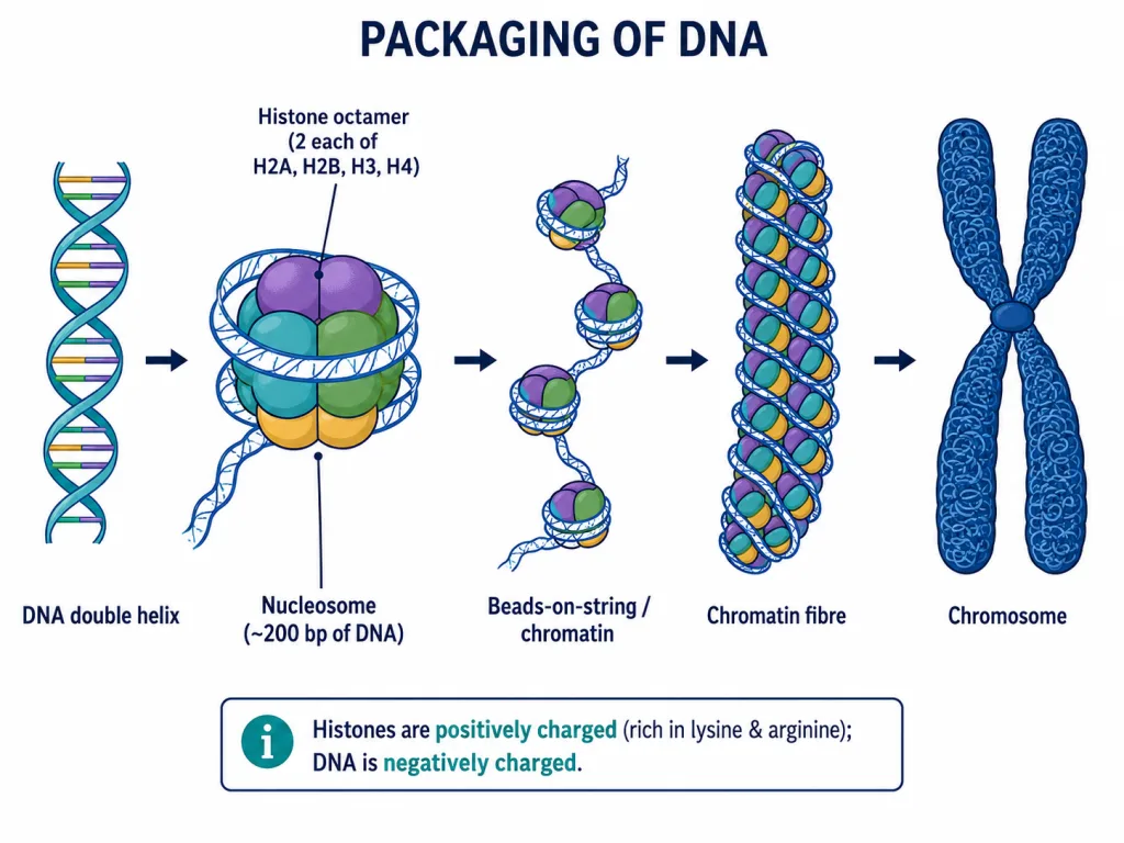
The DNA of a cell is enormously long (about 2.2 metres in a human cell) and must be packed into a tiny nucleus. Because DNA is negatively charged (phosphate groups), it is packaged with positively charged proteins.
In prokaryotes
E. coli has no nucleus; its DNA (held with some positively charged proteins) is organised into large loops in a region called the nucleoid.
In eukaryotes
- The positively charged basic proteins are histones, which are rich in the basic amino acids lysine and arginine.
- Eight histones (two each of H2A, H2B, H3 and H4) form a histone octamer; about 200 base pairs of DNA wrap around it to form a nucleosome.
- A chain of nucleosomes gives the 'beads-on-string' appearance, which coils further into the chromatin fibre and finally the chromosome.
Euchromatin vs heterochromatin
Chromatin is of two kinds: euchromatin is loosely packed, stains light and is transcriptionally active; heterochromatin is densely packed, stains dark and is inactive.
One-liners: DNA is negatively charged -> packed with histones (rich in lysine & arginine); histone octamer + ~200 bp = nucleosome; beads-on-string -> chromatin -> chromosome; euchromatin = loose, active; heterochromatin = dense, inactive.