
Two Layers of Defence
The body meets microbes with two layers of defence, and most questions turn on knowing which layer is doing the work. The first is innate immunity: inborn and non-specific, ready before the intruder arrives and treating every invader alike. The second is acquired immunity: built during life, specific to one pathogen at a time, and improving each time that pathogen returns.
The Kinds of Barrier
Each group is named for the agent doing the work, not for the tissue that agent comes from.
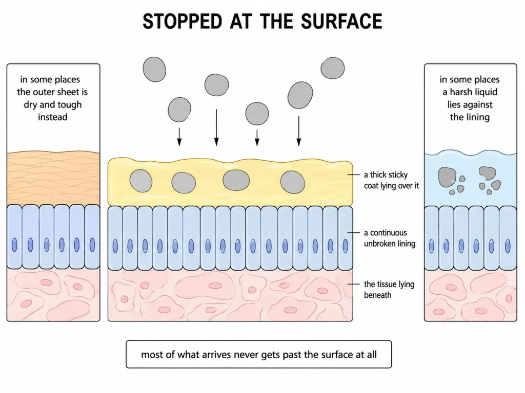
Physical. The skin covering the body is the main obstacle to entry. Alongside it, the mucus coating the epithelium lining the respiratory, gastrointestinal and urogenital tracts traps micro-organisms that get past the surface.
Physiological. The acid of the stomach, the saliva in the mouth and the tears from the eyes all prevent microbial growth.
Cellular. Certain leucocytes of the blood - polymorpho-nuclear leucocytes (neutrophils), monocytes and natural killer cells, a kind of lymphocyte - together with macrophages resident in the tissues, phagocytose and destroy microbes.
Cytokine. Cells that a virus has invaded secrete proteins called interferons. These do not rescue the cell that made them; they act on the healthy cells around it, putting them into a state that resists viral attack so the virus cannot spread.
[NEET Important] Each example belongs to exactly one group, and the group is named for the agent doing the work. The cytokine group is named for the protein that does the work, not for the cell that releases it.
The Second Layer: Specific and Remembering
Acquired immunity is pathogen specific and characterised by memory. The first encounter with a pathogen produces a response of low intensity, called the primary response. A later encounter with the same pathogen produces a highly intensified response, called the secondary or anamnestic response. That difference is what keeps you from falling ill twice with the same disease. The record of the first encounter is held by memory B and T cells, never by the phagocytic leucocytes of the first layer.
B lymphocytes produce an army of proteins in response to pathogens; those proteins are the antibodies that fight the pathogen. T lymphocytes do not themselves produce antibodies. What they do is help B lymphocytes to produce them, and run the other arm of the response in their own right.
Antibodies and Their Classes
An antibody molecule is built from four polypeptide chains, two light and two heavy, which is written H2L2, and disulfide bonds hold them together. Every chain carries a variable region at its N-terminal end and a constant region at its C-terminal end, and the site that binds the antigen is formed where the variable regions of a heavy chain and a light chain meet.
There are five classes and only five: IgA, IgD, IgE, IgG and IgM. Learn the roles with the names; the letters carry no clue. IgG is the most abundant antibody of the blood and the class that crosses the placenta to the foetus. IgA is the class abundant in colostrum. IgM is the first class to appear when a response begins. IgE is the class involved in allergic reactions.
Humoral and Cell-Mediated
Antibodies are found in the blood, so the antibody-based response is called the humoral immune response. The response mediated by T lymphocytes is called cell-mediated immunity. Underlying both is the body's ability to distinguish self from non-self. When an organ is grafted between unrelated persons the recipient's T lymphocytes act on the graft and destroy it, and graft rejection is the standard demonstration that this arm works in its own right. That is why tissue and blood group matching precede any transplant, and why the patient takes immuno-suppressants for life even after a good match.
[NEET Important] Do not read "humoral" and "cell-mediated" as rival theories. They are two arms of one system, running at the same time in the same person.
Getting Immunity: Two Routes
When a host is exposed to antigens - living microbes, dead microbes, or other proteins - antibodies are produced inside the host's own body. This is active immunity, and its drawback is that it takes time to become fully effective. The alternative is to give the body antibodies made elsewhere, ready to work at once. This is passive immunity.
A newborn lives on borrowed antibodies twice over. Antibodies pass from the mother to the foetus across the placenta during pregnancy, and the yellowish colostrum of the first days of lactation carries abundant IgA to the infant. Neither set is made by the infant, so both act from the moment they arrive.
Vaccination applies the first route deliberately. A preparation of antigenic proteins of a pathogen, or a weakened or inactivated pathogen, is introduced, and the body responds as it would to the pathogen itself - but without the disease. The response generates memory B and T cells, so a later infection meets an immediate massive response. A vaccine does not attack a pathogen already present; it prepares the body for one to come.
Where a fast response is essential and there is no time to build one, preformed antibodies, or antitoxin, are injected directly - in tetanus, and in snake bite, where the injection carries antibodies already raised against the venom in another host. Some vaccines are now produced by recombinant DNA technology: the antigenic polypeptides of a pathogen are made in bacteria or yeast, allowing large-scale production and greater availability. The hepatitis B vaccine produced in yeast is the standard example.
Lymphoid organs: two jobs, two categories
A lymphocyte has two separate needs, and the body builds two kinds of organ to meet them. Primary lymphoid organs - the bone marrow and the thymus - are where an immature lymphocyte becomes an antigen-sensitive, immunocompetent cell, able to recognise a foreign molecule long before it meets one. The bone marrow is the main lymphoid organ of all: every blood cell, lymphocytes included, is produced there. Secondary lymphoid organs - spleen, lymph nodes, tonsils, Peyer's patches and the appendix - are where that mature cell meets the antigen, is activated by it, and proliferates into an effector cell.
[NEET Important] Learn the two categories by the stage of lymphocyte life they serve, never by memorising which organ sits in which list.
Thymus, spleen, lymph nodes and the scattered tissue
The thymus is a lobed organ, sited near the heart and beneath the breastbone. Its behaviour with age is counter-intuitive and therefore examinable: it is quite large at the time of birth, shrinks as the years pass, and by puberty has been reduced to a very small size.
The spleen is a large bean-shaped organ, mainly containing lymphocytes and phagocytes. It sits in the path of the circulation and acts as a filter of the blood, trapping blood-borne micro-organisms, and holds a large reservoir of erythrocytes. Lymph nodes are small, solid structures placed at points along the lymphatic system, and they trap micro-organisms and other antigens that get into lymph and tissue fluid. An antigen held in a node activates the lymphocytes present there, and that activation is what produces the immune response.
[NEET Important] Fix the two fluids and most spleen-versus-node discriminations solve themselves. The spleen screens blood; a lymph node screens lymph and tissue fluid.
Mucosa-associated lymphoid tissue lies within the lining of the major tracts - respiratory, digestive and urogenital - and it holds about 50 per cent of the body's lymphoid tissue, though it forms no discrete organ. Tonsils, the appendix and the Peyer's patches of the small intestine are lymphoid tissue too.
When the immune system overreacts, and when it misfires
Allergy is the exaggerated response of the immune system to certain antigens present in the environment. The substances that provoke such a response are called allergens; mites in dust, pollens and animal dander are the everyday examples, and all of them come from outside the body, not from within. The antibodies produced against allergens are of the IgE type. The familiar symptoms - sneezing, watery eyes, a running nose, difficulty in breathing - are not produced by the allergen itself but by chemicals such as histamine and serotonin released from mast cells. Because the symptoms are chemically mediated, drugs like anti-histamine, adrenalin and steroids quickly reduce them. To find what a patient is reacting to, very small doses of possible allergens are given and the reactions studied. More and more children in the metro cities of India now suffer from allergies and asthma, and the protected environment provided early in life is the reason offered.
Autoimmunity is a different kind of failure altogether. Acquired immunity carries memory and normally distinguishes foreign molecules from the body's own cells. Sometimes, for genetic and other reasons that remain unknown, that distinction breaks down and the memory-based response is turned against self-cells. Notice carefully what has not failed. Antibody production is working perfectly well, and so is memory; the machinery is simply pointed at the wrong target. An auto-immune patient is not the same as an immune-deficient one.
Rheumatoid arthritis, in which the attack falls on the joints, is the standard example. Myasthenia gravis, in which it falls on the junction between nerve and muscle, and systemic lupus erythematosus, in which it is spread across many tissues at once, are two more standard examples. Two conditions of the same organs are not autoimmune at all: gout is a deposit of uric acid crystals in a joint, and muscular dystrophy is a genetic degeneration of muscle.
AIDS
AIDS stands for Acquired Immuno Deficiency Syndrome: a deficiency of the immune system, acquired rather than inherited, and presenting as a set of symptoms. The cause is the Human Immuno deficiency Virus, HIV, a retrovirus whose particle carries an RNA genome inside a protective protein coat. It was first reported in 1981.
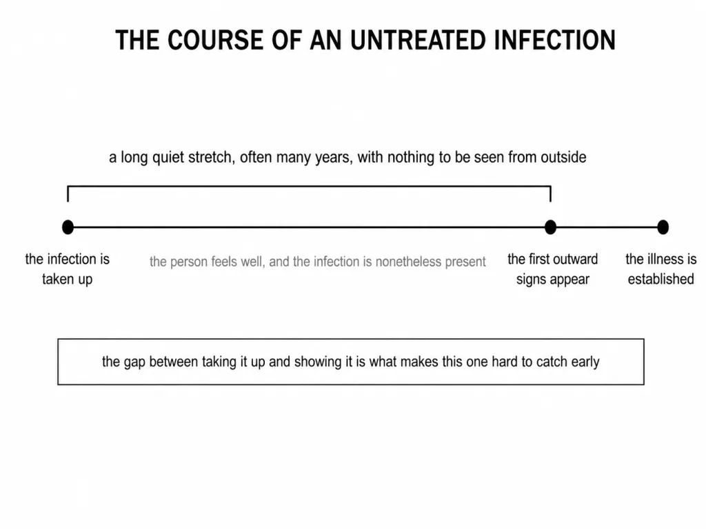
Inside the body the virus gets into macrophages, where the enzyme reverse transcriptase copies its RNA genome into DNA. That DNA enters the host cell's DNA and directs the infected cell to turn out virus particles, so the macrophage becomes an HIV factory. HIV also enters helper T-lymphocytes, replicates in them and destroys them, so their number falls progressively. The patient then suffers repeated bouts of fever, diarrhoea and weight loss, and becomes vulnerable to bacterial, viral, fungal and parasitic infections. The lag between infection and the appearance of symptoms runs from a few months to many years, which is why someone who looks entirely healthy may still be carrying and passing on the virus.
HIV travels in body fluids, by four routes: sexual contact with an infected person; transfusion of contaminated blood and blood products; sharing infected needles, as by intravenous drug abusers; and passage from an infected mother to her child through the placenta. It does not spread by touching, shaking hands, hugging, sharing food or living together, and no mosquito carries it. Diagnosis rests on ELISA, and anti-retroviral drugs are only partially effective. Control therefore rests on advocating safe sex, on making blood from blood banks safe, on disposable needles and syringes, on free distribution of condoms, on controlling drug abuse and on regular check-ups in susceptible populations, rather than on setting infected people apart.