
Structure of Actin and Myosin Filaments
Thin actin filament
Each thin filament contains two F-actin strands helically wound around each other. Every F-actin is a polymer of globular G-actin monomers. Two filaments of tropomyosin run close to the F-actin strands throughout their length. The complex protein troponin occurs at regular intervals on tropomyosin. In a resting muscle, one subunit of troponin masks the active binding sites for myosin on actin.
Thick myosin filament
A thick filament is a polymer of many monomeric proteins called meromyosins. Each meromyosin has a globular head with a short arm and a tail. The head and short arm together form heavy meromyosin (HMM), whereas the tail forms light meromyosin (LMM).
The HMM component projects out from the surface of the polymerised myosin filament at regular distances and angles as a cross arm. The globular head is an active ATPase enzyme and possesses binding sites for ATP as well as active sites for actin. These features prepare the thick filament for cross-bridge formation during contraction.
[NEET Important] Remember both contrasts: G-actin monomers polymerise into F-actin, while meromyosin units polymerise into the thick myosin filament; HMM is head plus short arm, and LMM is the tail.
From Neural Signal to Exposed Actin Sites
Skeletal-muscle contraction begins at the neuromuscular junction. A neural signal reaching the junction causes the motor-neuron ending to release acetylcholine. This neurotransmitter generates an action potential in the sarcolemma, and the electrical change spreads through the muscle fibre. It triggers the release of from the sarcoplasmic reticulum into the sarcoplasm.
The rise in sarcoplasmic is the switch that connects excitation with contraction. Calcium binds to a subunit of troponin on the actin filament. The troponin-tropomyosin arrangement changes, removing the mask from the active sites on actin. Energised myosin heads can now bind to those exposed sites.
[NEET Important] Keep the order exact: neural signal → acetylcholine release → action potential in sarcolemma → release → calcium binds troponin → active sites on actin become exposed. Calcium does not bind tropomyosin, and acetylcholine does not directly pull actin.
Sliding Filaments and the Cross-Bridge Cycle
The myosin head has an ATP-binding site and ATPase activity. Energy released by ATP hydrolysis energises, or cocks, the myosin head. The energised head binds an exposed active site on actin, forming a cross bridge. It then bends and pulls the attached thin filament towards the centre of the A band. This pulling step is the power stroke. As actin slides inward, the Z lines are drawn closer and the sarcomere shortens.
The filaments themselves do not become shorter; thin filaments slide past thick filaments. Therefore, during contraction, the I band becomes narrower and the H zone may narrow or disappear, while the length of the A band remains unchanged. This is the central prediction of the sliding-filament theory.
After the power stroke, the myosin head releases ADP and inorganic phosphate (). A fresh ATP molecule then binds to myosin and breaks the actin-myosin cross bridge. Hydrolysis of this ATP re-energises the head, allowing another cycle if the active sites remain exposed.
[NEET Trap] ATP has two distinct roles: ATP binding detaches myosin from actin, whereas ATP hydrolysis supplies energy to reset the head. Absence of ATP locks the cross bridges instead of releasing them, as seen in rigor mortis.
Relaxation, Fatigue and Motor Units
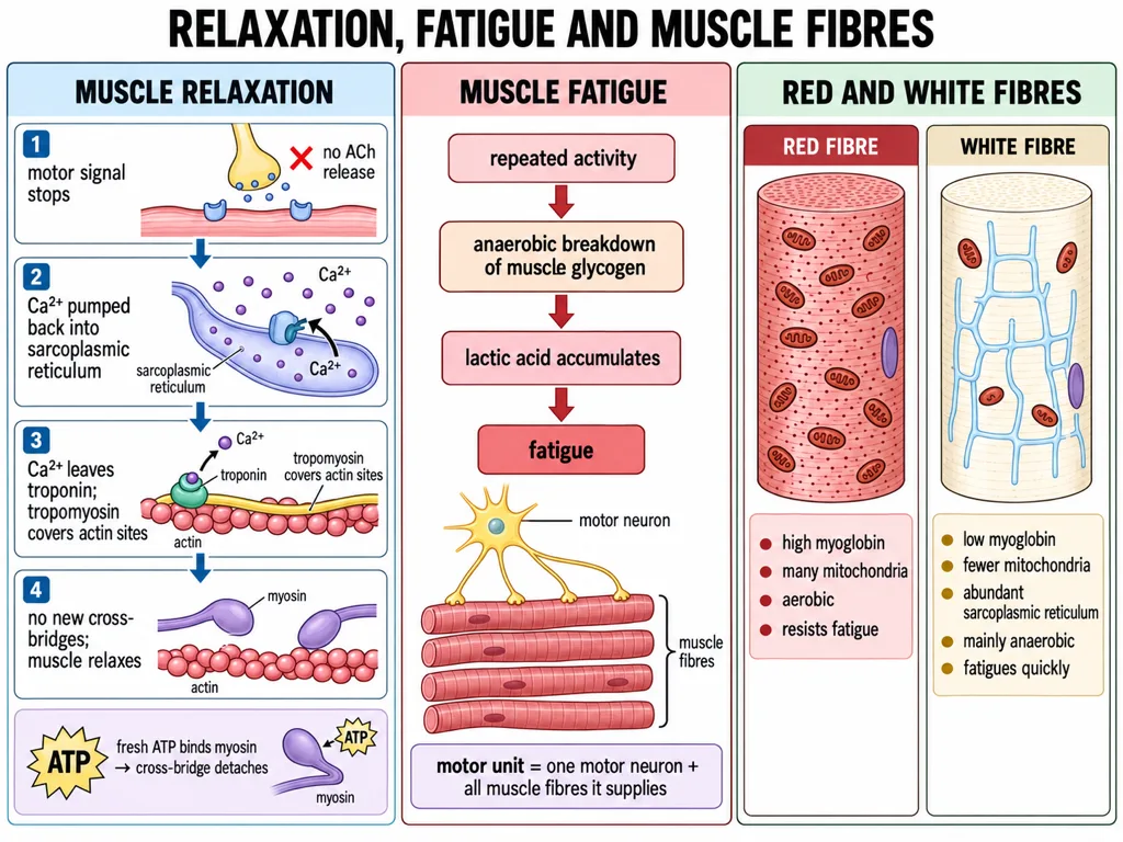
Cross-bridge cycling continues while stimulation maintains an elevated concentration in the sarcoplasm. When stimulation stops, calcium ions are actively pumped back into the sarcoplasmic reticulum. Sarcoplasmic calcium falls, troponin returns to its resting form, and tropomyosin again masks the myosin-binding sites on actin. New cross bridges cannot form, the Z lines return towards their original position, and the fibre relaxes.
Relaxation is therefore not simply the muscle running out of energy. Calcium reuptake is an active, ATP-dependent process. ATP is also needed to detach actin from myosin. This explains why ATP is necessary for both normal cycling and normal relaxation.
Repeated activation may lead to the accumulation of lactic acid due to anaerobic breakdown of glycogen in muscle; in the NCERT description, this causes muscle fatigue. Do not confuse fatigue with ordinary relaxation: relaxation follows removal of the stimulus, whereas fatigue is a decline in performance after sustained or repeated activity.
A motor neuron together with all the muscle fibres connected to it constitutes a motor unit. One motor neuron can therefore activate several fibres as a functional group.
Red and White Skeletal-Muscle Fibres
Red fibres contain abundant myoglobin, a red-coloured oxygen-storing pigment. They also contain many mitochondria and use large amounts of stored oxygen to produce ATP aerobically. This supports sustained activity and makes them relatively resistant to fatigue.
White fibres contain very little myoglobin and therefore appear pale. They have fewer mitochondria but an abundant sarcoplasmic reticulum. They depend mainly on anaerobic processes for energy, can support rapid forceful activity, and fatigue comparatively quickly as anaerobic products accumulate.
High-yield comparison
- Myoglobin: high in red fibres; low in white fibres.
- Mitochondria: numerous in red fibres; fewer in white fibres.
- Sarcoplasmic reticulum: especially abundant in white fibres.
- Major ATP pathway: aerobic in red fibres; predominantly anaerobic in white fibres.
- Fatigue resistance: higher in red fibres; lower in white fibres.
[NEET Important] Colour is determined primarily by myoglobin, not by mitochondria or glycogen. A white fibre can be poor in mitochondria yet rich in sarcoplasmic reticulum.
Skeletal-System Functions and Divisions
Bones and cartilages together constitute the skeletal system. Bone is hard because its matrix contains calcium salts, whereas cartilage has a more pliable matrix rich in chondroitin salts. In an adult human, 206 bones together with associated cartilages form the body framework.
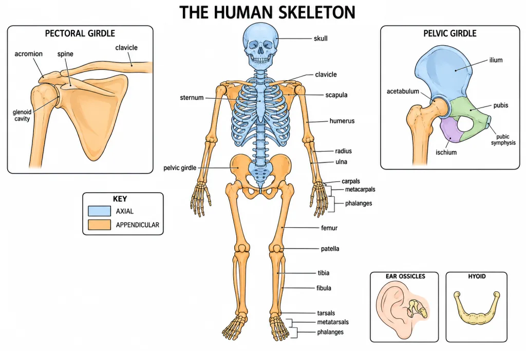
The skeleton supports the body, protects delicate organs, provides attachment to muscles and works with muscles and joints to produce movement. Bones also store minerals, especially calcium and phosphate, and bone marrow contributes to blood-cell formation.
The adult skeleton is divided into two unequal parts:
- Axial skeleton — 80 bones: skull, vertebral column, ribs and sternum, including the associated ear ossicles and hyoid in the standard count.
- Appendicular skeleton — 126 bones: the bones of the two forelimbs and two hindlimbs, plus the pectoral and pelvic girdles that attach them to the axial skeleton.
[NEET Important] Remember the complete check: 80 + 126 = 206. The appendicular skeleton has the larger share because four limbs alone contribute 120 bones.
Skull and Vertebral Column
The skull contains 22 bones: 8 cranial bones form the protective cranium, and 14 facial bones build the face. In addition, each middle ear has three ossicles — malleus, incus and stapes — giving 6 ossicles in all. A single U-shaped hyoid lies at the base of the buccal cavity. The hyoid and ear ossicles are counted separately from the 22 skull bones. The human skull articulates with the first cervical vertebra through two occipital condyles and is therefore dicondylic.
The adult vertebral column consists of 26 serially arranged units: 7 cervical, 12 thoracic, 5 lumbar, 1 sacral and 1 coccygeal. The sacral and coccygeal units represent groups of fused vertebrae in the adult. The first cervical vertebra is the atlas; it articulates with the occipital condyles. A neural canal runs through the vertebrae and encloses the spinal cord.
The vertebral column extends from the base of the skull, supports the head, protects the spinal cord and provides attachment for ribs and back muscles.
[NEET Trap] Do not mix the adult bone count of 26 with the developmental count of individual vertebrae before fusion. For NCERT's adult skeletal count, use 7 + 12 + 5 + 1 + 1 = 26.
Appendicular Skeleton: Girdles and Limbs
The pectoral girdle has two halves. Each half consists of a clavicle and a scapula, so both halves contribute 4 bones. The scapula is a large triangular flat bone on the dorsal thorax between the second and seventh ribs. Its spine expands into the acromion, which articulates with the clavicle. The glenoid cavity receives the head of the humerus.
Each forelimb has 30 bones: humerus 1, radius 1, ulna 1, carpals 8, metacarpals 5 and phalanges 14. Both forelimbs therefore contribute 60 bones.
The pelvic girdle consists of two coxal bones. Each coxal bone is formed by fusion of the ilium, ischium and pubis. Their fusion point bears the acetabulum, which receives the head of the femur. Ventrally, the two coxal bones meet at the pubic symphysis containing fibrous cartilage.
Each hindlimb also has 30 bones: femur 1, patella 1, tibia 1, fibula 1, tarsals 7, metatarsals 5 and phalanges 14. Both hindlimbs contribute 60 bones. Thus, 120 limb bones + 4 pectoral-girdle bones + 2 pelvic-girdle bones = 126 appendicular bones.
[NEET Important] The recurring count trap is 8 carpals versus 7 tarsals. Both hands and feet have 5 corresponding metacarpals/metatarsals and 14 phalanges.
Disorders of Joints and Bones
Arthritis
Arthritis is inflammation of joints. In a direct match-the-column question, do not add a different cause unless the question names a particular form of arthritis.
Osteoporosis
Osteoporosis is an age-related disorder characterised by decreased bone mass and an increased chance of fractures. Decreased levels of estrogen are a common cause, which is why the condition is especially important after menopause.
Gout
Gout is inflammation of joints caused by the accumulation of uric acid crystals. Keep the chemical clue specific: uric acid crystals belong to gout, not osteoporosis or general arthritis.
Rapid differential
- Myasthenia gravis: autoimmune neuromuscular junction disorder.
- Muscular dystrophy: progressive, usually genetic skeletal-muscle degeneration.
- Tetany: rapid muscle spasms due to low .
- Arthritis: inflammation of joints.
- Osteoporosis: reduced bone mass with greater fracture risk.
- Gout: uric acid crystal deposition causing joint inflammation.
[NEET Trap] Arthritis and gout both involve inflamed joints, but gout supplies the diagnostic crystal clue. Osteoporosis affects bone mass rather than producing a crystal deposit.