
Movement and Locomotion
Movement is a significant feature of living beings. It may involve a change in the position of a body part, as when we move the limbs, jaws, eyelids or tongue. Some movements produce a change in the place or location of the whole organism. Such voluntary movements are called locomotion. Walking, running, climbing, flying and swimming are familiar examples.
Therefore, every locomotory act is a movement, but every movement is not locomotion. Chewing and blinking involve movement without changing the organism's location. The same structure may also serve both locomotory and non-locomotory roles. In Paramoecium, cilia move food through the cytopharynx and also bring about locomotion. Hydra similarly uses its tentacles to capture prey as well as to locomote.
Animals locomote in response to their habitat and immediate needs, including finding food, shelter, mates, suitable breeding grounds or favourable climatic conditions, and escaping enemies or predators.
[NEET Important] Test the direction carefully: locomotion is a type of movement that changes location; movement of only a body part need not be locomotion.
Modes of Movement
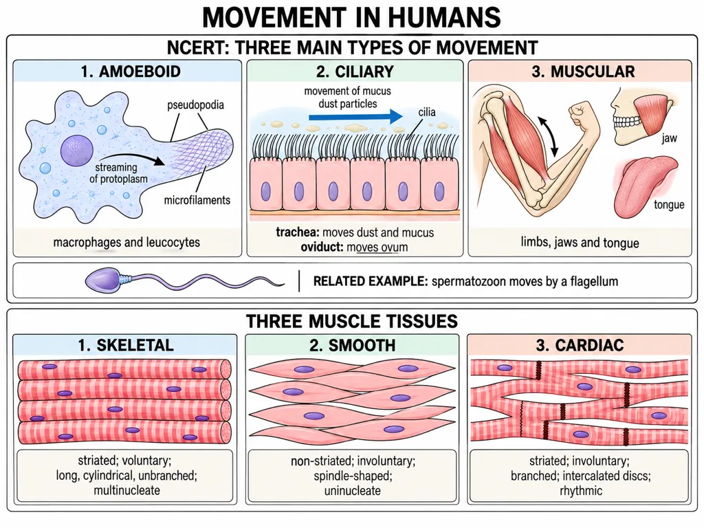
NCERT identifies three main types of movement in the cells of the human body: amoeboid, ciliary and muscular.
Amoeboid movement
Macrophages and leucocytes in blood show amoeboid movement. They form temporary cytoplasmic projections called pseudopodia, as in Amoeba. Cytoskeletal elements such as microfilaments participate in this movement.
Ciliary movement
Ciliary movement occurs in internal tubular organs lined by ciliated epithelium. Coordinated beating of cilia helps move the ovum through the female reproductive tract. In the trachea, cilia help remove dust particles and foreign substances carried in mucus.
Muscular movement
Movement of the limbs, jaws and tongue requires muscular movement. Locomotion in humans depends on coordinated activity of muscles, bones and neural signals.
Flagellar movement is a closely related cellular example: a human sperm is propelled by its flagellum. However, do not replace muscular movement with flagellar movement when asked for NCERT's three main types in human cells.
Syllabus–NCERT wording note: NCERT's chapter text groups amoeboid, ciliary and muscular movement, whereas the latest published official NEET-UG syllabus explicitly lists ciliary, flagellar and muscular movement. Learn both statements and their examples, and follow the source named in the question stem.
[NEET Important] Match each mechanism to its example: pseudopodia—leucocytes and macrophages; cilia—trachea and female reproductive tract; flagellum—spermatozoon; muscles—limbs, jaws and tongue.
Muscle Tissue and Its Properties
Muscle is a specialised tissue of mesodermal origin. About 40–50% of the body weight of a human adult is contributed by muscles. Muscle tissue consists of elongated cells called muscle fibres.
Muscles possess four important functional properties:
- Excitability: the ability to respond to a stimulus.
- Contractility: the ability to shorten and generate force.
- Extensibility: the ability to be stretched.
- Elasticity: the ability to return to the original resting length after being stretched.
Of these, contractility provides the force for muscular movement. Effective movement and locomotion require coordinated action of several muscles rather than an isolated contraction.
[NEET Important] The standard NCERT set is excitability, contractility, extensibility and elasticity. Conductivity is a tempting distractor but is not one of the four properties listed here.
Skeletal, Visceral and Cardiac Muscles
Muscles are identified as skeletal, visceral and cardiac according to their location and features.
Skeletal muscles
Skeletal muscles are closely associated with the skeletal components of the body. They show alternate light and dark bands, so they are striated. Their activity is generally under voluntary control, so they are also called voluntary muscles. They are primarily involved in locomotory actions and changes in body posture. The biceps is a typical example.
Visceral muscles
Visceral muscles occur in the inner walls of hollow visceral organs such as the alimentary canal and reproductive tract. They lack striations and appear smooth, so they are called smooth or non-striated muscles. Their activity is involuntary. They assist in transporting food through the digestive tract and gametes through the genital tract.
Cardiac muscles
Cardiac muscles form the muscular tissue of the heart. Cardiac muscle cells assemble in a branching pattern. They are striated in appearance but involuntary in action. This combination—striated and involuntary—is especially important for NEET.
Quick comparison: skeletal—striated and voluntary; visceral—non-striated and involuntary; cardiac—striated, branched and involuntary.
Organisation of a Skeletal Muscle
An organised skeletal muscle is built as a hierarchy. The whole muscle contains several muscle bundles called fascicles. A common collagenous connective tissue layer called fascia holds these bundles together. Each fascicle contains many muscle fibres, and each muscle fibre is a long muscle cell.
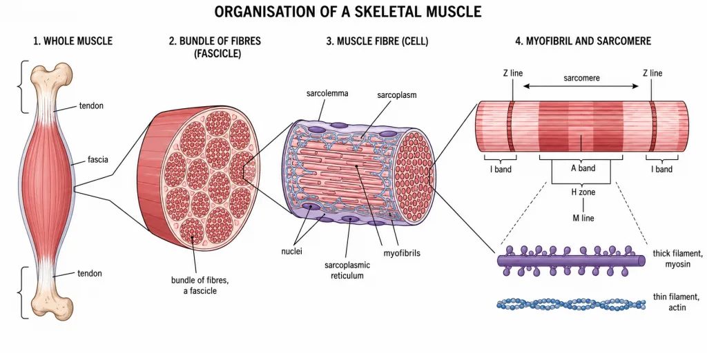
The plasma membrane of a muscle fibre is the sarcolemma, and the cytoplasm it encloses is the sarcoplasm. A skeletal muscle fibre is a syncytium because its sarcoplasm contains many nuclei. Its specialised endoplasmic reticulum, the sarcoplasmic reticulum, serves as a storehouse of calcium ions.
The sarcoplasm contains many parallel contractile structures called myofibrils; these are built from thin and thick myofilaments. The hierarchy from large to small is: whole muscle → fascicle → muscle fibre → myofibril → myofilaments.
[NEET Important] Do not confuse a muscle fibre with a myofibril. The fibre is the multinucleate cell; myofibrils are parallel contractile structures within it.
Striations, Bands and Lines
A myofibril displays alternating dark and light bands because actin and myosin filaments are arranged in an orderly pattern. The dark band is the A band or anisotropic band and contains the thick myosin filaments. The light band is the I band or isotropic band and contains thin actin filaments. Thin filaments also extend into part of the A band, producing an overlap with thick filaments.
Each I band is bisected by an elastic fibre called the Z line. Thin filaments are firmly attached to the Z line. At the centre of the A band, thick filaments are held together by a thin fibrous membrane called the M line.
A quick location map is useful:
- Z line: bisects the I band and anchors thin filaments.
- M line: lies in the middle of the A band and holds thick filaments together.
- A band: spans the length of the thick filament.
- I band: contains thin filaments without thick-filament overlap.
[NEET Important] The Z line and M line are structures, whereas A and I are bands. Questions often exchange their locations or the filament each anchors.
Sarcomere and the Resting Arrangement
The part of a myofibril between two successive Z lines is the sarcomere, the functional unit of contraction. A sarcomere contains one complete A band and half of an I band at each end. Its thin filaments project inward from the two Z lines, while thick filaments lie centrally around the M line.
In the resting state, the free ends of the thin filaments partly overlap the free ends of the thick filaments. However, they do not reach the very centre of the A band. This central portion, containing only thick filaments and no overlap with thin filaments, is the H zone. The M line passes through the centre of the H zone.
Visualise the resting sarcomere from one end to the other as: Z line → I-band half → overlap region of A band → H zone with M line → overlap region → I-band half → Z line.
[NEET Important] H zone means the non-overlap region of the A band; M line is the central fibrous line within that region. Do not treat them as synonyms.