
Neuron: the basic unit of the neural system
The neuron is the structural and functional unit of the neural system. It is an excitable cell specialised to receive a stimulus and transmit a neural signal. A nerve is different: it is a bundle of nerve fibres, whereas a neuron is a single cell. Keeping this cell-versus-bundle distinction clear prevents a common NEET error.
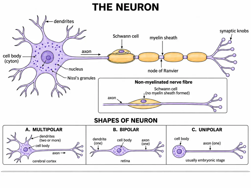
A typical neuron has three main regions: a cell body, several dendrites, and one axon. The cell body and dendrites mainly receive and integrate information; the axon carries the impulse away from the cell body towards another neuron, a muscle, or a gland.
Core map: dendrites → cell body → axon → terminal branches → synaptic knobs.
[NEET Important] Questions often exchange the properties of dendrites and the axon. Remember the contrast: dendrites are usually short, numerous, and repeatedly branched; the axon is single and generally long.
Cell body, dendrites, and axon
The cell body, also called the cyton, contains the nucleus, cytoplasm, and the usual cell organelles. Its cytoplasm also contains characteristic granular bodies called Nissl's granules. NCERT additionally states that dendrites contain Nissl's granules, so do not restrict them to the cell body in a statement-based question. They are not features of nodes of Ranvier or axon terminals.
Dendrites are short fibres that arise from the cell body and branch repeatedly. They carry impulses towards the cell body. Their extensive branching helps a neuron receive signals from several sources.
The axon is a long fibre whose distal end branches. Each terminal branch ends in a bulb-like synaptic knob containing synaptic vesicles filled with neurotransmitters. The axon carries impulses away from the cell body to a synapse or a neuromuscular junction. A node of Ranvier is not an axon ending; it is a gap in the myelin sheath along an axon.
[NEET Quick Check]
- Cyton: cell body containing Nissl's granules
- Dendrites: short, repeatedly branched, contain Nissl's granules, impulse towards cyton
- Axon: single and long, impulse away from cyton
- Synaptic knobs: bulb-like axon endings with neurotransmitter-filled vesicles
Myelinated and non-myelinated nerve fibres
In a myelinated nerve fibre, Schwann cells envelop the axon and form a myelin sheath around it. The sheath is interrupted between adjacent myelin-forming segments. These gaps are the nodes of Ranvier. NCERT identifies myelinated nerve fibres in spinal and cranial nerves.
A non-myelinated nerve fibre is not necessarily without a Schwann cell. A Schwann cell encloses the axon, but it does not form a myelin sheath around it. NCERT notes that such fibres are commonly found in the autonomic and somatic neural systems.
[NEET Important] Separate the cell from the material it forms: the Schwann cell encloses the axon; myelin is the sheath produced around a myelinated axon; the node of Ranvier is the gap between adjacent sheaths.
| Feature | Myelinated fibre | Non-myelinated fibre |
|---|---|---|
| Schwann cell | Envelops the axon | Encloses the axon |
| Myelin sheath | Present | Not formed |
| NCERT location cue | Spinal and cranial nerves | Autonomic and somatic neural systems |
Neuron types based on the number of processes
NCERT classifies neurons as multipolar, bipolar, and unipolar according to the number of axons and dendrites associated with the cell body.
A multipolar neuron has one axon and two or more dendrites. Its standard NCERT example is the cerebral cortex. A bipolar neuron has one axon and one dendrite and is represented by neurons of the retina. A unipolar neuron has a cell body with one axon only and is usually found in the embryonic stage.
| Neuron type | Processes stated by NCERT | Location cue |
|---|---|---|
| Multipolar | One axon, two or more dendrites | Cerebral cortex |
| Bipolar | One axon, one dendrite | Retina |
| Unipolar | Cell body with one axon only | Usually embryonic stage |
[NEET Memory Link] Multipolar–cortex, bipolar–retina, unipolar–embryo is a frequent match-the-column set. Do not infer the type from myelination: neuron shape and the presence of a myelin sheath are different classifications.
Organisation of the human neural system
The human neural system is divided into the central neural system (CNS) and the peripheral neural system (PNS). The CNS consists of the brain and spinal cord and is the site of information processing and control. The PNS comprises all the nerves of the body associated with the brain and spinal cord.
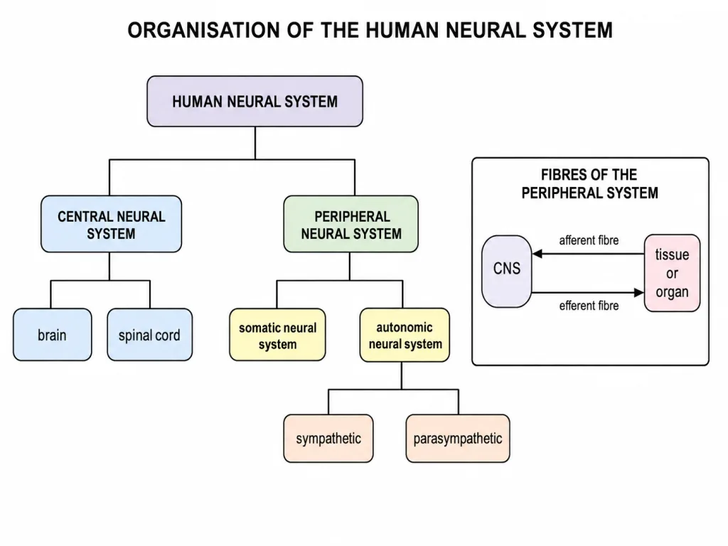
The spinal cord itself belongs to the CNS, but the nerves associated with or arising from the brain and spinal cord belong to the PNS. This boundary is frequently tested.
| Division | Components or description | Main role stated by NCERT |
|---|---|---|
| CNS | Brain and spinal cord | Information processing and control |
| PNS | All nerves associated with the brain and spinal cord | Connects the CNS with peripheral tissues and organs |
[NEET Important] Brain and spinal cord are central; cranial and spinal nerves are peripheral.
Afferent and efferent fibres: follow the direction
The nerve fibres of the PNS are of two types: afferent and efferent. Their names are determined by the direction in which they carry impulses relative to the CNS.
Afferent nerve fibres transmit impulses from tissues or organs to the CNS. They bring information towards the processing centre. Efferent nerve fibres transmit regulatory impulses from the CNS to peripheral tissues or organs. They carry instructions away from the processing centre.
A quick direction test is more reliable than memorising a long definition:
- Tissue or organ → CNS: afferent
- CNS → peripheral tissue or organ: efferent
[NEET Application] A signal travelling from a stimulated receptor towards the spinal cord is afferent. A command travelling from the spinal cord towards an effector is efferent. The destination, not whether the event feels voluntary, identifies the fibre in such questions.
Somatic and autonomic divisions of the PNS
The PNS is divided into the somatic neural system and the autonomic neural system. NCERT distinguishes them mainly by the peripheral structures to which impulses are relayed.
The somatic neural system relays impulses from the CNS to skeletal muscles. The autonomic neural system transmits impulses from the CNS to involuntary organs and smooth muscles of the body. The autonomic neural system is further classified into the sympathetic and parasympathetic neural systems.
| System | NCERT target cue |
|---|---|
| Somatic | Skeletal muscles |
| Autonomic | Involuntary organs and smooth muscles |
| Sympathetic and parasympathetic | Subdivisions of the autonomic neural system |
[NEET Important] Do not attach sympathetic and parasympathetic directly to the somatic system. They are subdivisions of the autonomic branch. Also, do not swap skeletal muscle with smooth muscle when matching targets.
Visceral nervous system
NCERT describes the visceral nervous system as the part of the PNS that comprises the whole complex of nerves, fibres, ganglia, and plexuses by which impulses travel from the CNS to the viscera and from the viscera to the CNS.
Two details make this definition highly testable. First, it belongs to the PNS, not the CNS. Second, its stated traffic is bidirectional: CNS → viscera and viscera → CNS. The four structural terms should be remembered as a fixed set: nerves, fibres, ganglia, and plexuses.
One-page hierarchy: Human neural system → CNS and PNS; PNS → somatic and autonomic; autonomic → sympathetic and parasympathetic. The visceral nervous system is described as the PNS complex connecting the CNS and viscera in both directions.
[NEET Precision] The visceral definition includes both incoming and outgoing paths. Therefore, do not reduce it to only efferent supply to smooth muscle, even though the autonomic system is described using an outward CNS-to-organ direction.
Ionic Distribution Across a Resting Axon
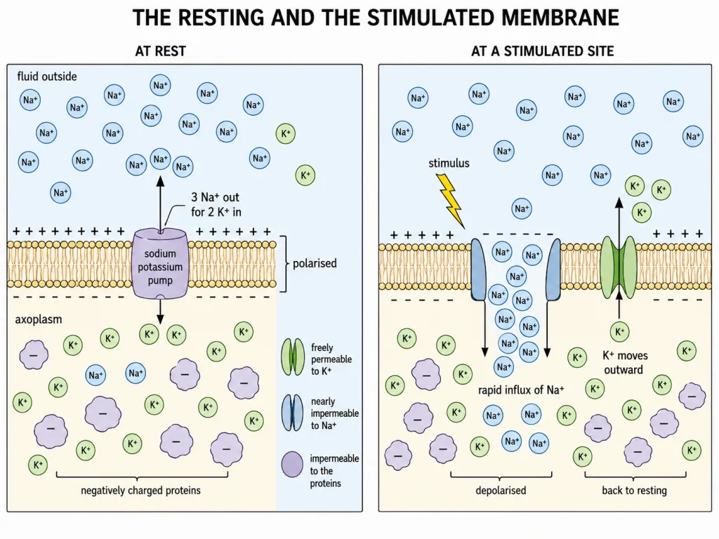
The axonal membrane separates the axoplasm from the extracellular fluid. In a resting neuron, the axoplasm contains a high concentration of K+, a low concentration of Na+ and negatively charged proteins. The extracellular fluid shows the reverse ionic pattern: Na+ is high and K+ is low.
The large negatively charged proteins remain inside because the resting membrane is impermeable to them. This unequal distribution is the starting condition for the resting membrane potential.
[NEET Important] Memorise the distribution as a pair: high K+ and negative proteins inside; high Na+ outside. Questions often preserve one half and reverse the other.
Selective Permeability and the Sodium-Potassium Pump
At rest, the axonal membrane is selectively permeable. It is more permeable to K+, nearly impermeable to Na+ and impermeable to the negatively charged intracellular proteins. Because the ions are unequally distributed and the membrane treats them differently, the two membrane surfaces carry different charges.
The sodium-potassium pump maintains the ionic gradients by transporting 3 Na+ out for every 2 K+ transported in. This active transport preserves low intracellular Na+ and high intracellular K+.
The resting membrane is therefore polarised: its outer surface is positively charged relative to the inner surface, which is negatively charged. The electrical potential difference across this resting membrane is the resting potential.
Quick recall: pump direction is Na+ out and K+ in; resting surface polarity is positive outside and negative inside.
Depolarisation and the Action Potential
When a stimulus reaches a site on the polarised membrane, that site becomes freely permeable to Na+. Because Na+ concentration is higher outside, Na+ rapidly enters the axoplasm. This influx reverses the local polarity: the outer surface becomes negatively charged and the inner surface becomes positively charged. The membrane at that site is now depolarised.
The electrical potential difference across the depolarised membrane is the action potential, also called a nerve impulse. Keep the cause-and-effect order clear: stimulation changes Na+ permeability first; Na+ influx follows; the influx reverses polarity; the reversed potential is the action potential.
[NEET Trap] The sodium-potassium pump does not suddenly reverse to generate an action potential. The immediate event is a short-lived rise in membrane permeability to Na+.
Repolarisation and Recovery
The increase in Na+ permeability is short lived. It is followed by an increase in permeability to K+, causing K+ to move out of the axon. This outward movement helps restore the original surface polarity, so the membrane repolarises and returns towards its resting state. The site can then become ready for another impulse.
One complete NEET sequence
- Resting membrane: high K+ inside, high Na+ outside; positive outside and negative inside.
- Stimulus: Na+ permeability rises sharply.
- Depolarisation: Na+ enters and local polarity reverses.
- Action potential: the potential across the depolarised site constitutes the nerve impulse.
- Repolarisation: K+ permeability rises and K+ moves outward.
- Gradient maintenance: the sodium-potassium pump continues to support the resting ionic gradients.
Do not confuse repolarisation with depolarisation: depolarisation is the reversal; repolarisation is the recovery.