
Propagation Along a Nerve Fibre
Generating an action potential at one point is only the beginning. For an impulse to travel, the depolarised region must stimulate the adjacent resting region. Suppose site A is depolarised while the neighbouring site B is still polarised. At A, the inner membrane surface is positive and the outer surface is negative; at B, the resting arrangement remains negative inside and positive outside.
This difference creates local currents. On the inner surface, current flows from A to B. On the outer surface, it flows from B to A, completing the circuit. The current makes site B permeable to Na+, so B becomes depolarised and a new action potential appears there. The same process repeats along the axon, allowing the impulse to move forward.
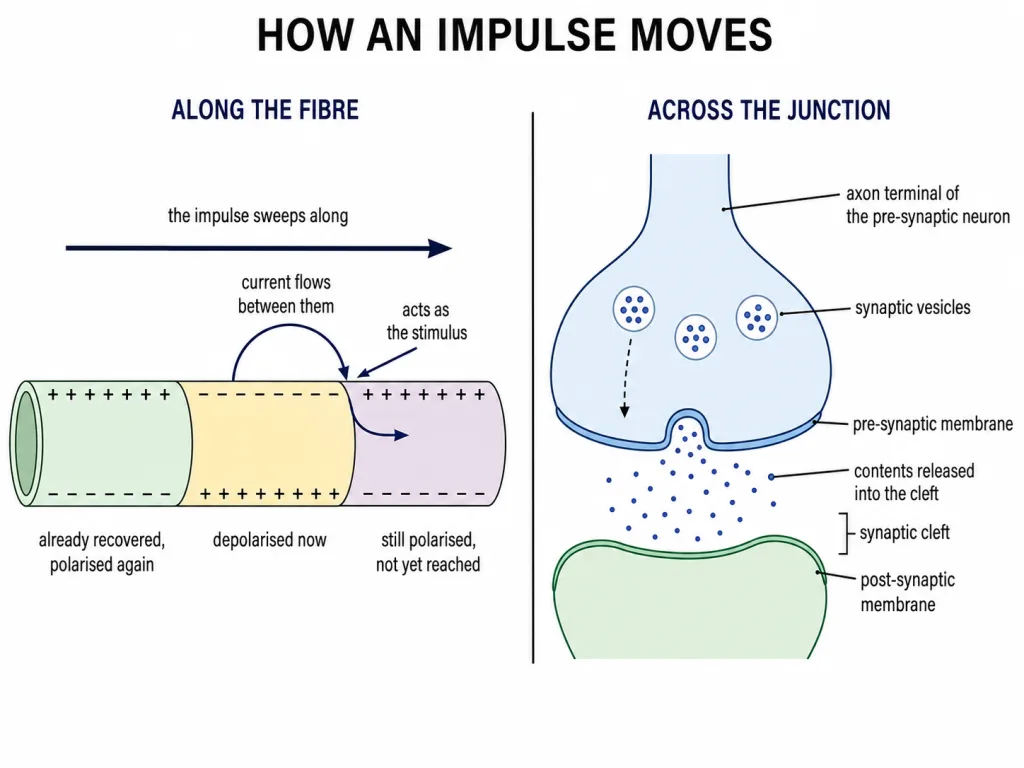
[NEET Important] Do not reverse the surface-current directions: inside A to B; outside B to A. The region behind the impulse repolarises, while the region ahead is depolarised next.
Synapses: The Junctions Between Neurons
A synapse is the junction formed by the membranes of a pre-synaptic neuron and a post-synaptic neuron. These membranes may or may not be separated by a gap, giving two functional types: electrical synapses and chemical synapses.
At an electrical synapse, the membranes of the two neurons lie in very close proximity. Electrical current can pass directly from one neuron to the next, so transmission resembles conduction along a single axon. It is always faster than transmission across a chemical synapse. NCERT also stresses that electrical synapses are rare in the human neural system.
[NEET Trap] A gap is not compulsory at every synapse. The conspicuous synaptic cleft belongs to a chemical synapse; electrical synapses have closely placed membranes that permit direct current flow.
Chemical Synaptic Transmission
In a chemical synapse, the pre-synaptic and post-synaptic membranes are separated by a fluid-filled synaptic cleft. Synaptic vesicles in the pre-synaptic terminal contain chemicals called neurotransmitters.
Follow the sequence carefully:
- An action potential reaches the pre-synaptic axon terminal.
- Synaptic vesicles move towards and fuse with the pre-synaptic membrane.
- Neurotransmitter is released into the synaptic cleft.
- It diffuses across the cleft and binds to specific receptors on the post-synaptic membrane.
- Receptor binding opens ion channels and produces a new potential in the post-synaptic neuron.
Transmission is from the pre-synaptic side to the post-synaptic side because transmitter-containing vesicles and transmitter receptors occupy different sides of the junction.
Excitation, Inhibition and High-Yield Comparisons
The new post-synaptic potential may be excitatory or inhibitory, depending on the nature of the neurotransmitter acting at that junction. An excitatory effect makes the post-synaptic neuron more likely to generate an impulse; an inhibitory effect makes it less likely. NCERT's essential test point is that chemical transmission does not always excite.
Electrical versus chemical synapse
- Path: direct current flow at an electrical synapse; neurotransmitter crosses a cleft at a chemical synapse.
- Speed: electrical transmission is faster.
- Structure: membranes are closely placed electrically; a fluid-filled cleft separates them chemically.
- Frequency in humans: electrical synapses are rare.
- Post-synaptic outcome: chemical transmission may produce an excitatory or inhibitory potential.
Quick recall: axon propagation uses local membrane currents; chemical synaptic transmission uses a neurotransmitter. Never treat these as the same mechanism.
Build the anatomical map first
The brain is the body's central information-processing organ and command-and-control system. It lies within the cranium and is covered by three cranial meninges. From outer to inner, their order is dura mater → arachnoid → pia mater. Pia mater lies closest to the brain surface.
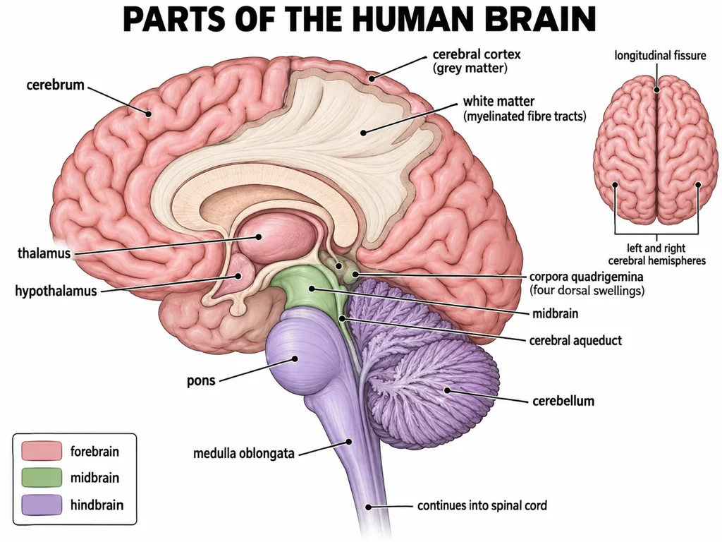
The three major divisions are the forebrain, midbrain and hindbrain. The forebrain includes the cerebrum, thalamus and hypothalamus. The hindbrain includes the pons, cerebellum and medulla oblongata. The brain stem is a different grouping: it consists of the midbrain, pons and medulla oblongata.
[NEET Important] Do not confuse a developmental division with the brain stem. The midbrain is itself a major division, but it also contributes to the brain stem; the pons and medulla belong to the hindbrain and also contribute to the brain stem; the cerebellum belongs to the hindbrain but not to the brain stem.
Forebrain: structure and function together
The cerebrum is the major part of the human brain. A deep longitudinal fissure separates its left and right hemispheres, whereas the corpus callosum, a tract of nerve fibres, connects the two hemispheres. The covering layer of neuronal cell bodies is the cerebral cortex or grey matter. Beneath it, tracts of myelinated nerve fibres form the white matter.
The cortex contains motor areas, sensory areas and association areas. Association areas support complex functions such as intersensory associations, memory and communication.
The cerebrum wraps around the thalamus, a major coordinating centre for sensory and motor signalling. The hypothalamus, located at the base of the thalamus, contains centres concerned with body temperature, hunger and thirst. It also contains neurosecretory cells that secrete hormones.
The inner parts of the cerebral hemispheres and associated deep structures such as the amygdala and hippocampus form the limbic lobe or limbic system. Together with the hypothalamus, this system participates in sexual behaviour, emotional reactions and motivation.
[NEET Trap] Longitudinal fissure separates, corpus callosum connects; cortex is grey matter outside, white matter lies beneath; thalamus coordinates signalling, while hypothalamus regulates homeostatic drives and links neural control with hormone secretion.
Midbrain and hindbrain: location-function pairs
The midbrain lies between the thalamus and hypothalamus of the forebrain and the pons of the hindbrain. The cerebral aqueduct passes through it. Its dorsal portion bears four round swellings called the corpora quadrigemina.
The pons consists mainly of fibre tracts that interconnect different regions of the brain. The cerebellum has a highly convoluted surface, providing additional space for many neurons, and is associated with posture and balance. The medulla oblongata continues into the spinal cord and contains centres controlling respiration, cardiovascular reflexes and gastric secretions.
[NEET Important] A reliable elimination map is: pons connects brain regions; cerebellum coordinates posture and balance; medulla regulates vital involuntary functions. Thermoregulation belongs to the hypothalamus, not the medulla.
How to solve integrated neural-control questions
Advanced questions usually combine a structural clue with a function. First identify the level: membrane, neuron, neural pathway or brain region. Then test direction and location before choosing a function. An afferent fibre carries input towards the central neural system; an efferent fibre carries output away from it. At a chemical synapse, the presynaptic terminal releases neurotransmitter and the postsynaptic membrane carries the specific receptors.
For lesion-style questions, use the most specific NCERT association. Disturbed posture and balance point to the cerebellum; altered body temperature, hunger or thirst point to the hypothalamus; disruption of respiration, cardiovascular reflexes or gastric secretion points to the medulla. A defect in interhemispheric connection points to the corpus callosum, not the longitudinal fissure.
[NEET Strategy] In statement questions, reject an option if even one membership, direction or location is wrong. Words such as outer, inner, dorsal, beneath, connects and continues are often the decisive clues.
NEET map 1: organisation and direction
Begin every pathway question by fixing the direction of information flow. A neuron's dendrites generally receive signals towards the cell body, while its axon conducts impulses away from the cell body to the terminal. Nissl's granules occur in the cell body and dendrites but not in the axon. In myelinated fibres, Schwann cells form the myelin sheath and adjacent sheaths are separated by nodes of Ranvier; impulses travel rapidly by jumping from node to node.
At the system level, the central neural system contains the brain and spinal cord. The peripheral neural system contains nerves associated with the central system. Afferent fibres carry impulses from tissues or organs towards the central system; efferent fibres carry regulatory impulses away from it. The somatic neural system carries impulses between the central system and skeletal muscles, while autonomic pathways regulate involuntary organs. The visceral neural system includes the nerves, fibres and ganglia connecting the central system with the viscera.
[NEET Trap] Afferent is arrival at the central system; efferent is exit from it. Somatic refers to skeletal-muscle control, whereas visceral or autonomic pathways concern internal organs.
NEET map 2: impulse and synapse sequence
At rest, the axonal membrane is more permeable to potassium ions and almost impermeable to sodium ions. Potassium concentration is higher inside and sodium concentration is higher outside. The sodium-potassium pump maintains these gradients by moving 3 sodium ions outward for every 2 potassium ions moved inward. The resting membrane is polarised: the outside is relatively positive and the inside relatively negative.
At a stimulated site, sodium permeability rises and sodium enters, reversing polarity and producing an action potential. This local reversal stimulates the adjacent membrane. Potassium movement outward then helps restore the resting polarity behind the advancing impulse. Do not credit the sodium-potassium pump with the rapid repolarising phase; the pump maintains the long-term gradients.
At an electrical synapse, closely placed membranes allow current to pass directly, so transmission is faster; such synapses are rare in humans. At a chemical synapse, an impulse reaches the presynaptic terminal, vesicles fuse, neurotransmitter enters the synaptic cleft, binds specific receptors on the postsynaptic membrane, opens ion channels and generates a new excitatory or inhibitory potential.
[NEET Trap] Presynaptic means vesicles and release; postsynaptic means receptors and response. Chemical transmission is slower and structurally one-way, whereas electrical transmission is direct and faster.
NEET map 3: brain structures that are repeatedly interchanged
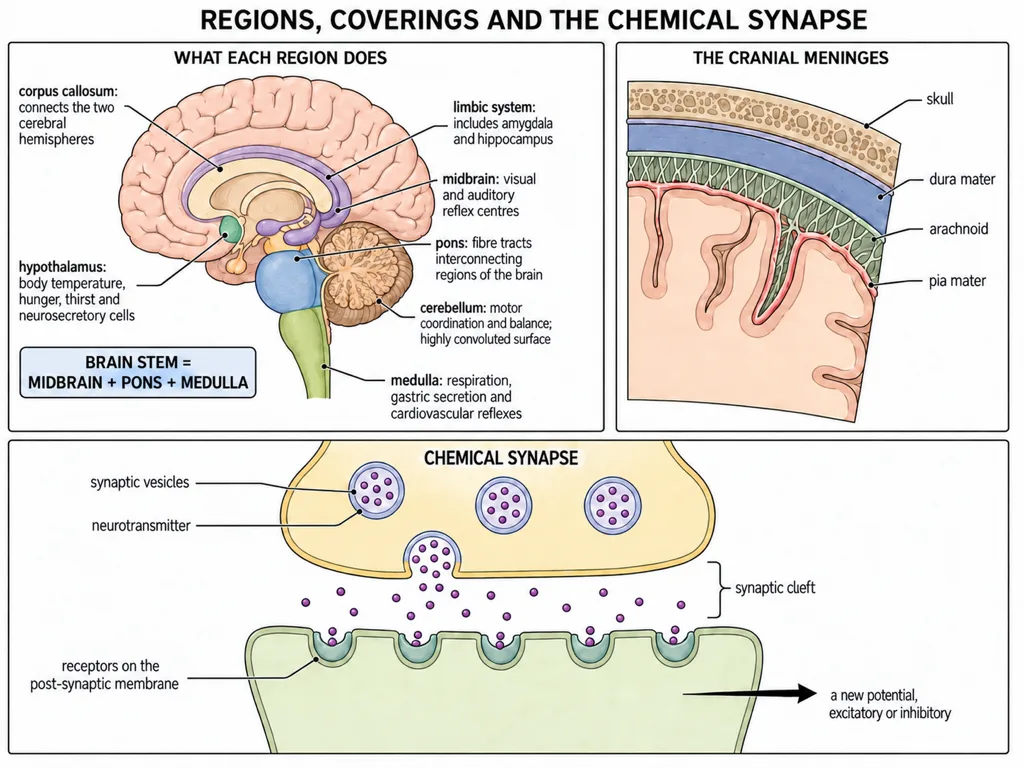
The forebrain contains the cerebrum, thalamus and hypothalamus. The cortex is outer grey matter; myelinated tracts beneath it form white matter. The longitudinal fissure separates the cerebral hemispheres.
The thalamus coordinates sensory and motor signalling.
The hypothalamus contains hunger and thirst centres plus neurosecretory cells. The limbic system with the hypothalamus participates in sexual behaviour, emotional reactions and motivation.
The midbrain carries the cerebral aqueduct and has four dorsal corpora quadrigemina. The midbrain, the pons and the medulla continue downward into the spinal cord.
[NEET Trap] Read every brain question by fixing the division first -- forebrain, midbrain or hindbrain -- and only then the structure, because most distractors move a structure into the wrong division rather than giving it the wrong job.