
The Chapter in One Read
According to the cell theory, cells arise from preexisting cells, and the process by which this occurs is called cell division. Any sexually reproducing organism starts its life cycle from a single-celled zygote, and cell division does not stop with the formation of the mature organism but continues throughout its life cycle. That is the whole reason this chapter exists.
The stages through which a cell passes from one division to the next is called the cell cycle. The cell cycle is divided into two phases - interphase, a period of preparation for cell division, and mitosis or M phase, the actual period of cell division. The split of time between the two is lopsided: cell division proper lasts for only about an hour of the 24 hour cycle of a human cell, and interphase lasts more than 95 per cent of the duration of the cell cycle. Interphase is further subdivided into , S and . phase is the period when the cell grows and carries out normal metabolism, and most of the organelle duplication also occurs during this phase. S phase marks the phase of DNA replication and chromosome duplication - the DNA per cell doubles from to while the chromosome number stays at . phase is the period of cytoplasmic growth. Cells that stop dividing exit the phase into , where they remain metabolically active but no longer proliferate.
Mitosis is also divided into four stages, namely prophase, metaphase, anaphase and telophase. Chromosome condensation occurs during prophase, and simultaneously the centrioles move to the opposite poles; the nuclear envelope and the nucleolus disappear and the spindle fibres start appearing. Metaphase is marked by the alignment of chromosomes at the equatorial plate, and it is here that the morphology of chromosomes is most easily studied, with spindle fibres attached to the kinetochores. During anaphase the centromeres divide and the chromatids start moving towards the two opposite poles. Once the chromatids reach the two poles, the chromosomal elongation starts, and the nucleolus and the nuclear membrane reappear - this stage is called the telophase. Nuclear division is then followed by the cytoplasmic division, and this is called cytokinesis - a furrow in an animal cell, a cell-plate growing from the centre outward in a plant cell. Mitosis, thus, is the equational division in which the chromosome number of the parent is conserved in the daughter cell.
In contrast to mitosis, meiosis occurs in the diploid cells which are destined to form gametes. It is called the reduction division since it reduces the chromosome number by half while making the gametes, and in sexual reproduction, when the two gametes fuse, the chromosome number is restored to the value in the parent. Meiosis is divided into two phases - meiosis I and meiosis II - on only a single cycle of DNA replication. In the first meiotic division the homologous chromosomes pair to form bivalents and undergo crossing over. Meiosis I has a long prophase, which is divided further into five phases - leptotene, zygotene, pachytene, diplotene and diakinesis. During metaphase I the bivalents arrange on the equatorial plate. This is followed by anaphase I, in which homologous chromosomes move to the opposite poles with both their chromatids, and each pole receives half the chromosome number of the parent cell. In telophase I the nuclear membrane and nucleolus reappear, giving a dyad. Meiosis II is similar to mitosis, and during anaphase II the sister chromatids separate. Thus at the end of meiosis four haploid cells are formed. Between the two divisions lies interkinesis, generally short lived, in which there is no replication of DNA.
The last idea is the one the whole chapter is built towards. Meiosis is the mechanism by which conservation of the specific chromosome number of each species is achieved across generations in sexually reproducing organisms, even though the process, per se, paradoxically, results in reduction of chromosome number by half. And meiosis also increases the genetic variability in the population of organisms from one generation to the next, which is very important for the process of evolution.
Master Quick Recap
The cell cycle
- Cell division is a very important process in all living organisms, and during the division of a cell, DNA replication and cell growth also take place, in a coordinated way to ensure correct division and formation of progeny cells containing intact genomes.
- The cell cycle is the sequence of events by which a cell duplicates its genome, synthesises the other constituents of the cell and eventually divides into two daughter cells.
- Cell growth, in terms of cytoplasmic increase, is a continuous process, but DNA synthesis occurs only during one specific stage in the cell cycle.
- The replicated chromosomes are distributed to daughter nuclei by a complex series of events during cell division, and these events are under genetic control.
- Human cells in culture divide once in approximately every 24 hours; yeast can progress through the cell cycle in only about 90 minutes.
- Two basic phases - interphase and M phase (mitosis phase). The M phase represents the phase when the actual cell division or mitosis occurs, and the interphase represents the phase between two successive M phases.
- Cell division proper lasts for only about an hour; the interphase lasts more than 95 per cent of the duration of the cell cycle.
- The M phase starts with the nuclear division, corresponding to the separation of daughter chromosomes - karyokinesis - and usually ends with division of the cytoplasm - cytokinesis.
- Interphase, though called the resting phase, is the time during which the cell is preparing for division by both cell growth and DNA replication in an orderly manner.
Interphase and the phase
- phase corresponds to the interval between mitosis and initiation of DNA replication. The cell is metabolically active and continuously grows but does not replicate its DNA.
- S phase or synthesis phase marks the period during which DNA synthesis or replication takes place. The amount of DNA per cell doubles - if the initial amount is it increases to - and there is no increase in the chromosome number, so a cell is still after S phase.
- In animal cells, during the S phase, DNA replication begins in the nucleus and the centriole duplicates in the cytoplasm.
- phase - proteins are synthesised in preparation for mitosis while cell growth continues.
- Some cells in adult animals do not appear to exhibit division, for example heart cells, and many others divide only occasionally to replace cells lost because of injury or cell death.
- These cells exit the phase to enter an inactive stage called the quiescent stage, or . Cells in this stage remain metabolically active but no longer proliferate unless called on to do so.
- In animals, mitotic cell division is only seen in the diploid somatic cells, with a few exceptions such as male honey bees; plants can show mitotic divisions in both haploid and diploid cells.
Mitosis - prophase and metaphase
- M phase is the most dramatic period of the cell cycle, involving a major reorganisation of virtually all components of the cell.
- Mitosis is called equational division because the number of chromosomes in the parent and progeny cells is the same.
- Cell division is a progressive process and very clear-cut lines cannot be drawn between the various stages.
- Karyokinesis has four stages - prophase, metaphase, anaphase and telophase.
- Prophase follows the S and phases, in which the new DNA molecules formed are not distinct but intertwined.
- Prophase is marked by the initiation of condensation of chromosomal material, which becomes untangled during chromatin condensation.
- Chromosomal material condenses to form compact mitotic chromosomes, seen to be composed of two chromatids attached together at the centromere.
- The centrosome, which had undergone duplication during interphase, begins to move towards opposite poles. Each centrosome radiates out microtubules called asters, and the two asters together with spindle fibres form the mitotic apparatus.
- At the end of prophase, cells do not show golgi complexes, endoplasmic reticulum, nucleolus and the nuclear envelope.
- The complete disintegration of the nuclear envelope marks the start of metaphase, so the chromosomes are spread through the cytoplasm.
- By metaphase, condensation of chromosomes is completed and this is the stage at which the morphology of chromosomes is most easily studied.
- Small disc-shaped structures at the surface of the centromeres are called kinetochores, and they serve as the sites of attachment of spindle fibres to the chromosomes.
- All the chromosomes come to lie at the equator, with one chromatid of each chromosome connected by its kinetochore to spindle fibres from one pole and its sister chromatid connected to fibres from the opposite pole.
- The plane of alignment of the chromosomes at metaphase is referred to as the metaphase plate.
Mitosis - anaphase, telophase and cytokinesis
- At the onset of anaphase, each chromosome arranged at the metaphase plate is split simultaneously, and the two daughter chromatids, now referred to as daughter chromosomes of the future daughter nuclei, begin their migration towards the two opposite poles.
- As each chromosome moves away from the equatorial plate, the centromere remains directed towards the pole and hence at the leading edge, with the arms of the chromosome trailing behind.
- Key events of anaphase - centromeres split and chromatids separate; chromatids move to opposite poles.
- At telophase, the chromosomes that have reached their respective poles decondense and lose their individuality, and each set of chromatin material tends to collect at each of the two poles.
- Key events of telophase - chromosomes cluster at opposite spindle poles and their identity is lost as discrete elements; a nuclear envelope develops around the chromosome clusters at each pole, forming two daughter nuclei; nucleolus, golgi complex and ER reform.
- Cytokinesis is the division of the cell into two daughter cells by the separation of cytoplasm, at the end of which cell division gets completed.
- In an animal cell, cytokinesis is achieved by the appearance of a furrow in the plasma membrane, which gradually deepens and ultimately joins in the centre, dividing the cell cytoplasm into two.
- Plant cells are enclosed by a relatively inextensible cell wall and therefore undergo cytokinesis by a different mechanism - wall formation starts in the centre of the cell and grows outward to meet the existing lateral walls.
- The new cell wall begins with a simple precursor called the cell-plate, which represents the middle lamella between the walls of two adjacent cells.
- At the time of cytoplasmic division, organelles like mitochondria and plastids get distributed between the two daughter cells.
- In some organisms karyokinesis is not followed by cytokinesis, giving a multinucleate condition called a syncytium - for example the liquid endosperm in coconut.
Significance of mitosis
- Mitosis, the equational division, is usually restricted to the diploid cells only, but in some lower plants and in some social insects, haploid cells also divide by mitosis.
- Mitosis usually results in the production of diploid daughter cells with identical genetic complement.
- The growth of multicellular organisms is due to mitosis.
- Cell growth results in disturbing the ratio between the nucleus and the cytoplasm, so it becomes essential for the cell to divide to restore the nucleo-cytoplasmic ratio.
- A very significant contribution of mitosis is cell repair - the cells of the upper layer of the epidermis, the cells of the lining of the gut and blood cells are being constantly replaced.
- Mitotic divisions in the meristematic tissues - the apical and the lateral cambium - result in a continuous growth of plants throughout their life.
Meiosis - why it exists, and its key features
- The production of offspring by sexual reproduction includes the fusion of two gametes, each with a complete haploid set of chromosomes, and gametes are formed from specialised diploid cells.
- Meiosis is the specialised kind of cell division that reduces the chromosome number by half and results in the production of haploid daughter cells.
- Meiosis ensures the production of the haploid phase in the life cycle of sexually reproducing organisms, whereas fertilisation restores the diploid phase.
- Meiosis is met with during gametogenesis in plants and animals, leading to the formation of haploid gametes.
- Key feature 1 - meiosis involves two sequential cycles of nuclear and cell division called meiosis I and meiosis II, but only a single cycle of DNA replication.
- Key feature 2 - meiosis I is initiated after the parental chromosomes have replicated to produce identical sister chromatids at the S phase.
- Key feature 3 - meiosis involves pairing of homologous chromosomes and recombination between non-sister chromatids of homologous chromosomes.
- Key feature 4 - four haploid cells are formed at the end of meiosis II.
Prophase I
- Prophase of the first meiotic division is typically longer and more complex when compared to prophase of mitosis, and is subdivided into leptotene, zygotene, pachytene, diplotene and diakinesis based on chromosomal behaviour.
- Leptotene - the chromosomes become gradually visible under the light microscope, and compaction continues throughout leptotene.
- Zygotene - chromosomes start pairing together, and this process of association is called synapsis; such paired chromosomes are called homologous chromosomes; synapsis is accompanied by the formation of a complex structure called the synaptonemal complex; the complex formed by a pair of synapsed homologous chromosomes is called a bivalent or a tetrad.
- Pachytene - the four chromatids of each bivalent chromosome become distinct and clearly appear as tetrads; recombination nodules appear, the sites at which crossing over occurs between non-sister chromatids of the homologous chromosomes.
- Crossing over is the exchange of genetic material between two homologous chromosomes, is enzyme-mediated, and the enzyme involved is called recombinase.
- Recombination between homologous chromosomes is completed by the end of pachytene, leaving the chromosomes linked at the sites of crossing over.
- Diplotene - the synaptonemal complex dissolves and the recombined homologues of the bivalents tend to separate from each other except at the sites of crossovers; these X-shaped structures are called chiasmata.
- In oocytes of some vertebrates, diplotene can last for months or years.
- Diakinesis - marked by terminalisation of chiasmata; the chromosomes are fully condensed and the meiotic spindle is assembled; by the end of diakinesis the nucleolus disappears and the nuclear envelope breaks down; diakinesis represents transition to metaphase.
Meiosis I - metaphase I to interkinesis
- Metaphase I - the bivalent chromosomes align on the equatorial plate, and the microtubules from the opposite poles of the spindle attach to the kinetochore of homologous chromosomes.
- Anaphase I - the homologous chromosomes separate, while sister chromatids remain associated at their centromeres. The centromere does not split.
- Each pole receives half the chromosome number of the parent cell, which is why meiosis I is the reductional division.
- Telophase I - the nuclear membrane and nucleolus reappear, cytokinesis follows, and this is called a dyad of cells.
- Although in many cases the chromosomes do undergo some dispersion, they do not reach the extremely extended state of the interphase nucleus.
- Interkinesis is the stage between the two meiotic divisions and is generally short lived. There is no replication of DNA during interkinesis. Interkinesis is followed by prophase II.
Meiosis II
- Meiosis II is initiated immediately after cytokinesis, usually before the chromosomes have fully elongated, and in contrast to meiosis I, meiosis II resembles a normal mitosis.
- Prophase II - the nuclear membrane disappears by the end of prophase II and the chromosomes again become compact. There is no synapsis, no crossing over and no chiasmata.
- Metaphase II - the chromosomes align at the equator and the microtubules from opposite poles of the spindle get attached to the kinetochores of sister chromatids.
- Anaphase II - it begins with the simultaneous splitting of the centromere of each chromosome, which was holding the sister chromatids together, allowing them to move toward opposite poles of the cell by shortening of microtubules attached to kinetochores.
- Telophase II - the two groups of chromosomes once again get enclosed by a nuclear envelope; cytokinesis follows, resulting in the formation of a tetrad of cells, that is four haploid daughter cells.
- Telophase I gives a dyad of two cells; telophase II gives a tetrad of four cells. The word tetrad is also used for a bivalent, whose four chromatids appear clearly at pachytene.
- Meiosis II is itself equational - the chromosome number stays , and the reduction already happened at meiosis I.
Chromosome number and DNA content
- N is the number of chromosomes per cell; C is the amount of DNA per cell, and they change at different moments.
- A chromosome counts as one whether it has one chromatid or two - in practice, count centromeres.
- C doubles only in S phase and is halved at each cytokinesis. N rises only when centromeres split, and falls only when a cell divides.
- is , ; S takes the DNA from to ; , prophase and metaphase are , ; anaphase is in the cell with the DNA still ; each daughter cell after cytokinesis is , .
- Meiosis starts at , ; prophase I, metaphase I and anaphase I are all , in the cell; each cell of the dyad is , ; interkinesis, prophase II and metaphase II are , ; anaphase II is in the cell with of DNA; each cell of the tetrad is , .
- Mitosis without DNA replication in S phase cannot give normal daughter cells - the daughter cells would carry half the DNA.
- DNA replication without cell division does occur, and gives a polyploid or polytene condition.
Significance of meiosis, and the comparison
- Meiosis is the mechanism by which conservation of the specific chromosome number of each species is achieved across generations in sexually reproducing organisms, even though the process, per se, paradoxically, results in reduction of chromosome number by half.
- When the two gametes fuse, the chromosome number is restored to the value in the parent.
- Meiosis also increases the genetic variability in the population of organisms from one generation to the next, and variations are very important for the process of evolution.
- Mitosis - one division, two daughter cells, chromosome number the same as the parent, equational, no pairing, no crossing over, no chiasmata, short prophase, daughter cells with identical genetic complement, in somatic cells, for growth, repair and replacement.
- Meiosis - two sequential cycles of nuclear and cell division on a single cycle of DNA replication, four daughter cells, chromosome number half that of the parent, reductional overall with meiosis II equational, pairing at zygotene, crossing over at pachytene, chiasmata from diplotene, long and complex prophase I with five sub-stages, genetically variable daughter cells, in diploid cells destined to form gametes, for gamete formation and variation.
Every Stage, Side by Side
This is the table to be able to reproduce from memory. Read across: the stage, the one or two events that define it, then the chromosome number and the DNA content. Every "name the stage" and every "give N and C" question in the paper is somewhere in these fifteen rows.
| Stage | Key events | N | C |
|---|---|---|---|
| The interval between mitosis and the initiation of DNA replication. The cell is metabolically active and continuously grows but does not replicate its DNA; most organelle duplication occurs here | |||
| S | DNA synthesis or replication. The amount of DNA per cell doubles but the chromosome number does not change. In animal cells the centriole duplicates in the cytoplasm | to | |
| Proteins are synthesised in preparation for mitosis while cell growth continues | |||
| Prophase | Chromosomal material condenses into compact mitotic chromosomes of two chromatids joined at the centromere. The centrosome, duplicated in interphase, begins moving to opposite poles; asters and spindle fibres form the mitotic apparatus. Golgi complex, ER, nucleolus and nuclear envelope are gone by the end | ||
| Metaphase | The nuclear envelope completely disintegrates. All chromosomes lie at the equator on the metaphase plate, spindle fibres attached to their kinetochores. The stage at which chromosome morphology is most easily studied | ||
| Anaphase | Each chromosome splits simultaneously - centromeres split and chromatids separate. Daughter chromosomes move to opposite poles, centromere at the leading edge and arms trailing | in the cell | |
| Telophase | Chromosomes decondense and lose their identity as discrete elements. A nuclear envelope develops around each cluster; nucleolus, golgi complex and ER reform. Cytokinesis follows - a furrow in an animal cell, a cell-plate from the centre outward in a plant cell | per daughter cell | per daughter cell |
| Prophase I | Long and complex, with five sub-stages. Leptotene - chromosomes become visible. Zygotene - synapsis, the synaptonemal complex, bivalents. Pachytene - tetrads, recombination nodules, crossing over by recombinase. Diplotene - the complex dissolves, chiasmata appear. Diakinesis - terminalisation of chiasmata, nucleolus and nuclear envelope go | ||
| Metaphase I | The bivalent chromosomes align on the equatorial plate, and microtubules from opposite poles attach to the kinetochore of homologous chromosomes | ||
| Anaphase I | Homologous chromosomes separate while sister chromatids remain associated at their centromeres. The centromere does not split; each pole receives half the chromosome number of the parent cell | in the cell, to each pole | in the cell, to each pole |
| Telophase I | The nuclear membrane and nucleolus reappear, cytokinesis follows, and a dyad of cells is formed. The chromosomes disperse somewhat but do not reach the extremely extended state of the interphase nucleus | per cell | per cell |
| Prophase II | Begins immediately after cytokinesis, before the chromosomes have fully elongated. The chromosomes again become compact and the nuclear membrane disappears by the end. No synapsis, no crossing over, no chiasmata | ||
| Metaphase II | The chromosomes align at the equator and microtubules from opposite poles attach to the kinetochores of sister chromatids | ||
| Anaphase II | Simultaneous splitting of the centromere of each chromosome, which was holding the sister chromatids together, allowing them to move to opposite poles by shortening of microtubules attached to kinetochores | in the cell, to each pole | in the cell, to each pole |
| Telophase II | The two groups of chromosomes get enclosed by a nuclear envelope; cytokinesis follows, giving a tetrad of cells, that is four haploid daughter cells | per cell | per cell |
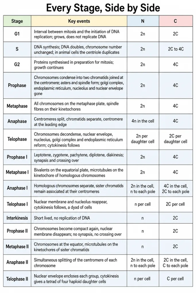
Interkinesis fits between telophase I and prophase II, and it changes nothing: it is generally short lived and there is no replication of DNA during it, so the cell sits at , throughout.
The Eight Comparisons Worth Memorising
- Mitosis against meiosis - one nuclear and cell division against two sequential cycles of nuclear and cell division, both on only a single cycle of DNA replication; two daughter cells against four; the same chromosome number as the parent, , against half that of the parent, ; equational against reductional overall, though meiosis II is itself equational; no pairing, no crossing over, no chiasmata against synapsis, crossing over and chiasmata; daughter cells with identical genetic complement against genetically variable daughter cells; in somatic cells, for growth, repair and replacement against in diploid cells destined to form gametes, for gamete formation and variation.
- Karyokinesis against cytokinesis - the nuclear division, corresponding to the separation of daughter chromosomes, which the M phase starts with and which has the four stages prophase, metaphase, anaphase and telophase, against the division of the cytoplasm, which the M phase usually ends with and at the end of which cell division gets completed. Karyokinesis without cytokinesis gives a syncytium, as in the liquid endosperm in coconut.
- Plant against animal cytokinesis - plant cells are enclosed by a relatively inextensible cell wall, so wall formation starts in the centre of the cell and grows outward to meet the existing lateral walls, beginning with a cell-plate that represents the middle lamella between the walls of two adjacent cells; an animal cell instead forms a furrow in the plasma membrane, which gradually deepens and ultimately joins in the centre. Centre outward against outside inward.
- Anaphase against anaphase I - the centromere of each chromosome splits and the sister chromatids separate, each pole receiving the parent chromosome number, against the homologous chromosomes separate while sister chromatids remain associated at their centromeres, the centromere not splitting, each pole receiving half the chromosome number of the parent cell. Equational against reductional. This is the single most examined contrast in the chapter.
- Meiosis I against meiosis II - preceded by a long, complex prophase with five sub-stages, with pairing, crossing over and chiasmata, separating homologous chromosomes and halving the chromosome number, hence reductional, against a division that resembles a normal mitosis, with a short prophase and no sub-stages, separating sister chromatids by splitting the centromere, and leaving the chromosome number unchanged at , hence equational. Between them lies interkinesis, with no replication of DNA.
- Bivalent against dyad against tetrad - a bivalent is the complex formed by a pair of synapsed homologous chromosomes, made at zygotene; a dyad is the two cells formed at telophase I; a tetrad is the four haploid daughter cells formed at telophase II - and, confusingly, also the name for a bivalent, because at pachytene the four chromatids of each bivalent chromosome become distinct and clearly appear as tetrads. Read the stem to see whether it means chromatids or cells.
- Synapsis against crossing over against chiasmata - synapsis is the pairing of chromosomes at zygotene, accompanied by the formation of the synaptonemal complex; crossing over is the exchange of genetic material between two homologous chromosomes at pachytene, at recombination nodules, enzyme-mediated by recombinase, completed by the end of pachytene; chiasmata are the X-shaped structures seen at diplotene where the recombined homologues stay linked after the synaptonemal complex dissolves, and they are terminalised at diakinesis. Three stages in order - zygotene, pachytene, diplotene.
- against - is a phase of the cycle, the interval between mitosis and the initiation of DNA replication, in which the cell is metabolically active, grows and is on its way to S phase; is an exit from the cycle, the quiescent stage reached from , in which cells remain metabolically active but no longer proliferate unless called on to do so - heart cells are the standard example.
Writing the Chapter-End Exercises Well
Class 11 has no board paper, but the sixteen chapter-end exercises are the best test of this chapter that exists. Between them they cover the timings, the stage names, the two diagrams, the two comparisons and the N and C analysis - which is very nearly the whole chapter. Marks here are lost cheaply, by naming a stage and never saying what happens in it.
The synapsis, bivalent and chiasmata diagram. One of the chapter-end exercises asks you to describe synapsis, bivalent and chiasmata and to draw a diagram to illustrate your answer, and the diagram is marked on its labels, not on its neatness. Draw two homologous chromosomes lying side by side, each with two chromatids, and carry these labels on the sheet:
- Homologous chromosomes - the two members of the pair.
- Synapsis - the process of association by which the chromosomes pair together, occurring at zygotene.
- Synaptonemal complex - the complex structure whose formation accompanies synapsis.
- Bivalent, or tetrad - the complex formed by a pair of synapsed homologous chromosomes, whose four chromatids become distinct at pachytene.
- Non-sister chromatids - the two chromatids, one from each homologue, between which crossing over occurs.
- Recombination nodule - the site at which crossing over occurs.
- Chiasma - the X-shaped structure at diplotene, where the recombined homologues remain linked at the site of a crossover; mark at least one, and say in the text that they are terminalised at diakinesis.
Then write the three definitions underneath in one line each. The examiner is checking that you can place synapsis at zygotene, crossing over at pachytene and chiasmata at diplotene without hesitating.
A full mitosis-versus-meiosis answer. Another exercise asks you to list the main differences between mitosis and meiosis, and the answer that scores is a two-column table, not a paragraph. Do not start with a random difference - start with the anchor: meiosis has two sequential cycles of nuclear and cell division on only a single cycle of DNA replication, and it pairs homologous chromosomes; mitosis does neither. Then hang the rows on it, in this order: number of divisions; number of DNA replications - one for each, which is the row most often got wrong; number of daughter cells, two against four; chromosome number of the daughter cells, against ; equational against reductional; pairing of homologues, absent against present at zygotene; crossing over, absent against present at pachytene; chiasmata, absent against present from diplotene; length of prophase, short against long with five sub-stages; the anaphase contrast; where it occurs, somatic cells against cells destined to form gametes; genetic outcome, identical against variable; and function, growth and repair against gamete formation and variation. A separate exercise asks only for anaphase of mitosis against anaphase I - write that one as what splits, what separates, what reaches each pole, and equational or reductional.
Laying out the N and C analysis. The exercise that asks you to analyse the events during every stage of the cell cycle and notice how the number of chromosomes (N) per cell and the amount of DNA content (C) per cell change is asking for a table, one row per stage, one column for N and one for C, and a reason column if you have room. Two habits make it a full-mark answer. First, say why each value moved or did not move - the DNA doubles only in S phase and is halved at each cytokinesis; the chromosome number rises only when centromeres split and falls only when a cell divides. Second, write the three rows students lose marks on, loudly: anaphase of mitosis is but still ; each cell of the dyad is , , not , ; each cell of the tetrad is , . The neighbouring exercises about mitosis without DNA replication in S phase and DNA replication without cell division are answered from the same table - the first would give daughter cells with half the DNA, so normal daughter cells are not possible, and the second does happen, giving a polyploid or polytene condition.
The habit all sixteen exercises reward. Name the stage, then the event, then the outcome, in that order. "Name the stage at which the centromere splits and the chromatids separate" is answered as anaphase - the centromeres split simultaneously and the chromatids separate - so each pole receives a full set and the division is equational. "Why is mitosis called equational division" is mitosis - the chromosome number of the parent is conserved - so the daughter cells are like the parent. Most marks lost in this chapter are lost by giving only the first of the three. The same habit answers the "find out" and "discuss" exercises: haploid cells divide by mitosis in male honey bees and in some lower plants, while haploid cells in higher plants such as the pollen grain nucleus and the egg do not divide further in that way; and the four daughter cells of meiosis are equal in size in male gametogenesis, as in the four sperms formed from one spermatocyte, and unequal in size in female gametogenesis, where one large egg and small polar bodies are formed.
NEET Strategy
What the paper asks. This chapter is almost pure stage-identification. There is nothing to work out, so speed comes from certainty. A question hands you an event and wants a stage, or hands you a stage and wants an event, a number or a pair of values. The time you save here is time you keep for the calculation-heavy chapters.
The marking is +4 and -1. On a pure-recall question, a coin-flip between two options loses marks on average. If both stage names have genuinely gone, skip it and bank the time.
Budget. Aim for 25 to 30 seconds per question in this chapter. If a question is still open at forty seconds, staring at it will not fix it - mark it and move.
The four shapes, in the order they are easiest to attempt.
- Give the chromosome number or the DNA content at a stage. These are the fastest of all if the table is in your head - is , ; the dyad is , ; the tetrad is , ; anaphase of mitosis is , . Answer in five seconds or move on.
- State what happens at a named stage. The stage name is given, so there is nothing to identify - just recall. Metaphase I - bivalents on the equatorial plate. Anaphase II - the centromere splits.
- Name the stage from an event. Read until one clue fixes the stage and then stop reading. "Pairing of homologous chromosomes" is zygotene and nothing else; "terminalisation of chiasmata" is diakinesis and nothing else.
- Spot the odd one out or the incorrect statement. Leave these for last. A "which is NOT" question forces you to check all four options, so it costs two to three times what the others cost.
Read the direction word before you read the options. NOT, INCORRECT and respectively are the three words that turn a fact you know into a mark you lose. Underline them as you read the stem. In a respectively question, match the first pair first - it usually eliminates two options in one step.
One last habit that fits this chapter in particular. When two options both look right, ask which stage each one actually belongs to. The wrong option in this chapter is almost never invented - it is a true statement about a neighbouring stage: centromere splitting offered for anaphase I, crossing over offered for prophase II, the cell-plate offered for an animal cell, an S phase offered for interkinesis.
The Mistakes That Cost the Most Marks
- Treating interphase as a rest. It is called the resting phase, but it lasts more than 95 per cent of the duration of the cell cycle, and cell growth and DNA replication both happen in it, while cell division proper lasts for only about an hour.
- Saying cells enter from . Cells that do not divide further exit the phase to enter the quiescent stage, and while there they remain metabolically active but no longer proliferate - not division-inactive and metabolically inactive both.
- Letting S phase change the chromosome number. The amount of DNA per cell doubles from to , and there is no increase in the chromosome number - stays , because each chromosome merely gains a second chromatid.
- Putting centriole duplication in prophase. In animal cells the centriole duplicates in the cytoplasm during the S phase. In prophase, the already duplicated centrosome only begins to move towards opposite poles.
- Naming prophase as the stage for studying chromosome morphology. It is metaphase, because by then condensation of chromosomes is completed and they can be observed clearly under the microscope.
- Saying the spindle attaches to the centromere. It attaches to the kinetochores - small disc-shaped structures at the surface of the centromeres which serve as the sites of attachment of spindle fibres.
- Confusing anaphase with anaphase I. In anaphase of mitosis the centromeres split and the sister chromatids separate. In anaphase I the homologous chromosomes separate while sister chromatids remain associated at their centromeres, and the centromere does not split. Sister chromatids separate only at anaphase II.
- Swapping synapsis and crossing over, or chiasmata and their terminalisation. Synapsis is at zygotene. Crossing over is at pachytene. Chiasmata appear at diplotene and are terminalised at diakinesis. Four stages, four events, in that order.
- Giving interkinesis an S phase, or calling meiosis two replications. There is no replication of DNA during interkinesis, and meiosis involves two sequential cycles of nuclear and cell division but only a single cycle of DNA replication.
- Mixing up dyad and tetrad, and the two senses of tetrad. Telophase I gives a dyad of two cells; telophase II gives a tetrad of four haploid cells. A bivalent is also called a tetrad, because at pachytene its four chromatids appear as tetrads - and while you are there, remember the cell-plate grows from the centre outward in a plant while an animal cell furrows from the outside inward.
A Short Revision Plan
First pass - one hour. Read sections 1 to 9 straight through without stopping to memorise. You are building the map: the cycle and its phases, then mitosis stage by stage, then what mitosis is for, then meiosis I with its long prophase, then meiosis II, then the N and C accounting, then what meiosis is for. Do not stop to learn stage names on this pass - just notice that events happen in a fixed order.
Second pass - one hour and fifteen minutes. Work the Solved Examples in each section, writing the answers out rather than reading them, then check your wording against the answer given and mark only the words you missed. The marking-scheme keywords are in bold in every answer for exactly this purpose. Pay particular attention to the anaphase items and to anything asking for N and C.
Third pass - forty minutes. Learn the fifteen-row table in this section and the eight comparisons above. Then say the stage-and-event list out loud in one run, in order: growth and metabolism, S DNA replication and centriole duplication, protein synthesis, prophase condensation and asters, metaphase alignment on the metaphase plate with kinetochores attached, anaphase centromeres split and chromatids move, telophase decondensation and two nuclei, cytokinesis furrow or cell-plate; leptotene visible, zygotene synapsis and synaptonemal complex, pachytene tetrads and crossing over by recombinase, diplotene chiasmata, diakinesis terminalisation, metaphase I bivalents, anaphase I homologues separate with chromatids attached, telophase I dyad, interkinesis no replication, prophase II compact again, metaphase II sister chromatids attached, anaphase II centromeres split, telophase II tetrad of four haploid cells. Then the numbers: 24 hours, 90 minutes, about an hour of division, more than 95 per cent interphase, four stages of mitosis, five sub-stages of prophase I, two divisions, one replication, four haploid cells.
Fourth pass - one hour. Sit the 45 NEET-Pattern Practice Questions under time, then go back only to the rows of the table your wrong answers came from. Redo the same questions three days later; the second attempt is what tells you whether the stage names stuck.
The night before. Read the fifteen-row table once, then the eight comparisons, then the ten mistakes. Nothing else, and no new material. If you have time for only one thing, read row by row down the N and C columns and say aloud why each value moved or did not move. This chapter rewards a clean recent pass far more than a long one.