
Hormones and Chemical Coordination
The nervous and endocrine systems coordinate body functions in different ways. Nerve impulses are rapid, short-lived and directed along neurons. Endocrine messages are chemical and may act more slowly, but their effects can be widespread and long-lasting.
NCERT defines hormones as non-nutrient chemicals that act as intercellular messengers and are produced in trace amounts. Keep all three parts together. Glucose and amino acids influence metabolism, but they are nutrients, not hormones.
An endocrine gland is ductless. Its hormones enter the surrounding tissue fluid and then the blood. An exocrine gland uses a duct to deliver a secretion to a surface or cavity; salivary and sweat glands are familiar examples.
NEET trap: A hormone is identified by its signalling role, not merely by being present in blood. The definition must include non-nutrient, intercellular messenger and trace amount.
High-Yield Map of the Syllabus Endocrine System
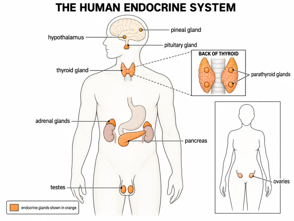
The NCERT chapter names eight components of the human endocrine system: hypothalamus, pituitary, pineal, thyroid, parathyroid, adrenal, pancreas and gonads. The hypothalamus, pituitary and pineal are associated with the brain. The thyroid and parathyroids lie in the neck. The adrenal glands lie at the anterior or upper ends of the kidneys, the pancreas lies in the abdomen, and the gonads are the testes or ovaries.
The hypothalamus is the neuroendocrine coordinating centre. It regulates the pituitary, which in turn controls several other endocrine glands. This hypothalamus-pituitary relationship is more useful for NEET than memorising the organs as an isolated list.
[NEET Important] For this chapter, prioritise the eight structures named in the NCERT chapter. Learn each structure with its position, hormone and principal action.
Hypothalamus: Link Between Nervous and Endocrine Control
The hypothalamus lies in the basal part of the diencephalon in the forebrain. It contains neurosecretory cells that produce hormones regulating the pituitary.
Hypothalamic hormones are of two functional types:
- Releasing hormones stimulate pituitary secretion. Gonadotrophin-releasing hormone (GnRH), for example, stimulates synthesis and release of gonadotrophins.
- Inhibiting hormones suppress pituitary secretion. Somatostatin, for example, inhibits growth hormone release.
These regulatory hormones reach the anterior pituitary through the hypothalamo-hypophyseal portal circulation. The short vascular route delivers them at an effective concentration without first diluting them through the systemic circulation.
The hypothalamus also synthesises oxytocin and vasopressin (ADH). These travel through axons to the posterior pituitary, where they are stored and released.
NEET trap: Portal blood carries releasing and inhibiting hormones to the anterior pituitary; axons carry ADH and oxytocin to the posterior pituitary.
Pineal Gland and Melatonin
The pineal gland lies on the dorsal side of the forebrain and secretes melatonin. Its best-known role is regulation of the body's 24-hour or diurnal rhythm, including the sleep-wake cycle.
NCERT also associates melatonin with regulation of body temperature and metabolism and with influences on pigmentation, the menstrual cycle and defence capability.
[NEET Important] Match the contrast precisely: hypothalamus - basal diencephalon; pineal - dorsal forebrain. Melatonin is the pineal hormone, not a pituitary hormone.
Target Cells, Receptors and Specificity
Hormones circulate through many tissues, but only cells with a specific receptor respond. Such cells are called target cells. Hormone binding forms a hormone-receptor complex, which initiates the cellular response.
Receptors are proteins located either:
- on the cell membrane, commonly for peptide and protein hormones and catecholamines; or
- inside the cell, commonly for steroid and thyroid hormones.
A cell without the appropriate receptor does not respond even if the hormone reaches it. Therefore, target specificity depends primarily on receptor expression, not simply on blood supply.
Rapid NEET Recall
- Hormone: non-nutrient + intercellular messenger + trace amount
- Endocrine gland: ductless
- GnRH: stimulates gonadotrophin release
- Somatostatin: inhibits GH release
- Melatonin: diurnal rhythm
- Target cell: possesses the matching receptor
Pituitary Location and Organisation
The pituitary gland lies in a bony cavity called the sella turcica and is connected to the hypothalamus by a stalk.
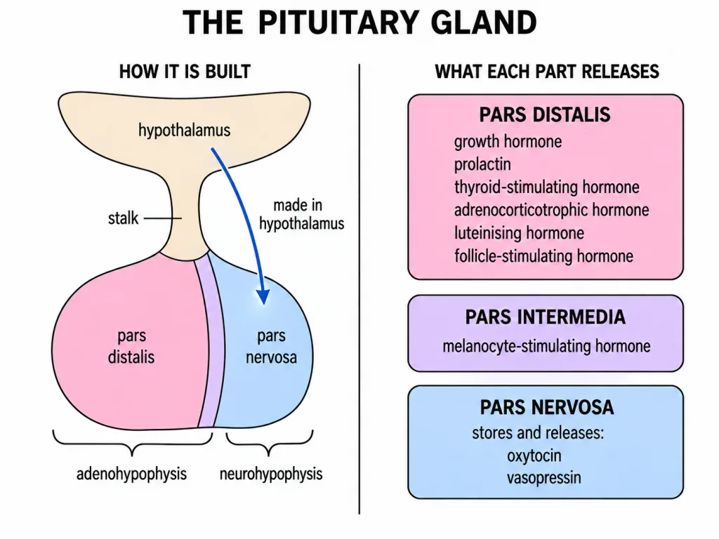
The gland has two major divisions:
- Adenohypophysis (anterior pituitary): consists of pars distalis and pars intermedia. In humans, the pars intermedia is almost merged with the pars distalis.
- Neurohypophysis (posterior pituitary): consists mainly of the pars nervosa and remains functionally connected with hypothalamic neurons.
NEET trap: Pars distalis and pars intermedia belong to the adenohypophysis; pars nervosa belongs to the neurohypophysis.
Hormones of Pars Distalis
Pars distalis secretes six major hormones: growth hormone (GH), prolactin (PRL), thyroid-stimulating hormone (TSH), adrenocorticotrophic hormone (ACTH), luteinising hormone (LH) and follicle-stimulating hormone (FSH).
- GH acts on many tissues and promotes body growth.
- PRL regulates growth of the mammary glands and formation of milk.
- TSH stimulates synthesis and secretion of thyroid hormones.
- ACTH stimulates synthesis and secretion of glucocorticoids from the adrenal cortex.
- LH and FSH are called gonadotrophins because they act on the gonads.
[NEET Important] Prolactin produces milk; oxytocin ejects milk. TSH acts on the thyroid, whereas ACTH acts on the adrenal cortex.
Gonadotrophins and Pars Intermedia
In males, LH stimulates Leydig cells to synthesise and secrete androgens, while FSH acts on Sertoli cells and supports spermatogenesis. In females, LH induces ovulation and formation of the corpus luteum, while FSH supports growth and development of ovarian follicles.
Pars intermedia secretes melanocyte-stimulating hormone (MSH). MSH acts on melanocytes and regulates pigmentation of the skin.
NEET trap: MSH is a product of pars intermedia, not of the pineal gland. Melatonin, a similarly named hormone, is secreted by the pineal gland.
Growth Hormone and Its Disorders
GH promotes growth of body tissues, especially bone and muscle, partly by supporting protein synthesis. The outcome of abnormal GH secretion depends strongly on age.
- GH deficiency during childhood: pituitary dwarfism; the individual is short but usually proportionate.
- GH excess before epiphyseal closure: gigantism; excessive linear growth produces unusual height.
- GH excess in adults: acromegaly; bones cannot lengthen normally, so the jaw, hands, feet and other extremities enlarge.
[NEET Important] Gigantism and acromegaly both involve GH excess. The timing - before or after completion of linear growth - separates them.
Posterior Pituitary: Storage and Release, Not Synthesis
The neurohypophysis stores and releases oxytocin and vasopressin or antidiuretic hormone (ADH). Both hormones are actually synthesised by the hypothalamus and transported along axons to the posterior pituitary.
Oxytocin causes vigorous uterine contractions during childbirth and milk ejection from mammary glands. ADH acts mainly on distal renal tubules and promotes reabsorption of water and electrolytes, thereby reducing water loss through urine.
Deficient ADH release causes diabetes insipidus, characterised by excessive production of dilute urine and intense thirst. It must not be confused with diabetes mellitus, which concerns glucose regulation.
Rapid NEET Recall
- Anterior pituitary: synthesises and secretes GH, PRL, TSH, ACTH, LH and FSH
- Pars intermedia: MSH
- Posterior pituitary: stores and releases hypothalamic ADH and oxytocin
- PRL: milk production; oxytocin: milk ejection
- ADH deficiency: diabetes insipidus