
Plant Tissues - Meristematic vs Permanent
Anatomy is the study of the internal structure of plants. A tissue is a group of cells with a common origin, usually performing a common function. Plant tissues are of two broad kinds: meristematic (dividing) and permanent (non-dividing).
Meristematic tissues (meristems)
A meristem is a group of actively dividing cells. Meristematic cells are immature, thin-walled (cellulose), living, with dense cytoplasm, a large prominent nucleus, and no (or small) vacuoles; intercellular spaces are absent.
Classified by position:
- Apical meristem - at the tips of roots and shoots; produces the primary plant body (increase in length, i.e. primary growth). The shoot apical meristem also gives rise to leaves and flowers. In the root, a region called the subapical meristem lies just behind the root cap.
- Intercalary meristem - at the base of internodes or leaves (e.g. grasses); a part of the apical meristem left behind during growth. Apical + intercalary meristems are primary meristems - they appear early and form the primary plant body.
- Lateral meristem - along the sides of the root/stem; cylindrical; responsible for secondary growth (increase in girth/thickness). The two lateral meristems are the vascular cambium and the cork cambium (phellogen). These are secondary meristems.
Trap: Apical & intercalary = primary (length); vascular cambium & cork cambium = lateral = secondary (girth). The promeristem/primordial meristem is the earliest meristem in a new organ.
The cells left behind
Cells derived from apical and lateral meristems divide, then differentiate and mature into permanent tissues - they lose the ability to divide, take on a specific size, shape and wall, and perform a specific function. Dedifferentiation is when living, differentiated (permanent) cells that had lost the ability to divide regain the capacity to divide under certain conditions - this is how the interfascicular cambium and the cork cambium arise from parenchyma. Redifferentiation is when the cells produced by such a dedifferentiated meristem again lose the ability to divide and mature into a permanent tissue - e.g. secondary xylem, secondary phloem and cork.
Simple Permanent Tissues
A simple tissue is made of only one type of cell. There are three: parenchyma, collenchyma and sclerenchyma.
1. Parenchyma - the fundamental, living packing tissue
- Cells are living, thin-walled (cellulose), isodiametric (roughly equal-sided), oval/round/polygonal; usually with intercellular spaces.
- The most abundant, least specialised tissue; forms the bulk of the cortex, pith, mesophyll etc.
- Functions: storage (starch, protein), photosynthesis (when it contains chloroplasts it is called chlorenchyma, e.g. leaf mesophyll), secretion, and buoyancy - parenchyma with large air cavities (in aquatic/hydrophytes) is called aerenchyma.
2. Collenchyma - the living mechanical tissue of young organs
- Cells are living, elongated, with walls thickened at the corners by deposition of cellulose, hemicellulose and pectin; little or no intercellular space.
- Occurs in layers below the epidermis - the hypodermis of dicot stems and in leaf petioles and margins.
- May contain chloroplasts and assimilate food.
- Function: provides mechanical support and flexibility to growing parts (young stems, petioles) so they bend without breaking. Absent in monocots.
3. Sclerenchyma - the dead mechanical tissue
- Cells are dead, long or short, with thick, lignified secondary walls and narrow cavities (lumen); often no protoplast at maturity.
- Two types:
- Fibres - long, narrow, thick-walled, pointed sclerenchymatous cells; occur in groups (e.g. in the pericycle, bundle caps, and as commercial fibres like jute/flax).
- Sclereids (stone cells) - spherical/oval/isodiametric, very thick-walled cells with a very narrow lumen; found in the hard parts - the shells of nuts, the pulp of guava, pear and sapota (the grittiness), and seed coats of legumes.
- Function: mechanical support; provides hardness and rigidity.
One-liner recap: Parenchyma = living, thin-walled, all-rounder. Collenchyma = living, corner-thickened, support of young/growing parts, hypodermis of dicot stem. Sclerenchyma = dead, lignified, support of mature parts (fibres + sclereids).
Visual - The Three Simple Tissues
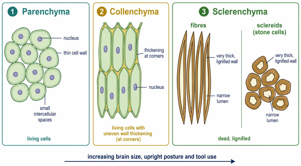
Parenchyma (thin-walled, living, intercellular spaces), collenchyma (living, thickened at the corners) and sclerenchyma (dead, uniformly thick lignified walls) - fibres and sclereids.