
Proteins - Structure and Function
Proteins are heteropolymers of amino acids linked by peptide bonds.
Peptide bond and chain direction
- A peptide bond forms between the carboxyl group of one amino acid and the amino group of the next. In the simplified condensation model, one water molecule is released per linkage.
- An unbranched chain of n residues contains n - 1 peptide bonds. Hydrolysis adds water across the linkages and releases amino acids.
- The primary sequence is written from the N-terminal free amino end to the C-terminal free carboxyl end.
Levels of protein organisation
- Primary: the linear amino-acid sequence. Proteins with the same composition can differ because order matters.
- Secondary: regular local folding such as the right-handed alpha-helix and beta-pleated sheet, stabilised mainly by hydrogen bonds.
- Tertiary: the overall three-dimensional fold of one polypeptide. For enzyme proteins, this fold creates the active-site pocket required for catalysis.
- Quaternary: the arrangement of two or more folded subunits. Adult human haemoglobin contains two alpha and two beta chains.
Sufficient heat or extreme pH can denature a protein by disrupting higher-order folding. Ordinary denaturation generally leaves the peptide-bond backbone and primary sequence intact.
Protein functions and abundance landmarks
- Trypsin is an enzyme; insulin is a hormone; antibodies participate in defence; receptors receive signals; GLUT-4 enables glucose transport; and collagen contributes to intercellular ground substance.
- Contractile proteins such as actin and myosin interact to generate force and shorten a sarcomere; the individual filaments do not themselves shorten.
- Collagen is the most abundant protein in the animal world. RuBisCO is the most abundant protein in the biosphere.
Biomacromolecules
Proteins, nucleic acids and polysaccharides are high-molecular-weight polymeric substances recovered in the acid-insoluble fraction. Polymer status does not require every sample to have one fixed molecular mass; polysaccharide preparations, for example, can contain chains of different lengths. Lipids are recovered in the same fraction because of membrane association but are not true macromolecular polymers.
Visual - Levels of Protein Structure
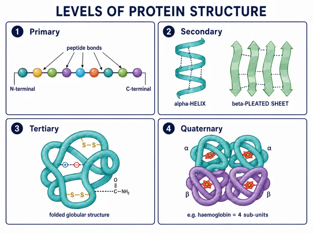
Primary through quaternary organisation, with adult haemoglobin shown as two alpha and two beta subunits.
Metabolites, Metabolic Flux and the Living State
Metabolism and metabolic pathways
- Metabolism is the sum of chemical reactions occurring in a living cell. Reactions are organised into pathways in which one product can become the substrate for a later step.
- Anabolic pathways build larger molecules and require energy; protein synthesis is an example.
- Catabolic pathways break molecules down and release usable energy; glucose respiration is an example.
Dynamic cellular pools
Cellular biomolecule pools undergo continuous turnover or metabolic flux. This does not mean that every individual molecule is synthesised and degraded at the same instant; it means concentrations are maintained through ongoing formation, conversion and breakdown.
Primary and secondary metabolites
- Primary metabolites have identifiable roles in normal physiology. Examples include amino acids, sugars, nucleotides and lipids.
- NCERT examples of secondary metabolites include pigments such as carotenoids and anthocyanins; alkaloids such as morphine and codeine; terpenoids; essential oils; toxins such as abrin and ricin; lectins; drugs such as vinblastine and curcumin; and polymeric substances such as rubber, gums and cellulose.
- Not every host function of every secondary metabolite is known, but many have ecological roles or human uses. Being secondary does not mean being useless waste, and structural function is not the defining criterion.
Living-state context
Living systems maintain a non-equilibrium steady state through continuous energy input, allowing work to be performed. This concept provides context for metabolic flux; true equilibrium is incompatible with the active living state.