
Tissues, organs and organ systems
A tissue is a group of cells with a common origin that work together to perform a specific function. Organising cells this way is the first level of division of labour in an animal body, and every structure is built from just four tissue types:
- Epithelial tissue
- Connective tissue
- Muscular tissue
- Neural tissue
An organ stands one level higher, because it is made of more than one type of tissue, and those tissues act together towards a shared purpose. Organs whose jobs are related are grouped into an organ system. The degree of complexity is not the same in all animals: different animals show different levels of organisation in their organ systems, and greater complexity brings a sharper division of labour and higher physiological efficiency.
What makes an epithelium an epithelium
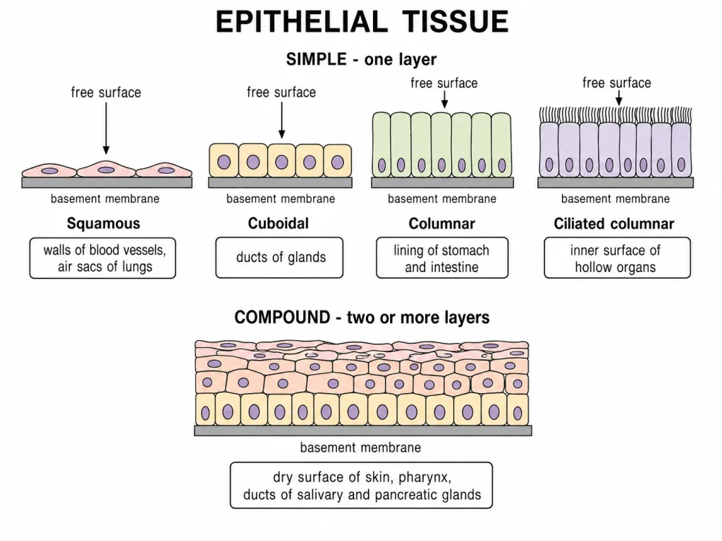
Epithelial tissue, or simply epithelium, is defined by one feature above all others: it always has a free surface, and that surface faces either a body fluid or the outside environment. The cells are compactly packed, with very little intercellular matrix separating them.
An epithelium rests on a basement membrane, a non-cellular layer that separates it from the tissue lying beneath. Epithelia are avascular, so nourishment must reach them from the tissue below that membrane. Two structural classes exist: simple epithelium, a single layer of cells, and compound epithelium, two or more layers.
Cell junctions
In nearly all animal tissues, specialised cell junctions provide structural and functional links between neighbouring cells. Tight junctions stop substances from leaking across a tissue. Adhering junctions perform a cementing role, holding adjacent cells firmly together. Gap junctions connect the cytoplasm of neighbouring cells and permit rapid transfer of ions and small molecules, so they serve communication between cells.
Simple epithelium: the three cell shapes
Simple epithelium, being a single layer of cells, lines body cavities, ducts and tubes. Its three forms are told apart by the shape of the cell.
Simple squamous epithelium is made of thin, flattened cells with irregular boundaries. It lines the walls of blood vessels and the air sacs of the lungs, and this thinness is the whole point: it forms a diffusion boundary, allowing gases to diffuse across it, and it takes part in filtration.
Simple cuboidal epithelium is built of cube-like cells. It is found in the ducts of glands and in the tubular parts of the nephrons in the kidneys, and its functions are secretion and absorption.
Simple columnar epithelium consists of tall, slender cells whose nuclei sit at the base of each cell. It forms the lining of the stomach and the intestine, and it too carries out secretion and absorption.
When columnar or cuboidal cells bear cilia on their free surface, the tissue is called ciliated epithelium. The cilia beat to move particles or mucus in a definite direction over the surface. This is the lining of the bronchioles and of the fallopian tubes.
Glandular epithelium and the two kinds of glands
Some columnar or cuboidal cells become specialised for secretion, and these are called glandular epithelium. A single secretory cell acting alone forms a unicellular gland - the goblet cells scattered through the lining of the alimentary canal are modified columnar epithelial cells of precisely this kind. Where many secretory cells are gathered together, as in the salivary glands, the gland is multicellular.
Glands are then separated by how they deliver what they make. Exocrine glands secrete through ducts, and their products include mucus, saliva, earwax, oil, milk and digestive enzymes. Endocrine glands are ductless; their products are hormones, which are poured directly into the fluid that bathes the gland rather than being carried away by any tube. The pancreas is a mixed gland: the islets of Langerhans form its endocrine portion and release hormones into the blood, while the acini form its exocrine portion and release digestive enzymes through ducts.
Compound epithelium
Compound epithelium is made of two or more layers of cells, and the extra thickness changes what the tissue is good for. Its main function is protection against chemical and mechanical stress; it has only a limited role in secretion and absorption, which is the reverse of the balance struck by the simple epithelia.
It covers the dry surface of the skin and the moist surface of the buccal cavity, and it also forms the lining of the pharynx and the inner lining of the ducts of the salivary glands and of the pancreatic ducts. Each of these places meets abrasion or strong secretions, and the deeper layers keep replacing whatever is worn away at the surface.
NEET Location–Function Checkpoint
| Tissue or structure | High-yield site | Main role |
|---|---|---|
| Simple squamous epithelium | Alveoli and blood-vessel walls | Diffusion and filtration |
| Simple cuboidal epithelium | Kidney tubules and gland ducts | Secretion and absorption |
| Simple columnar epithelium | Stomach and intestine | Secretion and absorption |
| Ciliated epithelium | Bronchioles and uterine tubes | Moves material over the epithelial surface |
| Compound epithelium | Skin, buccal cavity and pharynx | Protection from abrasion and stress |
Junctions are not interchangeable
- Tight junction: limits leakage across the epithelial sheet.
- Adhering junction: mechanically cements neighbouring cells.
- Gap junction: permits ions and small molecules to move directly between adjacent cells for rapid communication.
[NEET Important] “Endocrine pancreas” means the islets of Langerhans, not the entire pancreas. Its acini are exocrine and release enzymes into ducts.
The most abundant tissue in the body
Connective tissues are the most abundant and most widely distributed tissues in complex animals. The name states the job: they link and support the other tissues and organs of the body, holding the whole structure together rather than lining it, contracting it or wiring it.
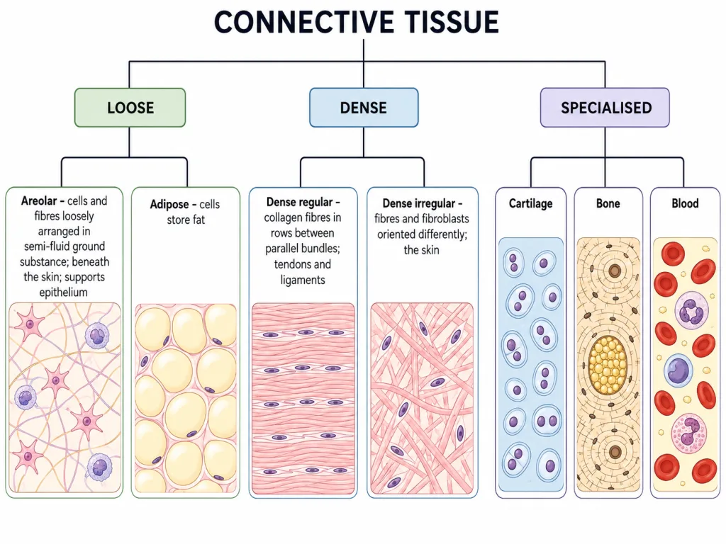
They are not one tissue but a family that ranges across a spectrum of firmness. At the soft end sits areolar tissue; further along come the dense connective tissues; and at the far end sit the three specialised ones, cartilage, bone and blood. A cartilage plate, a thigh bone and a drop of blood therefore belong to the same tissue category, which is exactly why blood is described as a specialised connective tissue rather than a fluid of its own kind.
Fibres and matrix
With the single exception of blood, connective tissues secrete fibres of structural proteins - either collagen or elastin. These fibres give the tissue its strength, elasticity and flexibility.
The same tissues also secrete modified polysaccharides, which accumulate between the cells and the fibres and act as the matrix, or ground substance. Whether a connective tissue turns out soft, dense, pliable or rock-hard depends almost entirely on what this matrix is made of and how the fibres are packed within it.
Loose connective tissue
In loose connective tissue, cells and fibres are loosely arranged in a semi-fluid ground substance. Areolar tissue is the classic example. It lies beneath the skin and frequently serves as a support framework for epithelium, filling the space between organs and binding them loosely to one another. Its residents include fibroblasts, which manufacture the fibres and matrix, macrophages, and mast cells.
Adipose tissue is the other loose connective tissue, located mainly beneath the skin. Its cells are specialised to store fat, and excess nutrients that are not used immediately are converted into fats and deposited here.
Dense connective tissue
In dense connective tissue, the fibres and the fibroblasts are compactly packed instead of loosely scattered. The subdivision turns on whether that packing is orderly.
Dense regular connective tissue has parallel bundles of collagen fibres, with fibroblasts arranged in rows between them; this alignment is built to resist pulling in one direction. Tendons are of this type, and they attach skeletal muscles to bones. Ligaments, also dense regular tissue, attach one bone to another.
Dense irregular connective tissue contains fibroblasts and many fibres, mostly collagen, that are oriented differently rather than in parallel. This tissue is present in the skin, where stress can arrive from any direction and a criss-crossed fibre arrangement resists all of them.
Cartilage
Cartilage is a specialised connective tissue in which the intercellular material is solid and pliable, so it resists compression without shattering. The cells of cartilage, the chondrocytes, are enclosed in small cavities within the matrix that they themselves secrete; these cavities are called lacunae.
Cartilage is found at the tip of the nose and in the pinna of the outer ear, at articular surfaces between adjacent limb bones, and between adjacent vertebrae. Most of the cartilage laid down in vertebrate embryos is later replaced by bone in the adult, so the material is as much a scaffold for development as a permanent tissue.
Bone
Bone has a hard and non-pliable ground substance rich in calcium salts and collagen fibres, and it is this combination that gives bone its strength. Bone provides the structural frame of the body, supporting and protecting the softer tissues and organs around it.
Like chondrocytes, the bone cells, the osteocytes, sit in spaces called lacunae. Some bones additionally house bone marrow, which is the site of production of blood cells. The limb bones, such as the long bones of the legs, bear the weight of the body and interact with skeletal muscles to bring about movement, so a bone is not merely a passive girder.
Blood
Blood is a fluid connective tissue, and it is the one connective tissue without fibres. It contains:
- Plasma, the fluid portion
- Red blood cells
- White blood cells
- Platelets
Blood is the main circulating fluid of the body, and its role is transport - it carries substances from where they are made or absorbed to where they are needed, which is how a tissue with no solid matrix still counts as one that connects everything.
Connective-Tissue Identification Grid
| Tissue | Diagnostic arrangement | Key cells or matrix | Typical role/site |
|---|---|---|---|
| Areolar | Loose fibres in semi-fluid ground substance | Fibroblasts, macrophages and mast cells | Packing and support beneath epithelia |
| Adipose | Closely packed fat-storing cells | Adipocytes | Energy storage and insulation beneath skin |
| Dense regular | Parallel collagen bundles | Fibroblasts in rows | Tendons and ligaments; one-directional pull |
| Dense irregular | Collagen fibres running in different directions | Fibroblasts | Dermis; stress from several directions |
| Cartilage | Solid but pliable matrix | Chondrocytes in lacunae | Nose, pinna, joints and between vertebrae |
| Bone | Hard matrix rich in calcium salts and collagen | Osteocytes in lacunae | Support, protection and movement |
| Blood | Fluid matrix called plasma | Formed elements | Transport |
[NEET Important] Mammalian platelets are anucleate cell fragments derived from megakaryocytes. RBCs, WBCs and platelets are grouped as the formed elements of blood; do not call all three complete cells.