
Transport of oxygen and carbon dioxide
Oxygen travels mostly on haemoglobin
Blood moves oxygen in two ways, and the split is heavily lopsided. About 97% of the O2 is transported by the RBCs bound to a pigment, and only the remaining 3% is carried in a dissolved state through the plasma. Hold that ratio steady in your head as "almost all bound, a sliver dissolved", because carbon dioxide behaves quite differently and these two sets of numbers get swapped more often than any other pair in the chapter.
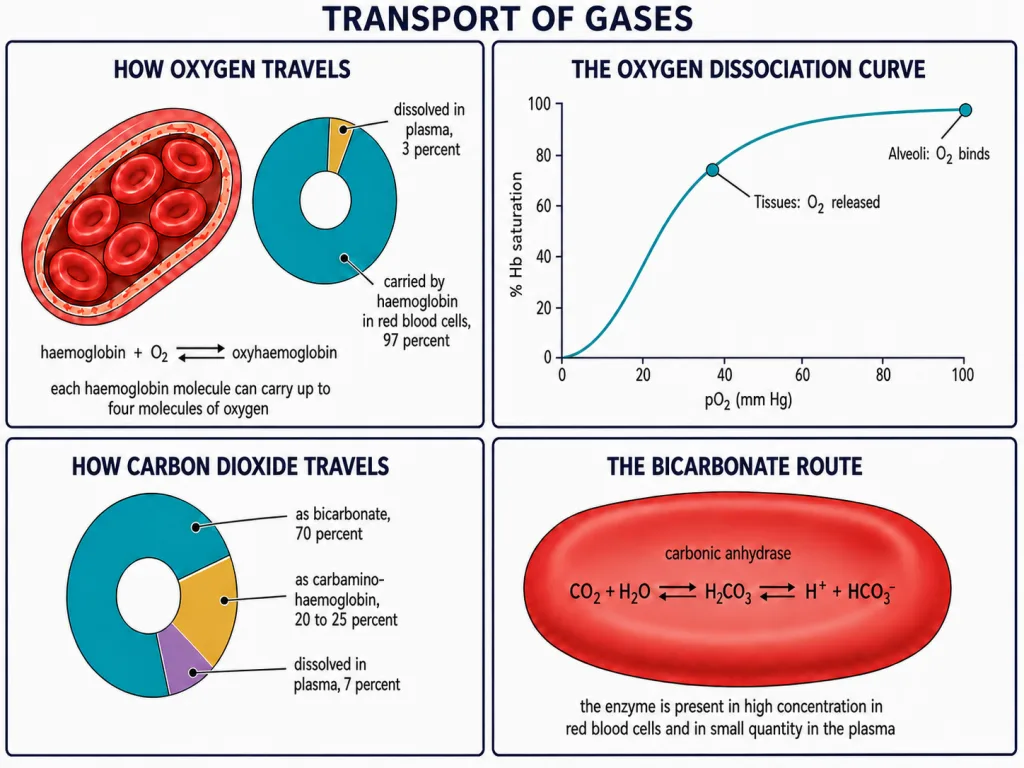
The pigment doing the work is haemoglobin, a red, iron-containing pigment present in the RBCs. Each haemoglobin molecule can carry a maximum of four molecules of O2 - four, not more - and it binds them reversibly to form oxyhaemoglobin. That reversibility is the entire point. A carrier that gripped oxygen permanently would be worthless; haemoglobin has to pick oxygen up in one place and let go of it in another.
What decides whether oxygen binds
Binding of O2 with haemoglobin is primarily related to the partial pressure of O2, written pO2. That is the master variable, and if you remember only one controlling factor, remember this one. Three others interfere with the binding: pCO2, hydrogen ion concentration and temperature.
If you plot the percentage saturation of haemoglobin with O2 against pO2, you obtain a curve with a characteristic S-shape. This is the sigmoid oxygen dissociation curve, and "sigmoid" is the word to write - not linear, not hyperbolic. The curve is a working tool rather than decoration: it is used to study the effect of factors like pCO2 and hydrogen ion concentration on oxygen binding with haemoglobin.
Loading at the lungs, unloading at the tissues
The same four factors point in opposite directions at the two ends of the circulation, and that opposition is what makes transport possible.
| Condition | At the alveoli | At the tissues |
|---|---|---|
| pO2 | high | low |
| pCO2 | low | high |
| Hydrogen ion concentration | less H+ | high H+ |
| Temperature | lower | higher |
| Effect on haemoglobin | oxyhaemoglobin forms | oxyhaemoglobin dissociates |
Read the last row carefully, because the direction is where marks are lost. In the alveoli, high pO2, low pCO2, lesser H+ concentration and lower temperature are all favourable for the formation of oxyhaemoglobin. In the tissues, low pO2, high pCO2, high H+ concentration and higher temperature are favourable for dissociation of oxygen from oxyhaemoglobin. So O2 gets bound to haemoglobin at the lung surface and gets dissociated at the tissues - one molecule, opposite behaviour, decided purely by where it happens to be.
Under normal physiological conditions, every 100 mL of oxygenated blood delivers around 5 mL of O2 to the tissues.
Carbon dioxide returns in three fractions
And every 100 mL of deoxygenated blood delivers approximately 4 mL of CO2 to the alveoli. Set those two sentences side by side and the contrast becomes obvious: 5 mL of oxygen going out to the tissues, 4 mL of carbon dioxide coming back to the alveoli.
Carbon dioxide is transported three ways:
- About 70% as bicarbonate.
- About 20-25% carried by haemoglobin in the RBCs as carbamino-haemoglobin. Keep that as a range, not a single figure.
- About 7% carried dissolved in plasma.
The carbamino fraction is governed by pressures, just as oxygen binding was. This binding of CO2 with haemoglobin is related to pCO2, and pO2 is a major factor which could affect this binding. At the tissues, where pCO2 is high and pO2 is low, more CO2 binds to haemoglobin. At the alveoli, where pCO2 is low and pO2 is high, CO2 dissociates from carbamino-haemoglobin.
The enzyme behind the bicarbonate route
The bicarbonate route carries the largest share, so the machinery behind it matters. RBCs contain a very high concentration of the enzyme carbonic anhydrase, and only minute quantities of it are present in the plasma. The enzyme facilitates this reaction:
Note the arrows: this reaction is facilitated in both directions. The same enzyme handles the forward and the reverse run, and the local pCO2 decides which way it goes.
At the tissue site, where partial pressure of CO2 is high because of catabolism, CO2 diffuses into blood and forms HCO3- and H+. At the alveolar site, where pCO2 is low, the reaction proceeds in the opposite direction, leading to the formation of CO2 and H2O. Carbon dioxide trapped as bicarbonate at the tissue level is thus carried to the alveoli and released out there as CO2.