
Thoracic chamber, mechanism of breathing and regulation
The thoracic chamber
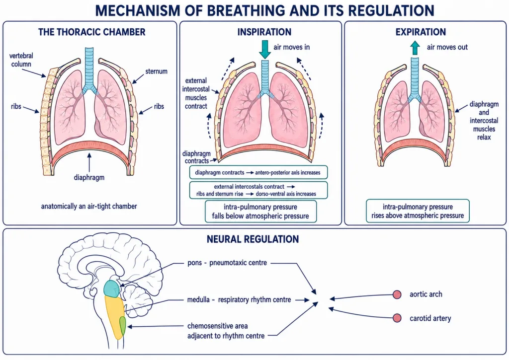
The lungs lie in an anatomically air-tight thoracic chamber. Its boundaries are:
- Dorsal: vertebral column
- Ventral: sternum
- Lateral: ribs
- Lower side: dome-shaped diaphragm
Because of this anatomical arrangement, a change in thoracic-cavity volume is reflected in lung or pulmonary volume. NCERT's key point is that pulmonary volume cannot be altered directly; it changes with thoracic volume.
Inspiration and the pressure gradient
Breathing has two stages: inspiration, during which atmospheric air is drawn in, and expiration, during which alveolar air is released. Air moves because a pressure gradient is created between the lungs and the atmosphere. NCERT identifies the diaphragm and both external and internal intercostal muscles as specialised muscles that help generate these gradients.
During normal inspiration, contraction of the diaphragm increases thoracic volume in the antero-posterior axis. Contraction of the external intercostal muscles lifts the ribs and sternum, increasing thoracic volume in the dorso-ventral axis. Pulmonary volume consequently increases and intra-pulmonary pressure falls below atmospheric pressure, so air moves into the lungs.
[NEET Important] Diaphragm → antero-posterior axis; external intercostals → dorso-ventral axis.
Expiration and assisted breathing
During quiet expiration, relaxation of the diaphragm and intercostal muscles returns the diaphragm and sternum to their normal positions. Thoracic and pulmonary volumes decrease, so intra-pulmonary pressure rises slightly above atmospheric pressure and air moves out. When airflow ends, the pressure gradient disappears.
Additional muscles in the abdomen can increase the strength of both inspiration and expiration. On average, a healthy human being breathes 12-16 times per minute. A spirometer estimates the air volumes involved in breathing and helps in clinical assessment of pulmonary functions.
[NEET Important] Air enters while intra-pulmonary pressure is below atmospheric pressure and leaves while it is above atmospheric pressure.
Neural regulation of respiration
The respiratory rhythm centre in the medulla is primarily responsible for regulating respiratory rhythm. The pneumotaxic centre in the pons moderates this rhythm centre; its signals can reduce the duration of inspiration and thereby alter respiratory rate.
A chemosensitive area adjacent to the rhythm centre is highly sensitive to carbon dioxide and hydrogen ions. An increase in these substances activates the area, which signals the rhythm centre to make adjustments that eliminate them. Receptors associated with the aortic arch and carotid artery also recognise changes in carbon dioxide and hydrogen-ion concentration and signal the rhythm centre.
For the NCERT/NEET statement, the role of oxygen in regulation of respiratory rhythm is quite insignificant. This does not mean oxygen is unimportant for cellular metabolism; it distinguishes the principal regulatory signals described here.