
Human excretory system
The human excretory system consists of a pair of kidneys, a pair of ureters, a urinary bladder and a urethra. Kidneys form urine, ureters carry it to the urinary bladder, the bladder stores it, and the urethra carries it outside.
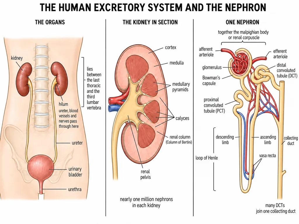
The kidneys are reddish-brown, bean-shaped organs situated close to the dorsal inner wall of the abdominal cavity, between the levels of the last thoracic and third lumbar vertebrae. Each adult kidney is approximately 10-12 cm long, 5-7 cm wide and 2-3 cm thick, with an average weight of 120-170 g.
[NEET Important] Pair the route correctly: kidney to ureter to urinary bladder to urethra.
Internal structure of the kidney
The outer surface is covered by a tough capsule. Internally, the kidney has an outer cortex and an inner medulla. The medulla is divided into conical medullary pyramids that project into the calyces. Cortical tissue extending between adjacent pyramids forms the renal columns or Columns of Bertini.
On the inner concave surface is a notch called the hilum, through which the ureter, blood vessels and nerves pass. Inner to the hilum lies the broad, funnel-shaped renal pelvis, whose projections are called calyces.
[NEET Important] Do not interchange the regions: cortex is outer, medulla is inner, and cortical extensions between pyramids are Columns of Bertini.
Nephron: renal corpuscle and tubule
Each kidney contains nearly one million nephrons, the functional units of the kidney. A nephron has two parts: the glomerulus and the renal tubule.
The glomerulus is a tuft of capillaries formed by an afferent arteriole, a branch of the renal artery. Blood leaves it through an efferent arteriole. The renal tubule begins as a double-walled cup, Bowman's capsule, which encloses the glomerulus. Together, the glomerulus and Bowman's capsule form the Malpighian body or renal corpuscle.
The tubular sequence is Bowman's capsule to PCT to descending limb of Henle's loop to ascending limb to DCT. DCTs of many nephrons open into a straight collecting duct. The Malpighian corpuscle, PCT and DCT lie in the cortex, whereas Henle's loop dips into the medulla.
Nephron types and blood vessels
In the majority of cortical nephrons, Henle's loop is short and extends only a little into the medulla. Juxtamedullary nephrons have long loops that run deep into the medulla.
The efferent arteriole forms peritubular capillaries around the renal tubule. A minute vessel from this network runs parallel to Henle's loop and forms the U-shaped vasa recta. Vasa recta is absent or highly reduced in cortical nephrons.
Many collecting ducts converge and open into the renal pelvis through the medullary pyramids in the calyces. From the pelvis, urine follows the route ureter to urinary bladder to urethra.
[NEET Important] Afferent arteriole enters the glomerulus; efferent arteriole leaves it and forms the peritubular network.