
Cilia and Flagella
Cilia (singular: cilium) and flagella (singular: flagellum) are hair-like outgrowths of the cell membrane.
Start with that word outgrowths. They are not stuck on to the cell - they grow out of the cell membrane itself, which is why electron microscopic study shows that they are covered with plasma membrane.
The two differ in size and in what they do:
| Cilia | Flagella | |
|---|---|---|
| Size | Small structures | Comparatively longer |
| How they work | Work like oars | Beat to drive the cell |
| What they move | Cause the movement of either the cell or the surrounding fluid | Responsible for cell movement |
One caution about bacteria. The prokaryotic bacteria also possess flagella, but these are structurally different from that of the eukaryotic flagella. The word is the same; the structure inside is not.
[NEET Important] Two wordings decide the marks here. Cilia move either the cell or the surrounding fluid - the "or the surrounding fluid" half is the part students drop, and it is the half that matters in tissues such as the lining of the respiratory tract. And a bacterial flagellum is not a small eukaryotic flagellum - it has no 9+2 axoneme at all.
Inside a Cilium - The Axoneme and the 9+2 Array
Their core, called the axoneme, possesses a number of microtubules running parallel to the long axis.
The axoneme usually has nine doublets of radially arranged peripheral microtubules, and a pair of centrally located microtubules. Such an arrangement of axonemal microtubules is referred to as the 9+2 array.
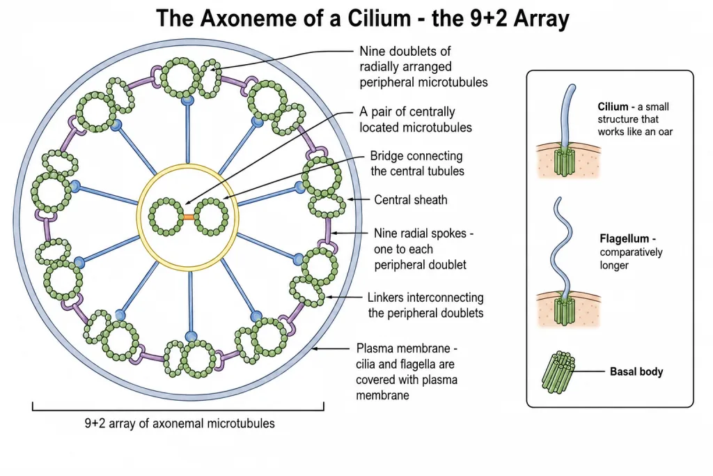
Now the parts that hold that array together, in the order the question paper asks for them:
- The central tubules are connected by bridges.
- They are also enclosed by a central sheath.
- The central sheath is connected to one of the tubules of each peripheral doublet by a radial spoke. Thus, there are nine radial spokes - one for each peripheral doublet.
- The peripheral doublets are also interconnected by linkers.
Both the cilium and the flagellum emerge from a centriole-like structure called the basal body.
| Part of the axoneme | What it is |
|---|---|
| Peripheral microtubules | Nine doublets, radially arranged |
| Central microtubules | A pair, centrally located |
| Bridges | Connect the two central tubules |
| Central sheath | Encloses the central tubules |
| Radial spokes | Nine, each joining the central sheath to one tubule of a peripheral doublet |
| Linkers | Interconnect the peripheral doublets |
| Basal body | The centriole-like structure the cilium or flagellum emerges from |
[NEET Important] Count carefully, because the numbers are all asked directly. Nine doublets, one central pair, nine radial spokes. The commonest wrong answer gives two radial spokes (one per central tubule) or eighteen (one per peripheral tubule). The spoke count follows the doublets, not the tubules.
Centrosome and Centrioles
Centrosome is an organelle usually containing two cylindrical structures called centrioles. They are surrounded by amorphous pericentriolar materials.
Both the centrioles in a centrosome lie perpendicular to each other, and each has an organisation like the cartwheel.
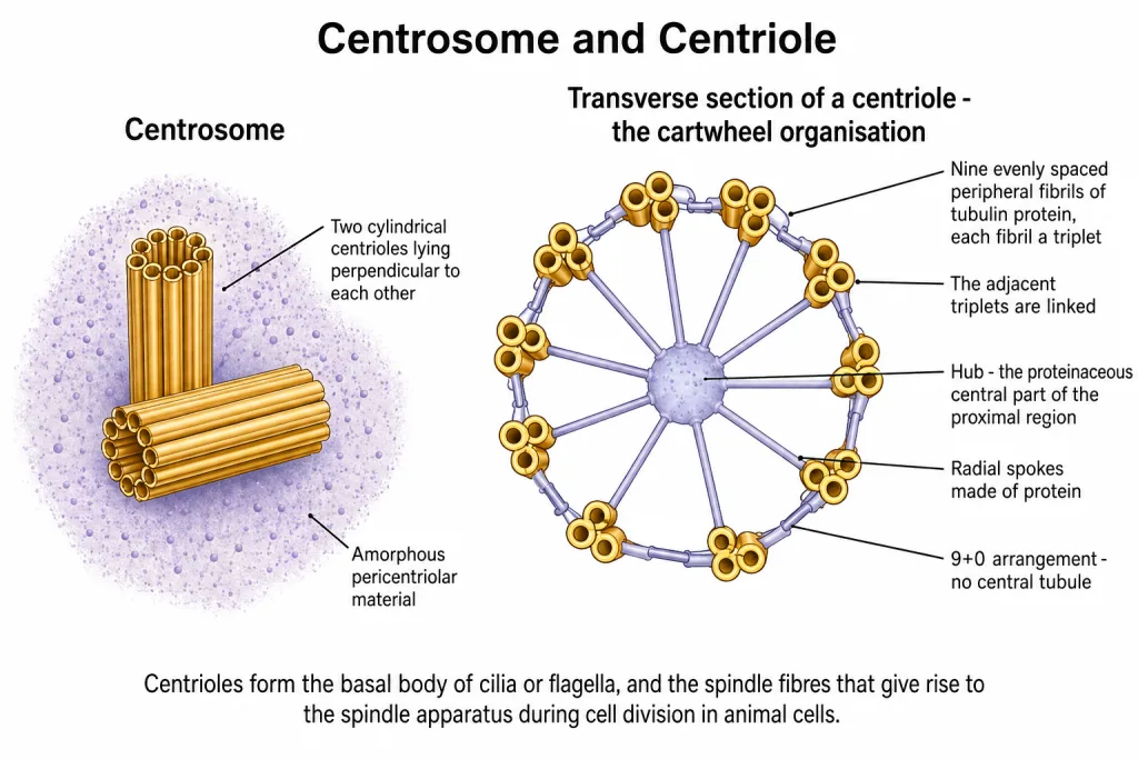
Read the cartwheel from the outside inwards:
- They are made up of nine evenly spaced peripheral fibrils of tubulin protein.
- Each of the peripheral fibrils is a triplet.
- The adjacent triplets are also linked.
- The central part of the proximal region of the centriole is also proteinaceous and is called the hub.
- The hub is connected with the tubules of the peripheral triplets by radial spokes made of protein.
What centrioles go on to build.
- The centrioles form the basal body of cilia or flagella.
- They form the spindle fibres that give rise to the spindle apparatus during cell division in animal cells.
[NEET Important] Note the material: the peripheral fibrils are of tubulin protein, and the hub and the radial spokes are proteinaceous too - a centriole is a protein structure, with no membrane anywhere in it. Also keep the two jobs of the centriole together, because a question that offers only one of them as "the function" is testing whether you remember the other.
9+2 versus 9+0 - The Comparison That Is Always Asked
Both the axoneme and the centriole are built on a ring of nine. That is where the resemblance stops, and the differences are the single most asked comparison in this chapter.
| Axoneme of cilium or flagellum | Centriole | |
|---|---|---|
| Peripheral units | Nine doublets | Nine triplets |
| Central microtubules | A pair present | Absent |
| Array | 9+2 | 9+0 |
| Central structure | Central sheath around the central pair | The proteinaceous hub |
| Radial spokes | Nine, from the central sheath to the peripheral doublets | From the hub to the peripheral triplets |
| Interconnections | Peripheral doublets joined by linkers | Adjacent triplets linked |
| Overall look | Nine doublets around a central pair | A cartwheel |
The relationship between them. The centriole forms the basal body, and the cilium or flagellum emerges from that basal body. So the 9+0 structure sits at the base and the 9+2 structure is the shaft that projects out.
[NEET Important] Learn the pairing as doublet with 9+2 and triplet with 9+0 and half the traps disappear. The wrong option almost always swaps them - "the centriole has nine doublets and a central pair" - or gives the centriole a central microtubule that it simply does not have.
Quick Recap
- Cilia (singular: cilium) and flagella (singular: flagellum) are hair-like outgrowths of the cell membrane.
- Cilia are small structures which work like oars, causing the movement of either the cell or the surrounding fluid.
- Flagella are comparatively longer and responsible for cell movement.
- Prokaryotic bacteria also possess flagella, but these are structurally different from eukaryotic flagella.
- Electron microscopy shows that both are covered with plasma membrane.
- Their core is called the axoneme, and it possesses a number of microtubules running parallel to the long axis.
- The axoneme usually has nine doublets of radially arranged peripheral microtubules and a pair of centrally located microtubules - the 9+2 array.
- The central tubules are connected by bridges and enclosed by a central sheath.
- The central sheath is connected to one tubule of each peripheral doublet by a radial spoke - there are nine radial spokes.
- The peripheral doublets are also interconnected by linkers.
- Both cilium and flagellum emerge from a centriole-like structure called the basal body.
- The centrosome is an organelle usually containing two cylindrical structures called centrioles, surrounded by amorphous pericentriolar materials.
- The two centrioles lie perpendicular to each other, each with an organisation like the cartwheel.
- A centriole has nine evenly spaced peripheral fibrils of tubulin protein; each peripheral fibril is a triplet; adjacent triplets are linked.
- The central part of the proximal region of the centriole is proteinaceous and is called the hub, connected with the tubules of the peripheral triplets by radial spokes made of protein.
- Centrioles form the basal body of cilia or flagella, and the spindle fibres that give rise to the spindle apparatus during cell division in animal cells.
- Axoneme = 9+2 (doublets, central pair present). Centriole = 9+0 (triplets, no central tubule).
Solved Examples
Question 1
Q. What are cilia and flagella?
Answer. They are hair-like outgrowths of the cell membrane. Because they grow out of the membrane, electron microscopy shows that both are covered with plasma membrane.
Question 2
Q. How do cilia differ from flagella?
Answer. Cilia are small structures which work like oars, causing the movement of either the cell or the surrounding fluid. Flagella are comparatively longer and are responsible for cell movement.
Question 3
Q. Bacteria have flagella too. Are they the same as eukaryotic flagella?
Answer. No. The prokaryotic bacteria also possess flagella, but these are structurally different from that of the eukaryotic flagella. A bacterial flagellum has no 9+2 axoneme.
Question 4
Q. What is the axoneme?
Answer. It is the core of a cilium or a flagellum. It possesses a number of microtubules running parallel to the long axis.
Question 5
Q. What is the 9+2 array?
Answer. It is the usual arrangement of axonemal microtubules - nine doublets of radially arranged peripheral microtubules, and a pair of centrally located microtubules.
Question 6
Q. How are the two central tubules of the axoneme held?
Answer. The central tubules are connected by bridges, and they are also enclosed by a central sheath.
Question 7
Q. What is a radial spoke in a cilium, and how many are there?
Answer. A radial spoke connects the central sheath to one of the tubules of each peripheral doublet. Since there are nine peripheral doublets, there are nine radial spokes.
Question 8
Q. What interconnects the peripheral doublets of the axoneme?
Answer. Linkers. The peripheral doublets are interconnected by linkers, while radial spokes run inwards to the central sheath.
Question 9
Q. From what structure do a cilium and a flagellum emerge?
Answer. Both the cilium and the flagellum emerge from a centriole-like structure called the basal body.
Question 10
Q. What is a centrosome?
Answer. It is an organelle usually containing two cylindrical structures called centrioles, which are surrounded by amorphous pericentriolar materials. The two centrioles lie perpendicular to each other.
Question 11
Q. Describe the fibrils of a centriole.
Answer. A centriole is made up of nine evenly spaced peripheral fibrils of tubulin protein. Each of the peripheral fibrils is a triplet, and the adjacent triplets are also linked. This gives the centriole an organisation like the cartwheel.
Question 12
Q. What is the hub of a centriole?
Answer. It is the central part of the proximal region of the centriole. It is proteinaceous, and it is connected with the tubules of the peripheral triplets by radial spokes made of protein.
Question 13
Q. State the two functions of centrioles.
Answer. They form the basal body of cilia or flagella. They also form the spindle fibres that give rise to the spindle apparatus during cell division in animal cells.
Question 14
Q. Distinguish clearly between the 9+2 array and the 9+0 arrangement.
Answer. The 9+2 array is found in the axoneme of a cilium or flagellum. It has nine peripheral doublets of microtubules plus a pair of central microtubules, which are enclosed by a central sheath and joined to the doublets by nine radial spokes.
The 9+0 arrangement is found in the centriole. It has nine peripheral fibrils, each a triplet, and no central microtubule at all. In place of a central pair there is the proteinaceous hub, joined to the triplets by radial spokes made of protein.
So the shortest way to hold it is: doublets and a central pair go with 9+2; triplets and no central tubule go with 9+0. And the two are directly related, because the centriole forms the basal body from which the cilium or flagellum emerges.
Question 15
Q. Describe the structure of the following with the help of labelled diagrams: (ii) Centrosome. This is one of the chapter-end exercises.
Answer. The centrosome is an organelle usually containing two cylindrical structures called centrioles, and these are surrounded by amorphous pericentriolar materials.
Position of the two centrioles. Both the centrioles in a centrosome lie perpendicular to each other, that is, at right angles - so a diagram must show one centriole drawn lengthwise and the second one turned across it.
Structure of a single centriole. Each centriole has an organisation like the cartwheel. Going from the rim inwards:
- It is made up of nine evenly spaced peripheral fibrils of tubulin protein.
- Each of the peripheral fibrils is a triplet, so there are nine triplets and no central tubule - the 9+0 arrangement.
- The adjacent triplets are also linked.
- The central part of the proximal region of the centriole is also proteinaceous and is called the hub.
- The hub is connected with the tubules of the peripheral triplets by radial spokes made of protein.
Functions. The centrioles form the basal body of cilia or flagella, and they form the spindle fibres that give rise to the spindle apparatus during cell division in animal cells.
The diagram must carry these labels: the two centrioles lying perpendicular to each other, the pericentriolar material around them, the nine peripheral triplet fibrils of tubulin protein in a transverse view of one centriole, the hub at the centre, the radial spokes running from the hub to the triplets, and the whole inner pattern marked as the cartwheel organisation.