
The Chapter in One Read
All organisms are made of cells or aggregates of cells, and cells vary in their shape, size and activities. Based on the presence or absence of a membrane bound nucleus and other organelles, cells - and hence organisms - can be named as eukaryotic or prokaryotic. That single test, the membrane bound nucleus, is the line the whole chapter is drawn along.
A typical eukaryotic cell consists of a cell membrane, a nucleus and cytoplasm, and plant cells have a cell wall outside the cell membrane. The plasma membrane is selectively permeable and facilitates the transport of several molecules - some of them freely down a gradient, some of them uphill at the cost of ATP.
The endomembrane system includes the ER, golgi complex, lysosomes and vacuoles, grouped together because their functions are coordinated. All the cell organelles perform different but specific functions. The endoplasmic reticulum contains tubules or cisternae and is of two types, rough and smooth; the ER helps in the transport of substances and in the synthesis of proteins, lipoproteins and glycogen. The golgi body is a membranous organelle composed of flattened sacs, and the secretions of cells are packed in them and transported from the cell. Lysosomes are single membrane structures containing enzymes for the digestion of all types of macromolecules. Ribosomes are involved in protein synthesis, and occur freely in the cytoplasm or associated with the ER.
Mitochondria help in oxidative phosphorylation and the generation of adenosine triphosphate. They are bound by a double membrane; the outer membrane is smooth and the inner one folds into several cristae. Plastids are pigment containing organelles of plant cells, and in plant cells the chloroplasts are responsible for trapping the light energy essential for photosynthesis. The grana, in the plastid, is the site of the light reactions and the stroma of the dark reactions. The green coloured plastids are chloroplasts, which contain chlorophyll, whereas the other coloured plastids are chromoplasts, which may contain pigments like carotene and xanthophyll.
Centrosome and centriole form the basal body of cilia and flagella that facilitate locomotion, and in animal cells centrioles also form the spindle apparatus during cell division. The nucleus is enclosed by the nuclear envelope, a double membrane structure with nuclear pores, and the inner membrane encloses the nucleoplasm and the chromatin material. The nucleus contains nucleoli and a chromatin network, and it not only controls the activities of the organelles but also plays a major role in heredity.
Thus, the cell is the structural and functional unit of life.
Master Quick Recap
Cell theory and the people
- The cell is the fundamental structural and functional unit of all living organisms; anything less than a complete structure of a cell does not ensure independent living.
- Antonie Von Leeuwenhoek - first saw and described a live cell. Robert Brown, 1831 - discovered the nucleus. Flemming - named chromatin.
- Schleiden, 1838, a German botanist - all plants are composed of different kinds of cells which form the tissues of the plant.
- Schwann, 1839, a German zoologist - animal cells have a thin outer layer, today the plasma membrane; a cell wall is a unique character of plant cells; the bodies of animals and plants are composed of cells and products of cells.
- Schleiden and Schwann together formulated the cell theory, which did not explain how new cells are formed.
- Virchow, 1855 - Omnis cellula-e cellula, new cells are formed from pre-existing cells.
- Cell theory today: (i) all living organisms are composed of cells and products of cells; (ii) all cells arise from pre-existing cells.
Cell size, shape and the great divide
- Mycoplasma 0.3 micrometre, the smallest cell. Bacteria 3 to 5 micrometre. Human RBC about 7.0 micrometre. Largest isolated single cell - the egg of an ostrich. Longest cells - nerve cells.
- Cells may be disc-like, polygonal, columnar, cuboid, thread like or irregular, and the shape may vary with the function performed.
- Eukaryotic = membrane bound nucleus present. Prokaryotic = membrane bound nucleus absent.
- Cytoplasm, a semi-fluid matrix, fills both kinds of cell and is the main arena of cellular activities.
- No organelles, like the ones in eukaryotes, are found in prokaryotic cells except for ribosomes.
- The centrosome is a non-membrane bound organelle of animal cells and helps in cell division.
Prokaryotic cells
- Represented by bacteria, blue-green algae, mycoplasma and PPLO (Pleuro Pneumonia Like Organisms); generally smaller and multiplying more rapidly than eukaryotic cells.
- Bacillus rod like, coccus spherical, vibrio comma shaped, spirillum spiral.
- A cell wall surrounds the cell membrane in all prokaryotes except mycoplasma.
- The genetic material is basically naked, not enveloped by a nuclear membrane; genomic DNA is the single chromosome, circular DNA.
- Plasmids are small circular DNA outside the genomic DNA; they confer characters such as resistance to antibiotics and are used to monitor bacterial transformation with foreign DNA.
- Cell envelope, outside inwards - glycocalyx, cell wall, plasma membrane; each performs a distinct function but they act as a single protective unit.
- Glycocalyx - a loose sheath, the slime layer, or thick and tough, the capsule. The cell wall determines the shape and prevents the cell from bursting or collapsing. The plasma membrane is selectively permeable.
- Gram positive bacteria take up the gram stain; Gram negative bacteria do not.
- Mesosome - extensions of the plasma membrane as vesicles, tubules and lamellae; a specialised differentiated form of cell membrane, characteristic of prokaryotes; helps in cell wall formation, DNA replication and distribution to daughter cells, respiration, secretion, and in increasing the surface area of the plasma membrane and its enzymatic content.
- Chromatophores - membranous extensions containing pigments, in cyanobacteria.
- Flagellum - filament, hook and basal body, the filament the longest portion. Pili and fimbriae are surface structures that do not play a role in motility; fimbriae help attach the bacteria to rocks in streams and to host tissues.
- Prokaryotic ribosomes are about 15 nanometre by 20 nanometre, made of 50S and 30S subunits forming 70S, associated with the plasma membrane, and are the site of protein synthesis. Many ribosomes on one mRNA form a polyribosome or polysome.
- Inclusion bodies store reserve material, lie free in the cytoplasm and are not bound by any membrane - phosphate granules, cyanophycean granules, glycogen granules. Gas vacuoles occur in blue green and purple and green photosynthetic bacteria.
The plasma membrane and transport
- Mainly lipids and proteins; phospholipids in a bilayer with polar heads outwards and hydrophobic tails inwards; cholesterol also present.
- The human erythrocyte membrane has approximately 52 per cent protein and 40 per cent lipids.
- Depending on the ease of extraction, proteins are peripheral (on the surface) or integral (partially or totally buried).
- Singer and Nicolson, 1972 - the fluid mosaic model. The quasi-fluid nature of lipid enables lateral movement of proteins within the bilayer, measured as fluidity. Fluidity matters for cell growth, formation of intercellular junctions, secretion, endocytosis and cell division.
- Passive transport - no energy, along the gradient. Neutral solutes by simple diffusion; water by osmosis; polar molecules through a carrier protein.
- Active transport - against the gradient, from lower to higher concentration, ATP utilised. Example: the Na+/K+ pump.
The cell wall
- A non-living rigid structure forming an outer covering for the plasma membrane of fungi and plants.
- Gives shape, protects from mechanical damage and infection, helps in cell-to-cell interaction, and is a barrier to undesirable macromolecules.
- Algae - cellulose, galactans, mannans and minerals like calcium carbonate. Other plants - cellulose, hemicellulose, pectins and proteins.
- Primary wall - of a young cell, capable of growth, which diminishes with maturity. Secondary wall - formed on the inner side, towards the membrane.
- Middle lamella - mainly calcium pectate, glues neighbouring cells together. Plasmodesmata connect the cytoplasm of neighbouring cells.
The endomembrane system
- ER, golgi complex, lysosomes and vacuoles, grouped because their functions are coordinated. Mitochondria, chloroplast and peroxisomes are excluded because their functions are not coordinated with these components.
- The ER divides the intracellular space into luminal (inside ER) and extra luminal (cytoplasm) compartments.
- RER bears ribosomes, is frequent in cells actively involved in protein synthesis and secretion, and is continuous with the outer membrane of the nucleus.
- SER has no ribosomes and is the major site for the synthesis of lipid; in animal cells lipid-like steroidal hormones are synthesised in SER.
- Camillo Golgi, 1898. Flat disc-shaped sacs or cisternae, 0.5 to 1.0 micrometre in diameter, stacked parallel, with a convex cis or forming face and a concave trans or maturing face, entirely different but interconnected.
- The golgi packages materials for delivery to intra-cellular targets or for secretion, and is the important site of formation of glycoproteins and glycolipids.
Lysosomes and vacuoles
- Lysosomes are membrane bound vesicular structures formed by the process of packaging in the golgi apparatus, rich in hydrolytic enzymes - lipases, proteases, carbohydrases - all optimally active at the acidic pH, and capable of digesting carbohydrates, proteins, lipids and nucleic acids.
- The vacuole is the membrane-bound space in the cytoplasm, containing water, sap, excretory product and other materials not useful for the cell, bound by a single membrane called the tonoplast, and occupying up to 90 per cent of the volume of a plant cell.
- The tonoplast transports ions and other materials against concentration gradients into the vacuole, so their concentration is significantly higher in the vacuole than in the cytoplasm.
- Contractile vacuole in Amoeba - osmoregulation and excretion. Food vacuoles in protists - formed by engulfing food particles.
Mitochondria
- Not easily visible unless specifically stained. The number per cell is variable, depending on the physiological activity of the cells.
- Sausage-shaped or cylindrical; diameter 0.2 to 1.0 micrometre, average 0.5 micrometre; length 1.0 to 4.1 micrometre.
- Double membrane bound, dividing the lumen into an outer compartment and an inner compartment filled with the matrix.
- The outer membrane is the continuous limiting boundary; the inner membrane forms cristae towards the matrix and the cristae increase the surface area. Both membranes carry their own specific enzymes.
- Sites of aerobic respiration; produce cellular energy as ATP, hence the power houses of the cell.
- The matrix has a single circular DNA molecule, a few RNA molecules, 70S ribosomes and the components required for protein synthesis. Mitochondria divide by fission.
Plastids
- Found in all plant cells and in euglenoides; large and easily observed; classified by the type of pigments into chloroplasts, chromoplasts and leucoplasts.
- Chloroplast - chlorophyll and carotenoid pigments, trapping light energy essential for photosynthesis.
- Chromoplast - fat soluble carotenoid pigments like carotene and xanthophylls, giving yellow, orange or red colour.
- Leucoplast - colourless, with stored nutrients: amyloplasts store carbohydrates (starch, as in the potato), elaioplasts oils and fats, aleuroplasts proteins.
- Chloroplasts lie mainly in the mesophyll cells of the leaves; lens-shaped, oval, spherical, discoid or ribbon-like; 5 to 10 micrometre long and 2 to 4 micrometre wide; 1 per cell in Chlamydomonas to 20 to 40 per cell in the mesophyll.
- Double membrane bound, the inner membrane relatively less permeable.
- Stroma - the space limited by the inner membrane; holds the enzymes for synthesis of carbohydrates and proteins, small double-stranded circular DNA and ribosomes; site of the dark reactions.
- Thylakoids - flattened membranous sacs in the stroma, enclosing a lumen, carrying the chlorophyll pigments; stacked like piles of coins as grana, or lying separately as intergranal thylakoids; stroma lamellae connect the thylakoids of different grana.
- Grana - site of the light reactions. Chloroplast ribosomes are 70S, smaller than the cytoplasmic 80S.
Ribosomes, cytoskeleton and microbodies
- George Palade, 1953 - ribosomes first observed under the electron microscope as dense particles.
- Composed of ribonucleic acid (RNA) and proteins, and not surrounded by any membrane.
- Eukaryotic 80S = 60S + 40S. Prokaryotic 70S = 50S + 30S. S is Svedberg's Unit, the sedimentation coefficient, indirectly a measure of density and size, which is why the subunit values do not add up arithmetically.
- Cytoskeleton - an elaborate network of filamentous proteinaceous structures in the cytoplasm: microtubules, microfilaments and intermediate filaments; mechanical support, motility and maintenance of the shape of the cell.
- Microbodies - membrane bound minute vesicles containing various enzymes, present in both plant and animal cells, and not part of the endomembrane system.
Cilia, flagella, centrosome and centrioles
- Cilia and flagella are hair-like outgrowths of the cell membrane, covered with plasma membrane.
- Cilia are small and work like oars, causing the movement of either the cell or the surrounding fluid; flagella are comparatively longer and are responsible for cell movement.
- Prokaryotic bacteria also possess flagella, but these are structurally different from eukaryotic flagella.
- Axoneme - the core, with microtubules running parallel to the long axis: nine doublets of radially arranged peripheral microtubules and a pair of centrally located microtubules, the 9+2 array.
- The central tubules are connected by bridges and enclosed by a central sheath; nine radial spokes join the sheath to one tubule of each peripheral doublet; linkers interconnect the peripheral doublets.
- Both cilium and flagellum emerge from a centriole-like structure called the basal body.
- Centrosome - an organelle usually containing two cylindrical centrioles lying perpendicular to each other, surrounded by amorphous pericentriolar materials.
- Centriole - a cartwheel of nine evenly spaced peripheral fibrils of tubulin protein, each a triplet, adjacent triplets linked; a proteinaceous hub in the central part of the proximal region joined to the peripheral triplets by radial spokes made of protein; 9+0.
- Centrioles form the basal body of cilia or flagella and the spindle fibres that give rise to the spindle apparatus during cell division in animal cells.
Nucleus, chromatin and chromosomes
- The interphase nucleus is the nucleus of a cell when it is not dividing, and it has chromatin, nuclear matrix and one or more nucleoli.
- The nuclear envelope is two parallel membranes with a perinuclear space of 10 to 50 nanometre between them, forming a barrier between the material inside the nucleus and that of the cytoplasm.
- The outer membrane usually remains continuous with the endoplasmic reticulum and bears ribosomes.
- Nuclear pores are formed by the fusion of the two membranes and are the passages through which RNA and protein molecules move in both directions.
- Normally one nucleus per cell, with variations frequently observed; erythrocytes of many mammals and sieve tube cells of vascular plants lack a nucleus when mature.
- The nucleoplasm contains the nucleolus and chromatin. The nucleolus is spherical, not membrane bound, and is a site for active ribosomal RNA synthesis; larger and more numerous in cells actively carrying out protein synthesis.
- Chromatin contains DNA, basic proteins called histones, some non-histone proteins and RNA. A single human cell has approximately two metre long thread of DNA in forty six (twenty three pairs) chromosomes.
- Every chromosome, visible only in dividing cells, essentially has a primary constriction or centromere, which holds the two chromatids; disc shaped kinetochores lie on the sides of the centromere.
- A few chromosomes have non-staining secondary constrictions at a constant location, giving a small fragment called the satellite.
- Metacentric - middle centromere, two equal arms. Sub-metacentric - slightly away from the middle, one shorter and one longer arm. Acrocentric - close to the end, one extremely short and one very long arm. Telocentric - terminal centromere.
Plant Cell and Animal Cell, Side by Side
| Feature | Plant cell | Animal cell |
|---|---|---|
| Cell wall | Present, outside the plasma membrane | Absent |
| Plastids | Present | Absent |
| Vacuole | A large central vacuole, up to 90 per cent of the volume of the cell | Small vacuoles, if any |
| Centrioles | Absent in almost all plant cells | Present, as the centrosome |
| Nucleus | Membrane bound, pushed to one side by the vacuole | Membrane bound, usually central |
| Mitochondria | Present | Present |
| Ribosomes | Present - 80S in the cytoplasm, 70S in mitochondria and chloroplasts | Present - 80S in the cytoplasm, 70S in mitochondria |
| Lysosomes | Present | Present |
| Reserve food | Starch, in amyloplasts | Glycogen |
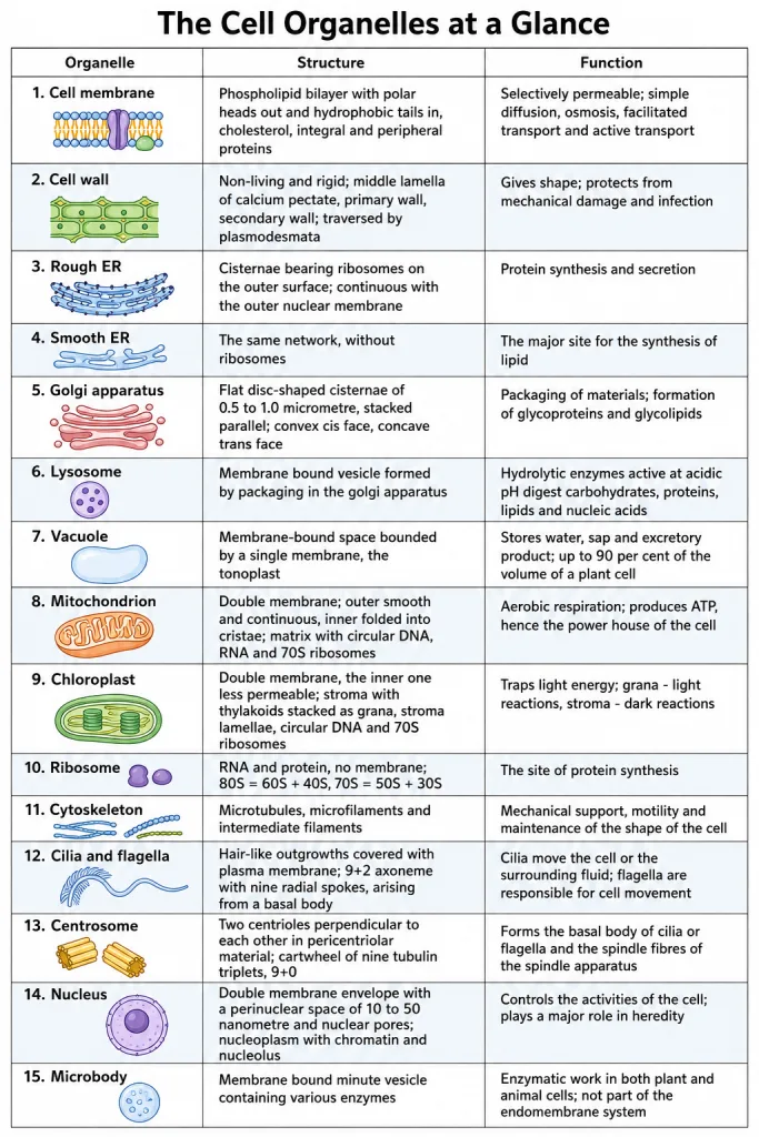
Now the organelles themselves, one row each. This is the table to be able to reproduce from memory.
| Organelle | Structure | Function |
|---|---|---|
| Plasma membrane | Phospholipid bilayer with polar heads out and hydrophobic tails in, cholesterol, and integral and peripheral proteins; fluid mosaic model of Singer and Nicolson, 1972 | Selectively permeable; transport of molecules by simple diffusion, osmosis, facilitated transport and active transport |
| Cell wall | Non-living and rigid; middle lamella of calcium pectate, primary wall capable of growth, secondary wall on the inner side; traversed by plasmodesmata | Gives shape, protects from mechanical damage and infection, helps in cell-to-cell interaction, barrier to undesirable macromolecules |
| Rough ER | Network of tubules and cisternae with ribosomes on the outer surface; continuous with the outer nuclear membrane | Protein synthesis and secretion |
| Smooth ER | The same network, without ribosomes | Major site for the synthesis of lipid; steroidal hormones in animal cells |
| Golgi apparatus | Flat disc-shaped cisternae, 0.5 to 1.0 micrometre, stacked parallel, with a convex cis face and a concave trans face | Packaging of materials for intra-cellular targets or secretion; formation of glycoproteins and glycolipids |
| Lysosome | Membrane bound vesicle formed by packaging in the golgi apparatus, filled with hydrolases active at acidic pH | Digests carbohydrates, proteins, lipids and nucleic acids |
| Vacuole | Membrane-bound space bounded by a single membrane, the tonoplast | Stores water, sap and excretory product; the tonoplast moves materials in against the gradient; osmoregulation and excretion in Amoeba |
| Mitochondrion | Double membrane; outer smooth and continuous, inner folded into cristae; matrix with circular DNA, RNA and 70S ribosomes | Aerobic respiration; produces ATP, hence the power house of the cell |
| Chloroplast | Double membrane, inner one less permeable; stroma with thylakoids stacked as grana, stroma lamellae, circular DNA and 70S ribosomes | Traps light energy for photosynthesis; grana - light reactions, stroma - dark reactions |
| Chromoplast and leucoplast | Chromoplast with fat soluble carotenoids; leucoplast colourless | Colour of flowers and fruits; storage of starch, oils and fats, or proteins |
| Ribosome | RNA and protein, no membrane; 80S = 60S + 40S, 70S = 50S + 30S | The site of protein synthesis |
| Cytoskeleton | Microtubules, microfilaments and intermediate filaments | Mechanical support, motility and maintenance of the shape of the cell |
| Microbody | Membrane bound minute vesicle with various enzymes | Enzymatic work in both plant and animal cells; not part of the endomembrane system |
| Cilium and flagellum | Hair-like outgrowths covered with plasma membrane; axoneme of 9+2, nine radial spokes, basal body | Cilia move the cell or the surrounding fluid; flagella are responsible for cell movement |
| Centrosome and centriole | Two cylindrical centrioles perpendicular to each other in pericentriolar material; cartwheel of nine tubulin triplets, 9+0, with a proteinaceous hub | Forms the basal body of cilia or flagella and the spindle fibres of the spindle apparatus |
| Nucleus | Double membrane envelope with a perinuclear space of 10 to 50 nanometre and nuclear pores; nucleoplasm with chromatin and nucleolus | Controls the activities of the organelles; plays a major role in heredity |
| Nucleolus | Spherical, not membrane bound, continuous with the nucleoplasm | Site for active ribosomal RNA synthesis |
The Eight Comparisons Worth Memorising
- Prokaryotic against eukaryotic cell - no membrane bound nucleus and no membrane bound organelles except ribosomes, 70S ribosomes, naked circular DNA, mesosome present against an organised nucleus with a nuclear envelope, compartmentalised cytoplasm with membrane bound organelles, 80S cytoplasmic ribosomes and genetic material organised into chromosomes.
- Plant against animal cell - cell wall, plastids and a large central vacuole present against absent; centrioles absent in almost all plant cells against present in animal cells.
- Rough ER against smooth ER - ribosomes on the outer surface, continuous with the outer nuclear membrane, protein synthesis and secretion against no ribosomes, the major site for the synthesis of lipid and of steroidal hormones.
- Cis against trans face of the golgi - cis is the forming face and is convex, and vesicles from the ER fuse with it against trans is the maturing face and is concave, and modified material is released from it.
- Lysosome against vacuole - a vesicle made by packaging in the golgi, full of hydrolases active at acidic pH, digesting all four classes of macromolecules against a membrane-bound space bounded by the tonoplast, storing water, sap and excretory product, and taking materials in against the gradient.
- Mitochondrion against chloroplast - both double membrane bound with their own circular DNA and 70S ribosomes, but cristae from the inner membrane and a matrix, doing aerobic respiration and making ATP against thylakoids stacked into grana within a stroma, trapping light energy for photosynthesis.
- 9+2 against 9+0 - the axoneme of a cilium or flagellum: nine peripheral doublets, a central pair, a central sheath and nine radial spokes against the centriole: nine peripheral triplets, no central tubule, a proteinaceous hub and a cartwheel organisation.
- Chromatin against chromosome - the same material in two states: highly extended and elaborate nucleoprotein fibres, loose and indistinct, in the interphase nucleus against structured bodies visible only during cell division, each with a primary constriction or centromere holding two chromatids.
Writing the Chapter-End Exercises Well
Class 11 has no board paper, but the fourteen chapter-end exercises are the best test of this chapter that exists, because between them they cover almost every fact that gets asked. Four of them ask for labelled diagrams, and diagrams are where marks are lost most cheaply. A labelled drawing scores on the labels, not on the artwork.
The nucleus. The drawing must carry, at the very least: the nuclear envelope shown as two parallel membranes, the perinuclear space between them, at least two nuclear pores drawn as places where the two membranes fuse, the outer membrane running on into the endoplasmic reticulum and bearing ribosomes, the nucleoplasm or nuclear matrix, the chromatin and the nucleolus. Two labels decide the mark here - the perinuclear space and the nuclear pore - and both are dropped by most students.
The centrosome. Draw two centrioles lying perpendicular to each other, inside the amorphous pericentriolar material. Then draw one of them in section as a cartwheel: nine evenly spaced peripheral fibrils, each a triplet, the adjacent triplets linked, the proteinaceous hub in the centre, and the radial spokes joining the hub to the peripheral triplets. Write 9+0 beside it and add the line that there is no central microtubule.
The mitochondrion. A longitudinal section, with the outer membrane as a smooth continuous limiting boundary, the inner membrane thrown into cristae projecting towards the matrix, the outer compartment between the two membranes, the inner compartment, and the matrix with circular DNA and 70S ribosomes marked in it. Add one line - the cristae increase the surface area - and one more - the site of aerobic respiration and of ATP production.
The chloroplast. A sectional view showing the double membrane, the stroma, the thylakoids, the grana as stacks of thylakoids like piles of coins, the stroma lamellae connecting the thylakoids of different grana, and the lumen inside a thylakoid. Mark grana - light reactions and stroma - dark reactions on the diagram itself; that pairing is worth as much as the drawing.
The habit all fourteen exercises reward: give the name, then the location, then the function, in that order, for every structure you mention. When an exercise asks for "the characteristics of these two organelles and their functions", it is asking for exactly that sequence three times over. Most of the marks lost in this chapter are lost by naming a structure correctly and then never saying where it sits or what it does.
Two more habits worth copying from the exercises. When a question says "comment" or "discuss in brief" - as the ones on lysosomes against vacuoles and on the cell as the basic unit of life do - answer in the shape of a comparison or a list of numbered points, not in one long paragraph. And when a question offers statements to be judged correct or not correct, read every option to the end; the correct-looking one is usually built by swapping two real names, as in "Robert Brown discovered the cell".
NEET Strategy
What the paper asks. One organelle, one number, one function, one odd-one-out - with a distractor pulled from a neighbouring organelle. There is nothing here to work out, so speed comes from certainty, not from technique. This chapter usually contributes more questions than any other in the unit, so the time you save here is time you keep for physiology.
The marking is +4 and -1. On a pure-recall question a coin-flip between two options loses marks on average. If the pair has genuinely gone, skip it and bank the time.
Budget. Aim for 25 to 30 seconds per question in this chapter. If a question is still open at forty seconds, it is not going to be solved by staring at it - mark it and move.
The four shapes, in the order they are easiest.
- Give the number or the size - 0.3 micrometre, 70S, nine radial spokes, 10 to 50 nanometre, 90 per cent, forty six chromosomes. These are instant if you know them and hopeless if you do not, so decide in five seconds.
- State the one function - power house, packaging, ribosomal RNA synthesis, digestion. Answer from the organelle's name and move on.
- Name the organelle from its description - here you can stop reading as soon as one clue identifies it: "infoldings towards the matrix" is the mitochondrion and nothing else.
- Spot the odd one out - leave these for last, since a "which is NOT" question forces you to check all four options.
Read the direction word before you read the options. NOT, INCORRECT and respectively are the three words that turn a fact you know into a mark you lose. Underline them as you read. In a respectively question, match the first pair first - it eliminates two options in one step more often than not.
The Mistakes That Cost the Most Marks
- Leeuwenhoek and Robert Brown swapped. Leeuwenhoek first saw and described a live cell; Robert Brown discovered the nucleus in 1831.
- Schleiden and Schwann swapped. Schleiden was the botanist, Schwann the zoologist, and it is Schwann who described the plasma membrane and said the cell wall is unique to plants.
- "Prokaryotes have no organelles" written without the exception. The correct sentence is "no organelles, like the ones in eukaryotes, are found in prokaryotic cells except for ribosomes".
- 70S and 80S subunits mixed. 70S = 50S + 30S. 80S = 60S + 40S. They do not add up because S is a sedimentation coefficient, not a mass, and trying to make the arithmetic work is what produces the wrong pairing.
- Cristae, thylakoids and cisternae confused. Cristae - infoldings of the inner mitochondrial membrane. Thylakoids - flat membranous sacs in the stroma. Cisternae - disc-shaped sacs of the golgi apparatus.
- Cis and trans faces reversed. Cis is the forming face and is convex; trans is the maturing face and is concave.
- Doublets and triplets swapped. The axoneme has nine doublets with a central pair, 9+2. The centriole has nine triplets with no central tubule, 9+0.
- Calling mitochondria, chloroplast or peroxisomes part of the endomembrane system because they are membrane bound. They are excluded because their functions are not coordinated with the ER, golgi, lysosomes and vacuoles.
- Saying the tonoplast moves materials along the gradient. It moves them against concentration gradients, into the vacuole.
- Naming an organelle and stopping there. In a written answer the function, and often the location, is worth as much as the name - and in a diagram, an unlabelled structure is worth nothing at all.
A Short Revision Plan
First pass - one hour and thirty minutes. Read sections 1 to 14 straight through without stopping to memorise. You are building the map: cell theory and cell size, then the prokaryote, then the membrane and the wall, then the organelles one by one, then the nucleus and the chromosome. Do not stop to learn numbers on this pass.
Second pass - one hour and thirty minutes. Work the Solved Examples in each section, writing the answers out rather than reading them, then check your wording against the answer given and mark only the words you missed. The marking-scheme keywords are in bold in every answer for exactly this purpose.
Third pass - forty minutes. Learn the two tables in this section - plant cell against animal cell, and the organelle, structure, function table - and the eight comparisons above. Then say the numbers out loud in one run: 0.3 micrometre, 3 to 5 micrometre, 7.0 micrometre, 52 and 40 per cent, 0.5 to 1.0 micrometre cisternae, 90 per cent, 0.2 to 1.0 and 1.0 to 4.1 micrometre, 5 to 10 and 2 to 4 micrometre, 70S and 80S, 50S and 30S, 60S and 40S, nine spokes, 10 to 50 nanometre, two metre, forty six.
Fourth pass - one hour. Sit the 45 NEET-Pattern Practice Questions under time, then go back only to the rows of the tables that your wrong answers came from. Redo the same questions three days later; the second attempt is what tells you whether the fact stuck.
The night before. Read the Master Quick Recap and the eight comparisons once, then the ten mistakes. Nothing else, and no new material. This chapter rewards a clean recent pass far more than a long one.