
Why We Need to Separate the Fragments
When DNA is cut with restriction endonucleases, the result is a mixture of fragments of different sizes. Before we can pick out and use the one we want, these fragments have to be separated from one another. The technique that does this is gel electrophoresis.
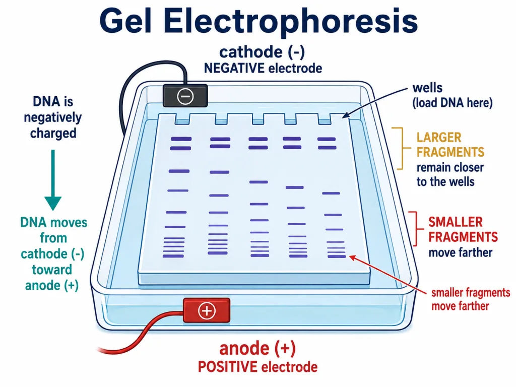
The idea rests on a simple property of DNA: it is a negatively charged molecule, because of the phosphate groups along its backbone. If we place the fragments in an electric field, they will move — and they will always move towards the positive electrode, the anode.
The Matrix — Agarose
The fragments do not move through open liquid; they are forced to travel through a matrix that acts like a molecular sieve. The most commonly used matrix today is agarose, a natural polymer extracted from seaweed.
Think of the agarose gel as a mesh full of tiny channels. As the fragments are driven towards the anode, they have to thread their way through this mesh. This sieving effect is exactly what separates them by size.
Smaller Fragments Travel Farther
Because the gel behaves as a sieve, larger fragments get held back more than smaller ones. A small fragment slips through the mesh with relative ease, while a bulky one is slowed down. The rule that follows is worth remembering: the smaller the fragment, the farther it moves in a given run.
So after electrophoresis, the fragments resolve into a series of positions along the gel, sorted by size — the largest nearest the well where the sample was loaded, the smallest closest to the anode.
Seeing the DNA and Recovering It
There is a catch: pure DNA is invisible in visible light and cannot be seen without staining. To make the separated fragments visible, the gel is stained with a compound called ethidium bromide and then exposed to UV radiation. Under UV light the DNA shows up as bright orange bands, each band a group of fragments of the same size.
Once the desired band has been located, it is cut out of the agarose gel and the DNA is extracted from that gel piece. This recovery step is called elution. The DNA purified in this way is then ready to be joined with cloning vectors to construct recombinant DNA.
Quick Recap
- Gel electrophoresis separates DNA fragments produced by restriction digestion.
- DNA is negatively charged, so under an electric field the fragments move towards the anode (positive electrode).
- They travel through a matrix — commonly agarose, a natural polymer from seaweed — which acts as a sieve.
- Fragments resolve by size: the smaller the fragment, the farther it moves.
- DNA is invisible unstained; staining with ethidium bromide and exposure to UV shows bright orange bands.
- The wanted band is cut out and the DNA extracted — a step called elution — then used with cloning vectors to make recombinant DNA.
Solved Examples — Section 6
Q1. Why do DNA fragments move towards the anode in gel electrophoresis?
Answer: DNA carries a net negative charge because of its phosphate groups, so under an electric field the fragments are attracted to the positive electrode, the anode.
Q2. What is the matrix commonly used, and where does it come from?
Answer: Agarose, a natural polymer extracted from seaweed.
Q3. In a gel, which fragments move farther — larger or smaller?
Answer: Smaller fragments move farther, because the sieving effect of the gel holds back the larger ones more.
Q4. How are the separated DNA fragments made visible?
Answer: The gel is stained with ethidium bromide and exposed to UV radiation, which makes the DNA appear as bright orange bands.
Q5. What is elution?
Answer: The step in which the desired DNA band is cut out of the agarose gel and the DNA is extracted from that gel piece.
Q6. What happens to the purified DNA fragments after elution?
Answer: They are used to construct recombinant DNA by joining them with cloning vectors.