
The Electrical Record, the Control of the Beat, and the Failing Circulation
What the tracing is, and how it is taken
An electrocardiograph writes a graphical record of the electrical activity of the heart through one cardiac cycle. The record itself is the electrocardiogram, or ECG: an ECG is a graphical record of the heart's electrical activity, never of its mechanical pumping. That single word electrical governs everything you can read off the paper. Pressure, volume, valve sounds and the force of a contraction are not drawn on it; they are inferred, and only loosely. What the pen follows is excitation spreading through muscle.
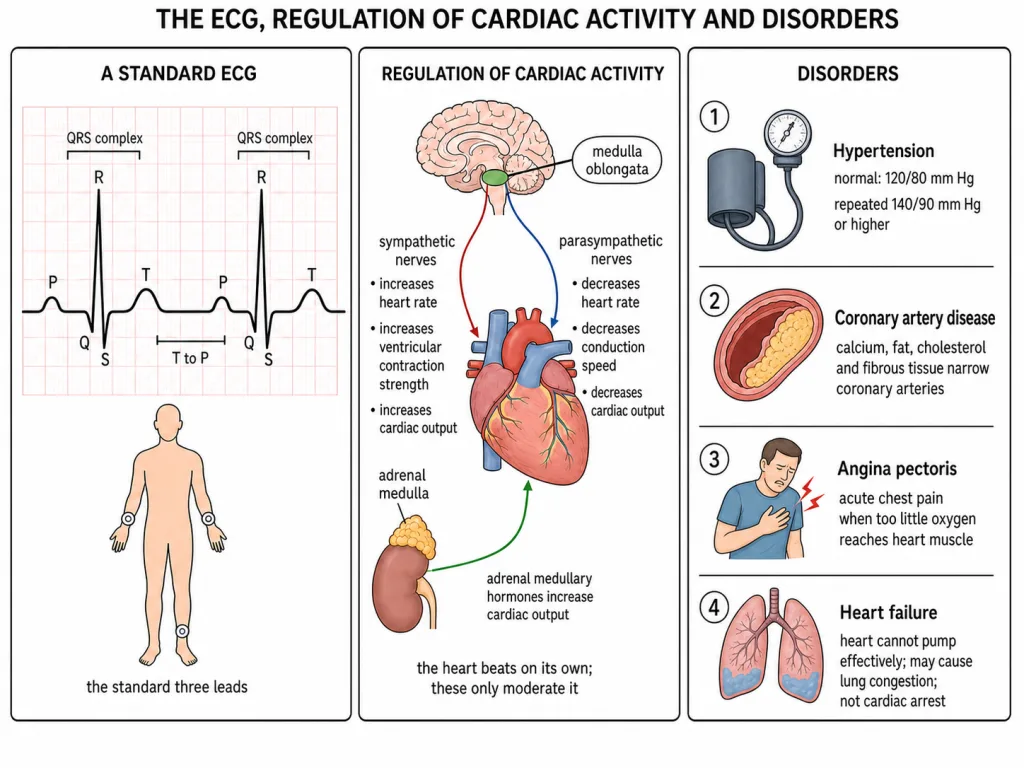
The standard technique is deliberately simple. The patient is connected to the machine by three electrical leads, one on each wrist and one on the left ankle. Nothing is placed on the right ankle in this arrangement, and the leads are not attached over the heart itself. Because the trace has a settled normal shape, any deviation from that standard shape indicates a possible abnormality or disease. That is the whole clinical logic: you compare a shape against a known shape, you do not read a diagnosis directly off a wave.
The waves, and the mechanics they bracket
The P wave is a small wave representing the depolarisation of the atria, and that excitation is what leads both atria to contract. The QRS complex represents the depolarisation of the ventricles. Its timing is the detail worth holding: ventricular contraction starts just after the Q wave, so the complex begins slightly ahead of the mechanical event it triggers. The T wave represents the return of the ventricles from their excited state to normal, that is, repolarisation. Here too the mechanical anchor matters more than the label: the end of the T wave marks the end of systole.
Between the end of one T wave and the beginning of the next P wave the trace runs flat. In that T-P gap the heart muscle is electrically silent: no depolarisation and no repolarisation is under way anywhere in it.
One practical use falls straight out of the trace. Since the ventricles fire once per beat, counting the number of QRS complexes in a known period of time gives the heart rate. You count complexes, not waves; a single beat contributes one P, one QRS and one T, so counting individual waves would inflate the figure.
The beat that needs no nerve
Normal activity of the heart is regulated intrinsically, that is, it is auto-regulated by the nodal tissue. This is what is meant by calling the heart myogenic: the impulse that drives each beat originates in the muscle tissue itself, so the heart does not require any nerve supply in order to beat at its normal rhythm.
Nerves are still able to moderate that rhythm. A neural centre in the medulla oblongata can modify cardiac function through the autonomic nervous system. Sympathetic signals increase three things: the rate of heart beat, the strength of ventricular contraction and therefore the cardiac output. Parasympathetic signals decrease the rate of heart beat, the speed of conduction of the action potential and therefore the cardiac output. Read the middle term of each triplet carefully, because that is where the two lists genuinely differ: sympathetic acts on contraction strength, parasympathetic on conduction speed. Adrenal medullary hormones can likewise increase cardiac output, and they reach the heart in the blood rather than along a nerve.
Pressure, and the arteries that feed the pump
Normal blood pressure is written 120/80 mm Hg, and the order is fixed: the first figure is the systolic, or pumping, pressure and the second is the diastolic, or resting, pressure. Repeated readings of 140/90 mm Hg or above amount to hypertension, or high blood pressure. It is not a nuisance number. Hypertension leads to heart diseases and also affects vital organs such as the brain and the kidney.
Coronary artery disease, often called atherosclerosis, affects the vessels that supply blood to the heart muscle itself. It is caused by deposits of calcium, fat, cholesterol and fibrous tissue in the wall of those arteries, and the consequence is that the lumen becomes narrower.
Angina, and the pump that falls behind
Angina pectoris, or simply angina, is the acute chest pain that appears when not enough oxygen is reaching the heart muscle. It can occur in men and women of any age, though it is more common among the middle-aged and the elderly.
Heart failure is the state in which the heart is not pumping blood effectively enough to meet the needs of the body. It is sometimes called congestive heart failure because congestion of the lungs is the main symptom of the condition, and that congestion is the reason for the name.
Two neighbouring terms are not the same thing, and the distinction is exact. Cardiac arrest is when the heart stops beating. A heart attack is when the heart muscle is suddenly damaged by an inadequate blood supply. Heart failure is a sustained shortfall in pumping while the heart continues to beat; the other two name a stopped heart and a damaged muscle respectively.