
Circulatory Patterns and Vertebrate Hearts
Open and closed circulation
In an open circulatory system, blood pumped by the heart passes through large vessels into open spaces or body cavities called sinuses. NCERT lists arthropods and molluscs for this pattern. In a closed circulatory system, blood remains within a closed network of vessels; annelids and chordates show this arrangement. Closed circulation is more advantageous because blood flow can be regulated more precisely.
Vertebrate heart patterns
All vertebrates possess a muscular, chambered heart:
- Fishes: two chambers - one atrium and one ventricle. The heart pumps deoxygenated blood to the gills; oxygenated blood then goes directly to the body before returning to the heart. Blood passes through the heart once per circuit, so this is single circulation.
- Amphibians and reptiles except crocodiles: three chambers - two atria and one ventricle. Oxygenated and deoxygenated blood mix in the common ventricle, producing incomplete double circulation.
- Crocodiles, birds and mammals: four chambers - two atria and two ventricles. Crocodiles are the exception to the usual reptilian chamber count. NCERT specifically describes the fully separated pathways of birds and mammals as double circulation without mixing.
Position, covering and chambers of the human heart
The human heart is a mesodermally derived, muscular organ in the thoracic cavity between the two lungs, tilted slightly to the left and about the size of a clenched fist. A double-walled membranous bag, the pericardium, encloses the heart and contains pericardial fluid.
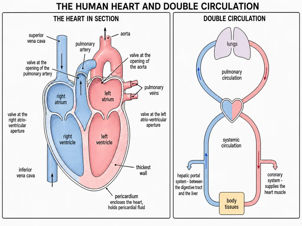
The heart has two relatively small upper chambers, the atria, and two larger lower chambers, the ventricles. A thin muscular inter-atrial septum separates the atria. A thick inter-ventricular septum separates the ventricles. On each side, a thick fibrous atrio-ventricular septum separates the atrium from the ventricle and contains the opening connecting those two chambers.
The ventricular walls are much thicker than the atrial walls because ventricles pump blood out of the heart. The left ventricular wall is the thickest because it generates the pressure needed for the systemic circuit.
Valves and the fixed direction of flow
The right atrio-ventricular opening is guarded by the tricuspid valve, which has three cusps. The left opening is guarded by the two-cusped bicuspid or mitral valve. Semilunar valves guard the openings of the right ventricle into the pulmonary artery and the left ventricle into the aorta. All these valves permit flow in only one direction and prevent backward flow.
Follow the route in order:
Body → venae cavae → right atrium → tricuspid valve → right ventricle → pulmonary artery → lungs → pulmonary veins → left atrium → bicuspid valve → left ventricle → aorta → body
An artery carries blood away from the heart and a vein carries blood towards it; oxygen content does not define the vessel. Therefore, the pulmonary artery carries deoxygenated blood away from the heart, whereas the pulmonary veins return oxygenated blood.
Pulmonary and systemic circulation
The route from the right ventricle through the lungs to the left atrium is the pulmonary circulation. The route from the left ventricle through the body tissues to the right atrium is the systemic circulation. Systemic blood delivers oxygen, nutrients and other essential substances and carries carbon dioxide and other harmful substances away. Together, the two pathways form double circulation: blood passes through the heart twice in one complete circuit. In humans, complete separation prevents oxygenated and deoxygenated blood from mixing.
According to NCERT, each artery and vein has three coats:
- Tunica intima: inner squamous endothelial lining
- Tunica media: smooth muscle and elastic fibres; comparatively thinner in veins
- Tunica externa: outer fibrous connective tissue containing collagen fibres
Do not extend this three-coat description to capillaries, whose walls are essentially a single endothelial layer.
Two special vascular routes are high-yield. The hepatic portal system carries blood from the intestine to the liver before it enters the systemic circulation. The coronary system carries blood to and from the cardiac musculature.