
Nodal Tissue and the Cardiac Cycle
The conducting system
Nodal tissue is specialised cardiac musculature distributed in the heart. The sino-atrial node (SAN) lies in the upper right corner of the right atrium. The atrio-ventricular node (AVN) lies in the lower left corner of the right atrium, close to the atrio-ventricular septum.
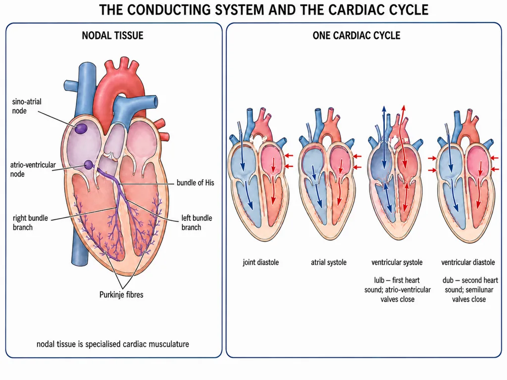
From the AVN, the atrio-ventricular bundle or bundle of His passes through the atrio-ventricular septum, reaches the top of the inter-ventricular septum and divides into right and left bundle branches. These branches give rise to Purkinje fibres throughout the ventricular musculature. The NCERT sequence is:
SAN → AVN → AV bundle (bundle of His) → right and left bundle branches → Purkinje fibres
Nodal tissue is autoexcitable: it generates action potentials without an external stimulus. The SAN generates the maximum number, about 70-75 per minute, so it initiates and maintains rhythmic contraction and is called the pacemaker. The normal heart rate is about 70-75 beats per minute, with an average of 72.
Joint diastole and atrial systole
A cardiac cycle is the sequence of events in the heart that repeats cyclically and includes systole and diastole of both atria and ventricles. At 72 cycles per minute, one cycle lasts about 0.8 second.
At the start, all four chambers are relaxed in joint diastole. The tricuspid and bicuspid valves are open, the semilunar valves are closed, and blood from the venae cavae and pulmonary veins passes through the atria into the ventricles.
The SAN then generates an action potential, causing both atria to contract simultaneously. During this atrial systole, the atrio-ventricular valves remain open and atrial contraction increases ventricular filling by about 30 per cent. The impulse then reaches the ventricles through the AVN and AV bundle.
Ventricular systole, ventricular diastole and heart sounds
During ventricular systole, ventricular pressure first rises enough to close the tricuspid and bicuspid valves because of attempted backflow towards the atria. Both sets of valves are briefly closed. As ventricular pressure rises further above the pressure in the pulmonary artery and aorta, the semilunar valves open and blood is ejected. The atria are in diastole during this phase.
During ventricular diastole, falling ventricular pressure first causes the semilunar valves to close, preventing arterial backflow. The atrio-ventricular valves do not open at that same instant. They open later, after ventricular pressure falls below atrial pressure, and filling resumes until joint diastole is restored.
Each prominent heart sound is associated with valve closure:
- First sound, lub: closure of the tricuspid and bicuspid valves at the beginning of ventricular systole
- Second sound, dub: closure of the semilunar valves as ventricular systole ends and ventricular relaxation begins
Both sounds have clinical diagnostic significance.
Stroke volume and cardiac output
The volume pumped by each ventricle during one cardiac cycle is the stroke volume, approximately 70 mL. Cardiac output is the volume pumped by each ventricle per minute:
At 70 mL per beat and 72 beats per minute, the calculated value is about 5040 mL per minute, rounded by NCERT to approximately 5000 mL or 5 litres per minute in a healthy individual.
The body can alter both stroke volume and heart rate, so cardiac output is not fixed. NCERT states that an athlete can have a cardiac output much higher than that of an ordinary person; it does not prescribe a fixed five-to-six-times multiplier.