
The Human Eye - Our Window to the World
The human eye is one of the most valuable and sensitive sense organs. It lets us see the wonderful, colourful world around us. With our eyes closed we can still recognise objects to some extent by their smell, taste, sound or touch - but it is impossible to identify colours without seeing them. That is what makes the eye so special.
The eye works very much like a camera. A camera has a lens that forms an image on a light-sensitive film or sensor. In the eye, the lens system forms an image on a light-sensitive screen at the back called the retina.
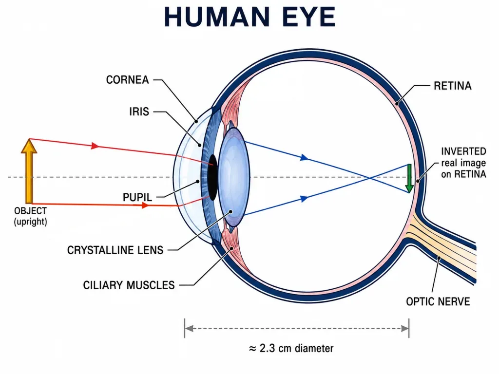
The eyeball is approximately spherical, with a diameter of about 2.3 cm.
The Main Parts of the Eye and What They Do
Cornea - Light first enters the eye through a thin transparent membrane called the cornea, which forms the transparent bulge on the front of the eyeball. Most of the refraction (bending) of light entering the eye happens at the outer surface of the cornea, not at the lens.
Crystalline lens - Behind the cornea is the eye lens, made of a fibrous, jelly-like material. It does not do most of the bending; it merely provides the fine adjustment of focal length needed to focus objects at different distances sharply on the retina.
Iris - Behind the cornea lies the iris, a dark muscular diaphragm that controls the size of the pupil. The iris is the coloured part of the eye.
Pupil - The pupil is the opening in the middle of the iris. It regulates and controls the amount of light entering the eye - widening in dim light and narrowing in bright light.
Retina - The retina is a delicate screen at the back of the eye with an enormous number of light-sensitive cells.
Optic nerve - Nerve that carries signals from the retina to the brain.
How the Eye Forms an Image
When light from an object enters the eye, the cornea and the eye lens together refract it and form an inverted, real image of the object on the retina - just like a convex lens forms a real image on a screen.
The light-sensitive cells of the retina get activated when light falls on them and generate tiny electrical signals. These signals travel to the brain through the optic nerves. The brain interprets the signals, and finally processes the information - correcting the inverted image - so that we perceive objects the right way up and as they really are.
Key Point: The image on the retina is real and inverted; it is the brain that lets us perceive the world upright.
[Exam Tip] Remember the light path in order: Cornea → Pupil (aperture set by iris) → Crystalline lens → Retina → Optic nerve → Brain.
Solved Examples
Example 1: The eye and the camera
In what way is the human eye similar to a camera, and what part plays the role of the film?
Solution: A camera uses a lens to form a real, inverted image on a light-sensitive film/sensor. Similarly, the eye's lens system (cornea + crystalline lens) forms a real, inverted image on the retina, which is the light-sensitive screen. So the retina plays the role of the camera film.
Example 2: Where does most bending happen?
A student thinks the crystalline lens does most of the focusing in the eye. Correct this idea.
Solution: Most of the refraction of incoming light actually occurs at the outer surface of the cornea. The crystalline lens only provides the fine adjustment of focal length (accommodation) needed to sharply focus objects at different distances. So the cornea is the main refracting surface, not the lens.
Example 3: Controlling light
Which part of the eye controls the amount of light entering it, and how?
Solution: The iris (a muscular diaphragm) controls the size of the pupil. In bright light the pupil contracts (becomes small) to let in less light; in dim light it dilates (becomes large) to let in more light. Thus the iris–pupil system regulates the light reaching the retina.