
Why Excretion Is the Final Life Process
Throughout this chapter we've followed materials coming in (food via digestion, oxygen via respiration) and moving around (via blood and xylem/phloem). Now the final step: what comes out, and how?
Every living process produces waste. If wastes accumulate, they become toxic and the organism dies. Removal of these wastes is excretion — the 6th life process and the finishing line of this chapter.
Why we make wastes
Wastes are not just leftover food. They are toxic by-products of normal cell metabolism:
1. Nitrogenous wastes — from breakdown of proteins and amino acids.
- When extra amino acids cannot be stored in our body, they are broken down in the liver.
- The amino group (-NH₂) is removed (deamination), and converted to urea (less toxic, water-soluble).
- Ammonia → Urea → Excreted in urine
2. CO₂ — from cellular respiration (Section 5). Already excreted through lungs.
3. Extra water and salts — diet provides more than we need; the excess must go.
4. Drugs, toxins, pigments — anything foreign or no-longer-needed.
Why we need a separate excretory system
In unicellular organisms (Amoeba, Paramecium), wastes simply diffuse out through the cell membrane — no special organs needed.
But in multicellular humans:
- Most cells are far from the body surface.
- Wastes must be collected (by blood) and filtered out (by kidneys).
- Hence: a dedicated excretory system is essential.
Different organisms, different nitrogenous wastes
The nitrogenous waste form depends on water availability:
| Organism type | Waste | Why |
|---|---|---|
| Fish (in water) | Ammonia (NH₃) | Very toxic but very water-soluble. Plenty of water around → just let it diffuse out via gills. |
| Mammals, amphibians | Urea | Less toxic than ammonia; needs less water to dilute. Suitable for land animals with limited water. |
| Birds, reptiles, insects | Uric acid | Almost insoluble. Excreted as a semi-solid paste — saves the most water. Crucial for flying animals and desert reptiles. |
Humans excrete UREA — a compromise between toxicity and water requirement.
The four 'excretory organs' in humans (not just kidneys!)
| Organ | What it excretes |
|---|---|
| Kidneys (main) | Urea, excess water, salts, drugs |
| Lungs | CO₂, some water vapour |
| Skin | Sweat (water, salts, small amounts of urea) |
| Liver | Bile pigments (broken-down haemoglobin) — into intestine via bile |
Most students forget that lungs and skin are also excretory! In a Board exam, list all four for full marks.
Excretion in plants — preview
Plants also excrete, but very differently — we'll cover this at the end of the section.
NCERT-canonical phrase: "The biological process involved in the removal of these harmful metabolic wastes from the body is called excretion."
The Human Urinary System
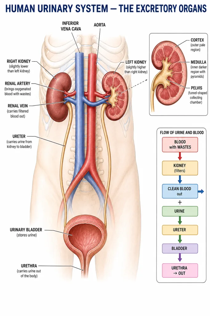
The urinary system is the main excretory system in humans. It has 4 main parts.
1. Kidneys — a pair of bean-shaped filters
Location: Two kidneys in the upper abdomen, one on each side of the spine, just below the rib cage. The right kidney is slightly lower than the left (because the liver pushes it down).
Size: About the size of your fist — roughly 10-12 cm long.
Colour: Dark reddish-brown (highly vascular — kidneys receive ~20% of all blood pumped by the heart, despite being only 0.5% of body weight!).
Function: The kidneys filter blood — removing wastes (urea, excess water, salts) and returning useful substances back to the blood.
Internal structure of a kidney
If you cut a kidney open, you'll see three distinct regions:
| Region | Description |
|---|---|
| Cortex | Outer pale layer — where most filtering units (nephrons) start |
| Medulla | Inner darker layer — contains the long tubules (Loop of Henle) and collecting ducts. Arranged in cone-shaped renal pyramids |
| Pelvis | Central funnel-shaped cavity that collects urine and channels it into the ureter |
Each kidney has about one million nephrons — the tiny functional units that do the actual filtering. We'll see one in detail in the next sub-section.
2. Ureters — the urine pipes
Two thin ureters (one from each kidney) carry urine from the kidneys down to the urinary bladder.
Length: ~25 cm each. Function: Transport urine via slow rhythmic muscular contractions (similar to peristalsis in the gut).
3. Urinary bladder — the storage sac
A hollow, muscular, sac-like organ that stores urine until it's convenient to release.
Capacity: Comfortable up to ~300-400 mL; can stretch to ~600 mL when full.
Muscles: The bladder wall has smooth muscle (involuntary) and a sphincter (ring muscle) at the exit. The sphincter is under voluntary control in adults — that's how we control when we urinate.
4. Urethra — the exit pipe
The urethra is a single tube that carries urine from the bladder to outside the body.
Length difference:
- In males: ~20 cm (passes through the penis; also carries semen).
- In females: ~4 cm (just exits below the genital opening).
This is why urinary tract infections (UTIs) are more common in females — bacteria have a shorter distance to travel to reach the bladder.
Blood supply — kidneys are super-filters
Each kidney receives:
- Renal artery — branches off the aorta. Brings blood with wastes into the kidney.
- Renal vein — joins the inferior vena cava. Carries the now-filtered (clean) blood out of the kidney.
Approximately 1,200 mL of blood flows through both kidneys per minute (≈25% of cardiac output).
Over 24 hours, the kidneys filter about 180 litres of fluid — but only 1.5 litres comes out as urine (because >99% of the filtered water is reabsorbed back into the blood).
Path of urine — memorise this flow
Always 4 parts in this order. Common 2-mark question.
Composition of urine
Normal urine contains:
- Water (~95%).
- Urea (~2%) — the main nitrogenous waste.
- Uric acid, creatinine (small amounts).
- Salts — Na⁺, K⁺, Cl⁻, etc.
- Pigments — urochrome (gives urine its yellow colour, from haemoglobin breakdown).
Urine should NOT contain: glucose, proteins, blood cells. Their presence indicates disease (diabetes, kidney damage, etc.).
[Board Important] "Name the parts of the human excretory system." — Always list all 4 in order: kidneys, ureters, urinary bladder, urethra. Common 2-mark question.
The Nephron — Structural and Functional Unit of the Kidney
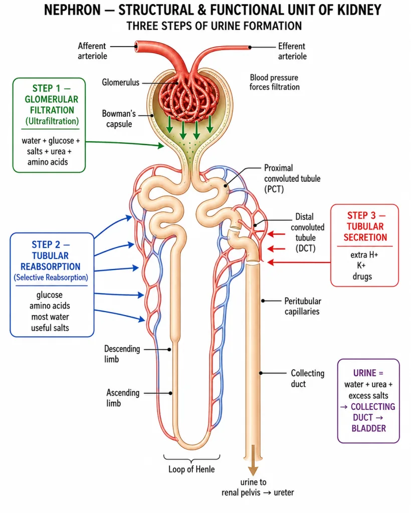
This is the most important diagram in the entire excretion section.
The nephron is the structural and functional unit of the kidney — meaning it is both the basic structural building block AND the unit that does the actual work of filtering.
Each kidney contains about 1 million nephrons. If you stretched all nephron tubules end-to-end, they would be ~85 km long!
Parts of a nephron
A nephron is essentially a long, twisted tubule connected to a cup-shaped capsule at one end and a collecting duct at the other. Let's trace it from start to finish.
1. Bowman's capsule
- A double-walled, cup-shaped structure at the top of the nephron.
- Located in the cortex.
- Encloses a tight ball of capillaries called the glomerulus.
- The capsule + glomerulus together are called the renal corpuscle or Malpighian body.
2. Glomerulus
- A tangled cluster of capillaries inside Bowman's capsule.
- Blood comes in via the afferent arteriole (wide).
- Blood leaves via the efferent arteriole (narrower — this creates pressure for filtration).
- Pressure here is very high — forces fluid out of blood into Bowman's capsule.
3. Proximal Convoluted Tubule (PCT)
- Highly coiled tubule emerging from Bowman's capsule.
- Located in the cortex.
- Site of most of the reabsorption.
4. Loop of Henle
- A long, hairpin-shaped loop that descends into the medulla and then comes back up.
- Two parts: descending limb (going down) and ascending limb (coming up).
- Concentrates the urine (makes it more concentrated than blood).
5. Distal Convoluted Tubule (DCT)
- A second coiled section.
- Back in the cortex.
- Site of fine-tuning of salt/water balance and the third step (tubular secretion).
6. Collecting Duct
- Multiple nephrons drain into one collecting duct.
- Passes through medulla → pelvis.
- Carries final urine into the renal pelvis → ureter.
Blood supply around the nephron — peritubular capillaries
After the efferent arteriole leaves the glomerulus, it doesn't go directly to a vein. Instead, it splits into a network of fine capillaries called the peritubular capillaries that wrap around the PCT, Loop of Henle, and DCT.
Function: These capillaries pick up the substances reabsorbed from the tubule (glucose, water, useful salts) and return them to the bloodstream. They also deliver substances to be secreted into the tubule (drugs, excess H⁺, etc.).
Cortical vs juxtamedullary nephrons
Two types exist (not very important for Class 10 but good to know):
- Cortical nephrons (~85%) — short Loop of Henle, mostly in cortex.
- Juxtamedullary nephrons (~15%) — long Loop of Henle dipping deep into medulla. These help concentrate urine in mammals living in dry environments.
Why is it called the 'structural and functional unit'?
- Structural — it is the basic building block (one million per kidney).
- Functional — each nephron performs the entire urine-formation process on its own (it filters blood, reabsorbs useful stuff, secretes wastes, and produces urine).
Compare: Neuron is the structural and functional unit of the nervous system; cell is the structural and functional unit of life.
Quick summary of nephron parts (memorise this order!)
Glomerulus (inside Bowman's capsule)
    ↓
Bowman's capsule
    ↓
Proximal Convoluted Tubule (PCT)
    ↓
Descending Loop of Henle
    ↓
Ascending Loop of Henle
    ↓
Distal Convoluted Tubule (DCT)
    ↓
Collecting Duct
    ↓
Renal pelvis → Ureter
Memory tip: G-B-P-L-D-C — Glomerulus, Bowman, PCT, Loop, DCT, Collecting.
[Board Important] "Draw a labelled diagram of a nephron." — Classic 5-mark Board question. Must label: Bowman's capsule, glomerulus, PCT, Loop of Henle, DCT, collecting duct, afferent and efferent arterioles.
Three Steps of Urine Formation
Now that we know the structure, let's see what actually happens inside a nephron. Urine formation occurs in three distinct steps, each at a different part of the nephron.
Step 1: Glomerular Filtration (= Ultrafiltration)
Site: The glomerulus (capillaries) and Bowman's capsule.
What happens:
- Blood enters the glomerulus through the afferent arteriole (wide).
- Blood leaves through the efferent arteriole (narrower).
- Because more blood enters than can leave, high pressure builds up in the glomerulus.
- This high pressure forces water and small dissolved molecules OUT of the blood, through the thin walls of the glomerular capillaries and into the Bowman's capsule.
What gets filtered through (into Bowman's capsule):
- Water.
- Glucose.
- Amino acids.
- Salts (Na⁺, K⁺, Cl⁻, etc.).
- Urea (the main waste).
- Vitamins.
What stays in blood (too big to pass through):
- Blood cells (RBCs, WBCs, platelets).
- Plasma proteins (albumin, globulins).
- Large molecules.
Volume: About 180 litres of filtrate per day! (Compare your daily water intake of ~2 L.)
Result: The fluid now in Bowman's capsule (called glomerular filtrate) is similar to blood plasma — but without the big proteins and cells.
Why is it called 'ULTRAfiltration'? Because it filters small molecules at a very high pressure — not just simple diffusion. The 'ultra' refers to the fineness of the filter and the speed.
Step 2: Tubular Reabsorption (= Selective Reabsorption)
Site: Mainly PCT, then Loop of Henle, then DCT.
What happens:
- As the filtrate flows down the long, twisty tubule, useful substances are reabsorbed back into the surrounding peritubular capillaries (and hence back into blood).
- This is selective — only the useful stuff goes back. Wastes stay in the tubule.
What gets reabsorbed (back into blood):
- Glucose — 100% reabsorbed (in PCT). None should be in urine.
- Amino acids — 100% reabsorbed (in PCT).
- Water — ~99% reabsorbed (in PCT, Loop of Henle, collecting duct).
- Useful salts — most reabsorbed (especially Na⁺, K⁺, Cl⁻, Ca²⁺).
- Vitamins — mostly reabsorbed.
What does NOT get reabsorbed (continues to urine):
- Urea (the waste!).
- Uric acid, creatinine.
- Excess salts.
- Excess water (depending on hydration).
Volume reduction: The 180 litres of filtrate gets reduced to about 1.5 litres of urine — that's a 99% reabsorption of water!
Why is it called 'SELECTIVE'? Because the body picks only useful substances to reabsorb — not random absorption. This is active transport in many cases (uses ATP).
Step 3: Tubular Secretion
Site: Mainly DCT (and to some extent collecting duct).
What happens:
- Some additional substances are actively secreted from the peritubular blood capillaries INTO the tubule — to be added to urine.
- This 'fine-tunes' the urine composition.
Substances secreted into urine:
- Excess H⁺ ions (to control blood pH).
- Excess K⁺ ions.
- Drugs and their metabolites (e.g., penicillin).
- Some creatinine.
- Ammonia.
Why is this needed? Because some wastes are present in such small amounts in blood that filtration alone won't remove them efficiently. Active secretion ensures they end up in urine.
Putting it all together — urine formation summary
| Step | Site | Direction | What moves |
|---|---|---|---|
| 1. Filtration | Glomerulus → Bowman's capsule | Blood → tubule | Water, small molecules (incl. urea) |
| 2. Reabsorption | PCT → Loop of Henle → DCT | Tubule → blood | Glucose, amino acids, most water, useful salts |
| 3. Secretion | DCT (mainly) | Blood → tubule | Excess H⁺, K⁺, drugs, creatinine |
Net result: Wastes + extra water + extra salts → urine. Useful stuff → back in blood.
How is the volume of urine controlled?
The amount of water reabsorbed depends on how much water is in your body:
- Body needs water (dehydrated) → kidney reabsorbs MORE water → urine is less in volume, more concentrated (darker yellow).
- Body has extra water (well-hydrated) → kidney reabsorbs LESS water → urine is more in volume, dilute (pale yellow).
This is controlled by a hormone called ADH (anti-diuretic hormone) released by the pituitary gland. (Brief mention only — details in Class 11/12.)
This is why the colour of your urine tells you whether to drink more water.
Why isn't filtration alone enough?
If the kidney only did filtration:
- It would lose 180 L of water/day → instant dehydration!
- It would lose all glucose, amino acids — starvation.
Reabsorption is what makes filtration practical — it recovers everything useful, leaving only true waste behind.
NCERT-canonical phrase
The NCERT textbook describes urine formation as: "The urine forming in each kidney eventually enters a long tube, the ureter, which connects the kidneys with the urinary bladder. Urine is stored in the urinary bladder until the pressure of the expanded bladder leads us to its expulsion through the urethra."
[Board Important] "Explain the process of urine formation." — Classic 5-mark Board question. Must cover ALL three steps: filtration, reabsorption, secretion — with sites and what moves at each step.
Dialysis — The Artificial Kidney
What if the kidneys fail? This is a real-life situation we must understand.
When do kidneys fail?
Kidney failure (also called renal failure) can happen due to:
- Diabetes — long-term high blood sugar damages glomeruli.
- Hypertension — high blood pressure damages kidney capillaries.
- Infections (kidney infections, repeated UTIs).
- Kidney stones blocking urine flow.
- Trauma (accidents).
- Toxins / drugs (some medications, heavy metals).
Effect of kidney failure:
- Wastes accumulate in blood — urea, creatinine, uric acid all rise.
- Excess water builds up → swelling (edema), high blood pressure.
- Electrolyte imbalance → heart and nerve problems.
- If untreated, kidney failure is fatal within days.
What is dialysis?
Dialysis = an artificial process that performs the kidney's filtration function outside the body, using a special machine called a dialyser or artificial kidney.
It is not a cure — it's a life-support treatment that keeps patients alive until a kidney transplant or recovery.
How does dialysis work?
The principle is selective diffusion across a semi-permeable membrane — similar to what nephron tubules do.
Setup:
- The patient's blood is drawn out through a tube (usually from a large vein in the arm).
- The blood flows into a dialyser — a chamber containing thousands of fine tubes made of a semi-permeable membrane.
- The tubes are bathed in a dialysing fluid — a fluid carefully prepared to have:
- Normal levels of glucose, salts.
- No urea, no toxic wastes.
What happens in the dialyser:
- Wastes (urea, etc.) in the blood are in higher concentration than in the fluid → they diffuse OUT of the blood into the dialysing fluid (down the concentration gradient).
- Useful substances (glucose, salts) are at normal levels in the fluid → no net loss from blood.
- Excess water can also be removed by adjusting pressure.
- The cleaned blood is returned to the patient's body (back into a vein in the arm).
Result: The patient's blood is filtered without using their failed kidneys.
Key differences from real kidneys
| Feature | Real Kidney | Dialysis Machine |
|---|---|---|
| Number of nephrons | 2 million (both kidneys) | None — uses semi-permeable tubes |
| Filtration | Pressure-driven (glomerulus) | Diffusion-driven (concentration gradient) |
| Active reabsorption/secretion | YES | NO — purely diffusion |
| Runs continuously | 24×7 | Only during sessions |
| Hormonal regulation | YES (ADH, etc.) | NO |
| Energy used | Cellular ATP | Machine pumps |
Real kidneys are vastly more sophisticated. Dialysis is a 'crude' substitute — but life-saving.
Practical aspects
- A patient on dialysis usually needs 3 sessions per week, each lasting 4-5 hours.
- Done at a hospital or specialised dialysis centre.
- Each session removes ~500 mL of urea-rich fluid.
- Strict diet (low salt, low protein, restricted water) helps reduce waste between sessions.
Long-term solution: kidney transplant
For permanent kidney failure, a kidney transplant is the only true solution:
- A healthy kidney from a donor (often a relative) is surgically implanted in the patient.
- The donor needs only ONE healthy kidney for normal life (humans can live with one kidney).
- The patient must take immunosuppressive drugs lifelong to prevent rejection.
Kidney donation is one of the most life-changing gifts a human can give.
Why we have TWO kidneys
This is a common 'curiosity' question. Reasons:
- Backup — if one fails or is damaged, the other can take over.
- Increased filtration capacity — twice as many nephrons = better waste removal.
- Surface area for blood supply — both kidneys receive ~25% of cardiac output combined.
A person can survive normally with just ONE kidney. The other is essentially a backup.
[Board Important] "What is dialysis? When is it needed?" — Standard 3-mark Board question. Always mention: kidney failure, semi-permeable membrane, dialysing fluid, removal of wastes by diffusion.
Excretion in Plants
Do plants excrete? Yes — but in a very different way from animals.
Unlike animals, plants do not have a specialised excretory system like kidneys. Why? Because:
- Plants produce much less waste per gram of body weight than animals.
- Many plant 'wastes' can be safely stored inside the plant itself (in old leaves, vacuoles, etc.) rather than removed.
- Plants grow slowly, so they have time to process or store wastes.
What wastes do plants produce?
Main plant wastes:
| Waste | Source |
|---|---|
| Oxygen (O₂) | By-product of photosynthesis |
| Carbon dioxide (CO₂) | By-product of respiration |
| Water vapour | From transpiration |
| Resin, gum, latex, alkaloids | Secondary metabolites |
| Tannins, pigments | Other secondary metabolites |
Note: O₂ and CO₂ are produced in opposite directions throughout day and night. We saw this in Section 5.
How plants get rid of wastes — five methods
1. Through stomata and lenticels
Gaseous wastes (CO₂ from respiration, O₂ from photosynthesis) are released through:
- Stomata in leaves.
- Lenticels in stems.
These are the same pores used for gas exchange in respiration.
2. Through transpiration
Excess water is released as water vapour through stomata — this is transpiration (we covered it in Section 7). It serves a dual function: water transport AND water excretion.
3. Storage in vacuoles
Many plant wastes are simply stored inside cells, in large central vacuoles, where they cause no harm. Examples:
- Calcium oxalate crystals — common in spinach, taro leaves.
- Tannins, pigments — in dead heartwood (old xylem).
- Anthocyanins — colour pigments stored in vacuoles.
The plant essentially 'imprisons' the waste in safe compartments.
4. Storage in old leaves (then shed)
Wastes are deposited in old leaves, which the plant then sheds in autumn.
This is why deciduous trees drop their leaves seasonally — partly to get rid of accumulated wastes (and partly to conserve water in winter).
Similarly:
- Old bark falls off (cork sheds in many trees).
- Old flowers and fruits drop off.
5. Secretion of gum, resin, latex
Some plants produce specific waste substances that are stored or secreted:
| Substance | Source | Example |
|---|---|---|
| Gum | Sticky exudate, often forms when bark is damaged | Acacia (gum arabic), Cherry |
| Resin | Sticky aromatic substance | Pine, fir trees |
| Latex | Milky white sap | Rubber tree (Hevea brasiliensis), banyan, papaya |
| Alkaloids | Bitter nitrogen-containing compounds | Nicotine (tobacco), caffeine (coffee), morphine (poppy) |
Many of these have commercial uses — but for the plant, they're wastes that the plant has cleverly stored.
Excretion in plants vs animals — comparison
| Feature | Plants | Animals (humans) |
|---|---|---|
| Specialised excretory organs? | NO | YES (kidneys, etc.) |
| Speed of metabolism | Slow | Fast |
| Waste production | Less | More |
| Main strategy | Storage or shedding | Active removal |
| Excretion of O₂/CO₂ | Through stomata/lenticels | Through lungs |
| Excretion of nitrogenous waste | Stored as alkaloids or shed in leaves | Urea via kidneys |
| Excretion of water | Transpiration | Sweat, urine |
Plants excrete more slowly and more passively than animals.
Why plants can 'store' waste but animals can't
If a human stored urea inside cells, it would quickly become toxic and the cells would die. But plants:
- Have large central vacuoles to safely contain wastes away from active cell parts.
- Can shed waste-laden parts (leaves, bark, flowers) without dying.
- Have slow metabolism so wastes don't accumulate fast.
Plants are masters of waste management without an excretory system.
Useful 'wastes' — commercial value
Many plant 'wastes' are extremely useful to humans:
- Rubber — from latex of Hevea brasiliensis.
- Quinine — alkaloid from cinchona bark, used to treat malaria.
- Caffeine — from coffee beans, tea leaves.
- Opium / morphine — from poppy.
- Resin — used in varnish, perfume, incense.
- Gum arabic — used in food, glue, ink.
One organism's waste is another's treasure!
[Board Important] "How do plants get rid of their waste products?" — Common 3-mark question. Must mention: gaseous via stomata/lenticels, water via transpiration, storage in vacuoles, shedding of leaves/bark, and secretion as gum/resin/latex.
Memory Capsule — Section 8
A compact recap of the whole excretion story before moving on to Section 9 (Solved Examples).
Definition
Excretion = removal of toxic metabolic wastes from the body.
Main waste in humans
Urea — formed in the liver from amino acid breakdown; removed by kidneys via urine.
Different organisms excrete different N-wastes
- Fish — ammonia (lots of water around).
- Mammals, amphibians — urea.
- Birds, reptiles, insects — uric acid (saves water).
Excretory organs in humans (all four)
- Kidneys — main; remove urea, water, salts.
- Lungs — CO₂, water vapour.
- Skin — sweat (water, salts, small urea).
- Liver — bile pigments via bile.
The human urinary system
Path of urine:
Four parts: Kidneys, Ureters, Urinary Bladder, Urethra.
Kidney structure (3 regions)
- Cortex (outer, pale) — where nephrons start.
- Medulla (inner, dark) — has the long tubules.
- Pelvis (centre, funnel) — collects urine.
Each kidney has ~1 million nephrons.
Nephron — structural and functional unit of kidney
Parts (in order):
Glomerulus (inside Bowman's capsule)
    ↓
Proximal Convoluted Tubule (PCT)
    ↓
Loop of Henle (descending + ascending)
    ↓
Distal Convoluted Tubule (DCT)
    ↓
Collecting Duct
Memory tip: G-B-P-L-D-C (Glomerulus, Bowman, PCT, Loop, DCT, Collecting).
Glomerulus is fed by afferent arteriole (wide) and drained by efferent arteriole (narrow) — pressure difference drives filtration. Surrounding tubule: peritubular capillaries.
Three steps of urine formation
| Step | Site | Direction | Substance moves |
|---|---|---|---|
| 1. Ultrafiltration | Glomerulus → Bowman's capsule | Blood → tubule | Water, small molecules (incl. urea) |
| 2. Selective Reabsorption | PCT, Loop, DCT | Tubule → blood | Glucose, amino acids, water (~99%), salts |
| 3. Tubular Secretion | DCT (mainly) | Blood → tubule | Excess H⁺, K⁺, drugs |
Volume: 180 L filtered per day → 1.5 L urine excreted (>99% water reabsorbed).
Glucose should NEVER be in urine — 100% reabsorbed in healthy person. Presence indicates diabetes.
Composition of urine
~95% water, ~2% urea, plus uric acid, creatinine, salts, urochrome (yellow pigment).
Dialysis — artificial kidney
When kidneys fail — dialysis machine filters blood outside the body using:
- Semi-permeable membrane.
- Dialysing fluid (similar to plasma but no urea).
- Diffusion down concentration gradient removes wastes.
Difference from real kidney: no active reabsorption/secretion, no hormones, just diffusion.
Permanent solution: kidney transplant (humans can live with one kidney).
Excretion in plants (5 ways)
- Gaseous wastes through stomata and lenticels (O₂ from photosynthesis, CO₂ from respiration).
- Excess water via transpiration.
- Storage in vacuoles (calcium oxalate crystals, tannins, pigments).
- Shedding of old leaves, bark, flowers (wastes go with them).
- Secretion of gum, resin, latex, alkaloids (some commercially valuable — rubber, quinine, caffeine).
Why plants don't need a specialised excretory system: slow metabolism, less waste, ability to store safely in vacuoles, ability to shed waste-laden parts.
NCERT-canonical phrases
- "The biological process involved in the removal of these harmful metabolic wastes from the body is called excretion."
- "The nephron is the basic filtration unit of the kidney."
- "In case of kidney failure, an artificial kidney can be used."
- "Plants use entirely different strategies for excretion than animals do."
Why we have TWO kidneys
Backup + extra filtration capacity. One kidney is sufficient for survival.
One-line takeaway
Humans excrete urea via 1 million nephrons in each kidney through three steps — ultrafiltration (pressure-driven), selective reabsorption (recovers useful stuff), and tubular secretion (adds final wastes) — producing 1.5 L of urine per day. Plants, lacking specialised excretory organs, use storage, shedding, and secretion strategies instead.
Solved Examples
Example 1: Explain the process of urine formation in three steps.
Describe each step with the location in the nephron and the substances involved.
Solution:
This is the most-asked 5-mark Board question of this section. Always cover ALL three steps with structure.
Step 1: Glomerular Filtration (Ultrafiltration)
Site: Glomerulus → Bowman's capsule.
Mechanism:
- Blood enters the glomerulus via the afferent arteriole (wide).
- Blood leaves via the efferent arteriole (narrower).
- This creates high pressure inside the glomerulus.
- The pressure forces fluid through the thin glomerular capillary walls into Bowman's capsule.
What passes through:
- Water.
- Glucose, amino acids.
- Salts.
- Urea, uric acid (wastes).
- Vitamins.
What stays behind in blood:
- Blood cells.
- Plasma proteins.
Volume: ~180 litres per day of glomerular filtrate (composition similar to blood plasma, minus the proteins).
Why 'ultra'? Because of the high pressure and fine filter — much finer than ordinary filtration.
Step 2: Tubular Reabsorption (Selective Reabsorption)
Site: Mainly PCT; also Loop of Henle, DCT.
Mechanism:
- As the filtrate flows through the long tubule, useful substances are reabsorbed back into the surrounding peritubular capillaries (and hence back into the bloodstream).
- Uses both passive (diffusion/osmosis) and active transport (ATP).
What gets reabsorbed:
- Glucose — 100% (in healthy people, NONE should appear in urine).
- Amino acids — 100%.
- Water — ~99% (90% in PCT, rest in Loop and collecting duct).
- Useful salts — most.
- Vitamins.
What stays in the tubule (will be excreted):
- Urea (the waste).
- Excess salts.
- Excess water.
Why 'selective'? Because the body picks only useful substances — not random absorption. The kidney 'decides' what to keep based on body's needs.
Volume reduction: 180 L of filtrate → only ~1.5 L of urine. That's >99% water recovery!
Step 3: Tubular Secretion
Site: Mainly DCT, some in collecting duct.
Mechanism:
- Some additional substances are actively secreted from the peritubular capillaries INTO the tubule.
- This is the opposite direction of reabsorption (blood → tubule).
- Adds final touches to the urine composition.
Substances secreted:
- Excess H⁺ ions (to control blood pH).
- Excess K⁺ ions.
- Drugs (penicillin, etc.) and their metabolites.
- Some creatinine, ammonia.
Why needed? Because some wastes are present in such small amounts in blood that simple filtration would miss them. Secretion ensures they go into urine.
Summary table
| Step | Location | Direction | Driving force | Examples |
|---|---|---|---|---|
| 1. Filtration | Glomerulus → Bowman's capsule | Blood → tubule | Pressure | Water, small molecules including urea |
| 2. Reabsorption | PCT, Loop of Henle, DCT | Tubule → blood | Active + passive | Glucose, amino acids, water, salts |
| 3. Secretion | DCT | Blood → tubule | Active | H⁺, K⁺, drugs, creatinine |
Final urine path
Quantitative insight
With 1 million nephrons per kidney and 2 kidneys = 2 million nephrons. Each filters ~125 mL/min × 60 × 24 = ~180 L/day total. Reabsorption rescues ~178.5 L. Final excretion: ~1.5 L/day.
Answer: Urine formation occurs in three steps. (1) Glomerular filtration (ultrafiltration) at the glomerulus and Bowman's capsule — high blood pressure (created by the afferent arteriole being wider than the efferent) forces water and small dissolved molecules (including glucose, amino acids, salts, urea) out of the glomerular capillaries into the Bowman's capsule; blood cells and large proteins stay in blood. About 180 L of filtrate is produced per day. (2) Tubular reabsorption (selective reabsorption) along the PCT, Loop of Henle, and DCT — useful substances (100% of glucose and amino acids, ~99% of water, most useful salts) are actively or passively reabsorbed back into the peritubular capillaries. Volume reduces from 180 L to ~1.5 L. (3) Tubular secretion at the DCT — additional wastes (excess H⁺, K⁺, drugs, creatinine) are actively secreted from blood into the tubule. The final fluid is urine, which travels through the collecting duct to the renal pelvis → ureter → bladder → urethra → out.
[Board Important] Classic 5-mark question. Must cover all three steps with sites, direction, and examples.
Example 2: Why is the nephron called the 'structural and functional unit' of the kidney?
Justify the term with reference to nephron's structure and function.
Solution:
Understanding the term
The phrase 'structural and functional unit' has two parts:
- Structural — the basic building block from which the organ is made.
- Functional — the smallest unit that can carry out the organ's main function on its own.
For the kidney:
- It is built up of nephrons (structural).
- Each nephron can filter blood and make urine (functional).
Hence: nephron = the structural and functional unit of the kidney.
Structural justification
The kidney is essentially a collection of about ONE MILLION nephrons.
- All nephron Bowman's capsules sit in the cortex (outer pale layer).
- All Loops of Henle dip down into the medulla (inner darker layer).
- All collecting ducts drain into the renal pelvis.
If you removed all the nephrons, very little kidney tissue would be left — they make up the bulk of the kidney structure.
Hence nephrons are the structural unit.
Functional justification
Each nephron, by itself, can perform the entire process of urine formation:
1. Filtration — at its own glomerulus + Bowman's capsule.
2. Reabsorption — along its own PCT, Loop of Henle, DCT.
3. Secretion — at its own DCT.
The output of one nephron = a tiny stream of urine, which combines with output from other nephrons in the collecting duct.
So even a single nephron can produce urine — though obviously not enough to support a whole body.
Hence nephrons are the functional unit.
Why both terms apply simultaneously
For most cells/units in biology, structural and functional roles align:
| Organ/System | Structural & Functional Unit |
|---|---|
| Kidney | Nephron |
| Nervous system | Neuron |
| Muscle | Muscle fibre/cell |
| Lungs | Alveolus (functional only — structural unit debated) |
| All life | Cell |
Whenever the smallest 'building block' can also perform the whole function, we call it 'structural and functional unit'.
Quantitative scale
- Number of nephrons per kidney: ~1,000,000.
- Length of one nephron: ~5 cm (if straightened).
- Total length of all nephron tubules in two kidneys: ~85 km.
- Filtration capacity per nephron: ~0.06 mL/min.
- Combined filtration: ~125 mL/min = 180 L/day.
Analogy
Think of a factory:
- A factory makes 1 million cars per year.
- Each car comes off ONE assembly line.
- One assembly line is both a 'structural unit' (it's a physical line) and a 'functional unit' (it can make a complete car).
The nephron is like one assembly line in the kidney 'factory' — capable of making one drop of urine on its own.
What happens if many nephrons fail?
If a kidney loses many nephrons (e.g., due to diabetes, infection):
- Total filtration capacity falls.
- Wastes accumulate in blood.
- This is chronic kidney disease (CKD).
- If too few nephrons remain → dialysis or transplant needed.
Once a nephron dies, it CAN'T be replaced (humans have a fixed number from birth).
Answer: The nephron is called the structural and functional unit of the kidney because: (a) Structural — each kidney is built from approximately one million nephrons; remove them and the kidney would have very little tissue left. Bowman's capsules form the cortex; Loops of Henle form the medulla; collecting ducts drain into the pelvis. (b) Functional — each individual nephron can carry out the entire urine-forming process on its own — filtration at its glomerulus, reabsorption along its PCT/Loop of Henle/DCT, and secretion at its DCT. Even a single nephron produces urine independently. Hence, both as a building block and as a functional unit, the nephron is the 'structural and functional unit of the kidney'.
[Board Important] 3-mark conceptual question. Always justify with BOTH structural and functional reasons.
Example 3: What is dialysis? Why and how is it done?
Explain dialysis with reference to the function it replaces.
Solution:
What is dialysis?
Dialysis = an artificial process that filters waste products from blood when the kidneys can no longer do so. It uses a machine called a dialyser (or 'artificial kidney') to perform this function outside the body.
Why is dialysis needed?
Dialysis is needed when kidneys fail — they can no longer filter blood properly. Causes:
- Chronic diabetes (most common cause worldwide).
- Hypertension (long-term high blood pressure).
- Repeated kidney infections.
- Polycystic kidney disease.
- Kidney stones blocking urine flow.
- Trauma or toxins.
When kidneys fail:
- Urea, creatinine, uric acid build up in blood → can reach toxic levels.
- Excess water accumulates → swelling, high blood pressure, heart strain.
- Salt imbalance → muscle and nerve dysfunction.
- Acid-base imbalance → blood becomes too acidic.
Without intervention, kidney failure is fatal within days.
How is dialysis done?
Step 1: Blood is drawn from the patient
A needle is inserted into a large vein in the patient's arm (typically connected to a fistula — a surgically widened vessel for repeated access). Blood is pumped out through tubes.
Step 2: Blood enters the dialyser
The dialyser is a chamber containing thousands of tiny tubes (called hollow fibres) made of a semi-permeable membrane. The patient's blood flows through these tubes.
Step 3: Dialysing fluid surrounds the tubes
Around the tubes flows a special fluid called dialysing fluid (or dialysate). It is carefully prepared to contain:
- Normal levels of glucose (so glucose doesn't leave the blood).
- Normal levels of salts (so salts don't drop too low).
- NO urea or wastes (so wastes can diffuse into the fluid).
Step 4: Diffusion occurs
Across the semi-permeable membrane:
- Urea, creatinine, excess salts — high in blood, low in dialysate → diffuse OUT into the fluid.
- Glucose — same in blood and fluid → no net loss.
- Excess water — removed by adjusting pressure (called ultrafiltration in dialysis terminology).
The fluid carrying wastes is constantly drained and replaced with fresh fluid, maintaining the concentration gradient.
Step 5: Cleaned blood is returned
The now-filtered blood is returned to another vein in the patient's arm. The cycle continues for 4-5 hours, processing the patient's entire blood volume multiple times.
Practical aspects
- Frequency: Usually 3 sessions per week.
- Duration: 4-5 hours per session.
- Location: Hospital or specialised dialysis centre.
- Diet: Patients must restrict salt, water, and protein between sessions.
Dialysis vs real kidneys
| Feature | Real Kidney | Dialyser |
|---|---|---|
| Filtration mechanism | Pressure-driven (in glomerulus) | Diffusion-driven (across membrane) |
| Reabsorption | Active (selective) | NO — just preventing loss via similar concentrations |
| Secretion of specific wastes | Yes (active) | NO |
| Hormonal regulation | Yes (renin, EPO, etc.) | NO |
| Vitamin D activation | Yes | NO |
| Runs continuously | 24×7 | Only during sessions |
| Number of filtering units | 2 million nephrons | Thousands of hollow fibres |
Dialysis is a 'crude' approximation — it removes wastes but doesn't do the other kidney jobs (hormones, vitamin D activation). Hence patients on long-term dialysis often need other medications.
Long-term solution
The permanent solution for kidney failure is a kidney transplant — a healthy kidney from a donor is surgically implanted. The patient must take immunosuppressive drugs lifelong to prevent rejection. One healthy kidney is enough for a normal life — that's why one person can donate a kidney to another and both can live normally.
Two types of dialysis
- Haemodialysis (most common, what we described) — blood is filtered outside the body in a dialyser.
- Peritoneal dialysis — dialysing fluid is introduced into the abdominal cavity through a tube. The peritoneal lining (a thin membrane lining the abdomen) acts as the semi-permeable membrane. The fluid (with wastes) is later drained out.
Peritoneal dialysis can be done at home — a major advantage.
Answer: Dialysis is an artificial process that filters waste products (urea, creatinine, excess salts, water) from the blood when the kidneys can no longer do so, using a machine called a dialyser (artificial kidney). It is needed in cases of kidney failure — caused by diabetes, hypertension, infections, etc. The process: the patient's blood is drawn from a vein and pumped through tubes made of a semi-permeable membrane in the dialyser. Around the tubes flows a dialysing fluid that has the same concentration of useful substances (glucose, salts) as normal blood but NO urea or wastes. Wastes diffuse from the blood (high concentration) into the fluid (low concentration) across the membrane, while useful substances stay in the blood. The cleaned blood is returned to the patient. Sessions typically last 4-5 hours and are done 3 times per week. The permanent solution is a kidney transplant.
[Board Important] 5-mark Board question staple. Must mention: kidney failure, semi-permeable membrane, dialysing fluid, diffusion of wastes, return of cleaned blood.
Example 4: How do plants get rid of their waste products?
List and explain the different methods of excretion in plants.
Solution:
Why plant excretion is different from animal excretion
Unlike animals, plants:
- Have no specialised excretory organs (no kidneys, no lungs, no sweat glands).
- Have slow metabolism → produce less waste per gram.
- Can store wastes safely in cells (large vacuoles).
- Can shed waste-laden parts (leaves, bark, flowers).
So plants excrete indirectly — through five major strategies.
Strategy 1: Gaseous excretion through stomata and lenticels
Gaseous wastes from metabolism:
- Oxygen (O₂) — by-product of photosynthesis (released during daytime).
- Carbon dioxide (CO₂) — by-product of respiration (released throughout day and night).
- Water vapour — from transpiration.
All three escape through:
- Stomata — tiny pores in leaves (controlled by guard cells).
- Lenticels — small openings in the bark/stems.
These are the same structures used for gas exchange in respiration (Section 5).
Strategy 2: Excess water via transpiration
Plants absorb large amounts of water from the soil, but use only a small fraction for photosynthesis and turgor. The rest is lost through transpiration — water vapour escaping through stomata.
We saw in Section 7 that transpiration serves multiple roles — water transport, cooling, mineral transport. It is also a form of water excretion.
Strategy 3: Storage in vacuoles
Many metabolic wastes can be safely stored inside plant cells, particularly in their large central vacuoles.
Examples of stored wastes:
- Calcium oxalate crystals — found in spinach, taro, and other plants. (This is why uncooked taro leaves are itchy!)
- Tannins — astringent compounds, e.g., in tea, oak bark.
- Anthocyanins — colour pigments (purple, red, blue) in flowers and fruits.
- Alkaloids — bitter nitrogen-containing compounds.
The waste is essentially 'locked away' from active cell processes — no harm done.
Strategy 4: Shedding of leaves, bark, and flowers
Wastes that have accumulated in old plant parts can be removed simply by dropping the part. The plant grows new parts to replace them.
Examples:
- Deciduous trees shed their leaves in autumn — partly to discard accumulated wastes, partly to conserve water in winter. Examples: oak, maple, neem, peepal (in summer).
- Trees shed old bark — cork is regularly replaced; old bark falls off.
- Flowers drop after fertilisation — wastes go with them.
- Fruits fall when ripe.
This is a one-way ticket for wastes — once shed, they're gone.
Strategy 5: Secretion of gum, resin, latex, alkaloids
Some plants secrete waste substances in specialised cavities or ducts:
| Substance | Source | Use to humans |
|---|---|---|
| Gum | Damaged bark of trees | Gum arabic (food, glue) |
| Resin | Pine, fir trees | Varnish, perfume, incense |
| Latex | Rubber tree, banyan, papaya | Rubber, chewing gum |
| Alkaloids | Tobacco, coffee, opium poppy | Nicotine, caffeine, morphine |
| Essential oils | Rose, mint, eucalyptus | Perfumes, medicines |
Many of these are commercially valuable — but for the plant, they're wastes.
Plant excretion summary table
| Waste | Method | Where |
|---|---|---|
| O₂ | Diffusion through stomata | Leaves |
| CO₂ | Diffusion through stomata, lenticels | Leaves, stems |
| Water | Transpiration | Stomata in leaves |
| Calcium oxalate | Stored in vacuoles | Many plants |
| Tannins, alkaloids | Stored in vacuoles or secreted | Heartwood, bark, fruits |
| Gum, resin, latex | Secreted in ducts | Bark mainly |
| Mixed wastes | Shedding | Old leaves, bark, flowers |
Why this works for plants but not animals
Plants can store wastes safely because:
- Their cells have large central vacuoles (animal cells have only small ones).
- They can shed waste-laden parts without dying.
- Their slow metabolism means slow waste accumulation.
Animals can't do this because:
- Animal cells lack large vacuoles.
- Animals can't shed body parts easily.
- Fast metabolism means rapid waste buildup.
Hence animals evolved active excretory organs; plants evolved storage and shedding strategies.
A clever twist — useful waste
Many plant 'wastes' are extremely valuable to humans:
- Rubber (from latex) — used in tyres, balloons, gloves.
- Quinine (alkaloid from cinchona) — anti-malarial drug.
- Caffeine (alkaloid in coffee/tea) — stimulant.
- Opium and derivatives (poppy) — painkillers.
- Perfumes (essential oils) — cosmetics.
- Cork (from cork oak bark) — bottle stoppers, insulation.
Plant waste = human wealth.
Answer: Plants get rid of waste products in five main ways: (1) Gaseous wastes (O₂ from photosynthesis, CO₂ from respiration, water vapour) are released through stomata in leaves and lenticels on stems. (2) Excess water is excreted via transpiration through stomata. (3) Many wastes are stored inside plant cells, especially in large central vacuoles — calcium oxalate crystals, tannins, anthocyanins, alkaloids. (4) Old leaves, bark, and flowers are shed along with their accumulated wastes (e.g., deciduous trees in autumn). (5) Gum, resin, latex, and alkaloids are secreted in specialised cavities (e.g., rubber from Hevea, caffeine from coffee, quinine from cinchona). Plants don't need specialised excretory organs because their slow metabolism produces less waste, and they can safely store or shed wastes.
[Board Important] 3-mark Board question. Always give 4-5 distinct methods. Don't just say 'transpiration' alone.
Example 5: Compare the urinary system of a human with that of a fish in terms of nitrogenous waste removal.
Explain why different organisms excrete different nitrogen wastes.
Solution:
The puzzle
If you've ever wondered: Why do fish excrete ammonia, mammals excrete urea, and birds excrete uric acid — the answer reveals one of biology's elegant compromises.
The three nitrogenous wastes
Protein → amino acids → deaminated → ammonia (NH₃).
From this point, different organisms convert ammonia into different final products:
| Final waste | Chemical | Toxicity | Water needed |
|---|---|---|---|
| Ammonia | NH₃ | Very toxic | LOTS (must be diluted heavily) |
| Urea | (NH₂)₂CO | Moderately toxic | Some |
| Uric acid | C₅H₄N₄O₃ | Almost non-toxic | Very little (insoluble paste) |
More toxic = more water needed to dilute it = more weight to carry around.
Trade-off: toxicity vs water
Less toxic wastes are better, but they require more energy (ATP) to convert from ammonia. Each strategy is a compromise based on the organism's environment:
1. Fish — ammoniotelic (excretes ammonia)
Strategy: Don't convert ammonia at all — just dump it out as fast as possible.
Why? Fish live IN water — they have unlimited water around them. Their gills are constantly bathed in water. Ammonia can simply diffuse out of the gills directly into the surrounding water, where it gets diluted instantly.
Cost: NONE — saves energy (no ATP needed to convert ammonia). Benefit: Less metabolic load, can grow fast.
Used by: Fish, tadpoles, aquatic invertebrates.
2. Mammals (including humans) — ureotelic (excretes urea)
Strategy: Convert ammonia → urea in liver; excrete urea via urine.
Why? Land mammals can't waste large amounts of water diluting ammonia (would dehydrate). Urea is less toxic, so it can be excreted in a more concentrated solution.
Cost: Requires ATP to convert NH₃ → urea (the urea cycle, mainly in the liver). Benefit: Saves a lot of water compared to ammonia.
Used by: Mammals, amphibians (adults), cartilaginous fish.
Volume: A typical adult human excretes ~25-30 g of urea per day in about 1.5 L of urine.
3. Birds, reptiles, insects — uricotelic (excretes uric acid)
Strategy: Convert ammonia → uric acid; excrete as semi-solid paste.
Why? Maximum water conservation. Uric acid is virtually insoluble — it forms a paste, not a solution. So:
- Birds: Need to be light for flight. Carrying watery urine is wasteful weight. Uric acid paste is dry → lightweight.
- Desert reptiles: Live in extreme drought; can't afford ANY water loss.
- Insects: Tiny bodies — even tiny water loss is fatal.
Cost: Most ATP-expensive — needs many enzymatic steps. Benefit: Almost ZERO water loss in excretion.
Used by: Birds, reptiles, insects, snails (land), pupae.
Observation: Ever notice the white droppings of birds? That white part is uric acid. Below it is faeces — the white and brown together are bird poop.
Comparison — fish vs human urinary system
| Feature | Fish (e.g., freshwater fish) | Human |
|---|---|---|
| Main N-waste | Ammonia (NH₃) | Urea |
| Excreted via | Gills (mainly), some via kidneys | Kidneys via urine |
| Kidneys present? | Yes (simpler) | Yes (complex) |
| Urine produced? | Yes — but very dilute, large volume | Yes — concentrated, smaller volume |
| Toxicity of waste | Very toxic | Moderate |
| Water in environment | Unlimited | Limited |
| Water conservation | Not needed | Highly needed |
Fish have kidneys too — but they mainly serve to balance salts and water, NOT to remove nitrogenous waste (gills do that).
Freshwater fish vs marine fish (a fascinating side point)
Different fish have different problems:
- Freshwater fish — surrounded by dilute water; water tends to enter their bodies by osmosis. They produce lots of dilute urine to expel excess water.
- Marine fish — surrounded by salty water; they tend to lose water. They produce little, concentrated urine and drink lots of seawater (then excrete the salt through gills).
Different evolution, same constraint: water balance.
Why is human urine sometimes dilute, sometimes concentrated?
Even within the same species, urine concentration varies:
- After drinking lots of water → pale, dilute urine.
- After dehydration → dark yellow, concentrated urine.
This is controlled by ADH (anti-diuretic hormone) from the pituitary, which adjusts how much water is reabsorbed in the collecting duct of the nephron.
Evolutionary insight
The choice of nitrogenous waste is one of the clearest examples of how environment shapes biology. Fish 'choose' ammonia because they have water; we 'choose' urea because we don't have unlimited water; birds choose uric acid because they need to be light. The chemistry of metabolism reflects the physics of habitat.
Answer: Fish and humans differ in nitrogen excretion because of water availability in their environments. Fish (especially freshwater fish) excrete ammonia (NH₃) directly through their gills — ammonia is highly toxic but very water-soluble, and fish have unlimited surrounding water to dilute it. Humans excrete urea through kidneys via urine — urea is moderately toxic, requires less water to dilute, and is suited for land animals with limited water. Humans have a complex urinary system (kidneys → ureters → bladder → urethra) with ~1 million nephrons per kidney, while fish have simpler kidneys mainly for salt/water balance (the actual nitrogen excretion happens at the gills). This difference reflects the evolutionary trade-off between toxicity of waste vs. water cost of excretion — fish in water can dump toxic ammonia freely; land animals like humans evolved a less-toxic alternative (urea) at an energy cost, to conserve water.
[NEET-foundation] Conceptual question testing understanding of ammoniotelism, ureotelism, uricotelism and the relationship between habitat and metabolism.
Example 6: Why is glucose NOT normally present in urine? What does its presence indicate?
Explain the kidney's handling of glucose and its diagnostic significance.
Solution:
The normal situation
In a healthy person, glucose enters the kidney's nephron during filtration but is 100% reabsorbed before urine forms. Hence, no glucose should appear in urine.
Let's trace what happens to glucose at each step:
Step 1: Filtration (glomerulus)
Glucose is a small molecule (180 Da). At the glomerulus, it passes freely through the filtration membrane into the Bowman's capsule.
Filtrate composition includes:
- Water, salts, urea, glucose, amino acids, vitamins, etc.
So glucose IS present in the filtrate, at the same concentration as in blood (~90 mg/dL normally).
Step 2: Reabsorption (PCT)
In the proximal convoluted tubule (PCT), glucose is actively reabsorbed back into the peritubular blood capillaries. This uses:
- Carrier proteins (specifically SGLT2 — Sodium-Glucose Linked Transporter).
- ATP (active transport).
Result: All the glucose that entered the filtrate is returned to blood.
This is why glucose normally does NOT appear in urine.
Why is the reabsorption 100%?
Glucose is too valuable to lose. The body works hard to make and store glucose; throwing it away in urine would be wasteful.
The carrier proteins in the PCT have a maximum capacity (called the renal threshold) — about 180 mg/dL. Below this, ALL glucose is reabsorbed. Above this, the carriers get saturated → excess glucose 'spills' into urine.
When DOES glucose appear in urine? — Glycosuria
Glucose in urine is called glycosuria (glyco = glucose, uria = urine).
The main cause is diabetes mellitus — a disease in which:
- The body can't produce enough insulin (Type 1 diabetes) OR
- The body's cells don't respond to insulin properly (Type 2 diabetes).
Result: Blood glucose level rises above normal (~180 mg/dL).
When blood glucose exceeds the renal threshold:
- The nephron's carriers are overwhelmed.
- Excess glucose remains in the tubule.
- It exits in the urine.
Hence: glucose in urine is a key sign of diabetes.
Other consequences of glycosuria
When glucose is in the tubule but unreabsorbed:
- It holds water (osmotic effect).
- This means less water is reabsorbed than normal.
- Result: frequent urination, large urine volumes, intense thirst — classic diabetes symptoms.
This is why people with uncontrolled diabetes:
- Pass lots of urine (polyuria).
- Feel constantly thirsty (polydipsia).
- Are hungry but lose weight (since cells can't use the glucose).
Other things that should NOT appear in urine
In a healthy person:
| Substance | Should be in urine? | If present, indicates |
|---|---|---|
| Glucose | NO | Diabetes |
| Proteins (albumin) | NO | Kidney damage (glomerular leak) |
| Blood cells (RBCs) | NO | Kidney/urinary tract injury, infection, stones |
| WBCs in large numbers | NO | Infection (UTI) |
| Bilirubin | NO | Liver disease (jaundice) |
| Ketones | NO | Uncontrolled diabetes (DKA), prolonged fasting |
This is why doctors order 'urine routine' tests — urine acts as a window into the body's internal chemistry.
Diagnostic significance
A simple urine test (using a dipstick) can detect glucose in seconds. This is one of the cheapest, fastest screening tests for diabetes — and the original way it was diagnosed for centuries.
Historical fact: In ancient times, doctors actually tasted urine to check for sweetness! Diabetes is from Greek diabaínein (to pass through) and mellitus (Latin for 'sweet') — meaning 'sweet pee that passes through'.
Modern management
Diabetes can be controlled by:
- Insulin injections (for Type 1 and severe Type 2).
- Oral medications (for Type 2).
- Diet (low carbohydrate, regular meals).
- Exercise (improves insulin sensitivity).
Well-controlled diabetics keep blood glucose below the renal threshold → no glycosuria → much less long-term damage to kidneys and other organs.
Long-term effects of uncontrolled diabetes on kidney
Ironically, prolonged glycosuria itself damages the kidney:
- High glucose in nephron damages tubular cells.
- Glucose-related changes in blood vessels damage the glomerulus.
- Eventually leads to diabetic nephropathy — a leading cause of kidney failure worldwide!
So diabetes can cause kidney damage via its effect on urine glucose — a feedback loop. This is why diabetics need regular kidney check-ups.
Answer: Glucose is normally NOT present in urine because, although it freely passes through the glomerular filter into Bowman's capsule (along with water and other small molecules), it is 100% reabsorbed in the proximal convoluted tubule (PCT) by active transport using ATP and specific carrier proteins (SGLT2). The carriers have a maximum capacity (renal threshold ~180 mg/dL). In a healthy person, blood glucose stays below this threshold, so all filtered glucose is recovered. The presence of glucose in urine — called glycosuria — usually indicates diabetes mellitus, where blood glucose rises above the renal threshold because of insufficient insulin or insulin resistance. The carriers in the PCT cannot keep up, and the excess glucose spills into urine. Other signs of diabetes include increased urination (because unreabsorbed glucose holds water osmotically), excessive thirst, and weight loss. Glycosuria is a simple, classical diagnostic indicator of diabetes.
[Board Important] Conceptual + diagnostic question. Common in 3-mark Board questions. Must mention: active reabsorption, renal threshold, diabetes as cause.
Quiz — Section 8
This is a 15-question Board + NEET-foundation drill on Excretion. Try each before reading the answer.
Q1. The structural and functional unit of the kidney is:
A. Glomerulus B. Nephron C. Bowman's capsule D. Renal pelvis
Answer: B. Nephron.
Why: Each kidney is made of ~1 million nephrons, and each nephron can independently filter blood and produce urine — hence structural AND functional unit.
Q2. The main nitrogenous waste in humans is:
A. Ammonia B. Uric acid C. Urea D. Creatinine
Answer: C. Urea.
Why: Humans are ureotelic. Liver converts ammonia (toxic) to urea (less toxic) for safe excretion via kidneys. Fish use ammonia, birds use uric acid.
Q3. The cup-shaped structure that surrounds the glomerulus is called:
A. Loop of Henle B. Distal convoluted tubule C. Bowman's capsule D. Collecting duct
Answer: C. Bowman's capsule.
Why: Bowman's capsule is double-walled, cup-shaped, located in the cortex, and encloses the glomerulus. The capsule + glomerulus together = Malpighian body (renal corpuscle).
Q4. The first step of urine formation is called:
A. Selective reabsorption B. Tubular secretion C. Ultrafiltration D. Active transport
Answer: C. Ultrafiltration (Glomerular filtration).
Why: In the glomerulus, high pressure forces water and small molecules out of blood into Bowman's capsule. About 180 L of filtrate is produced per day.
Q5. Glucose is 100% reabsorbed in the:
A. Bowman's capsule B. Proximal convoluted tubule (PCT) C. Loop of Henle D. Distal convoluted tubule (DCT)
Answer: B. Proximal convoluted tubule (PCT).
Why: The PCT is the main site for reabsorption of glucose, amino acids, and most water using active transport (carriers + ATP). Healthy urine should contain NO glucose.
Q6. Approximately how much filtrate does the human kidney produce per day?
A. 1.5 litres B. 18 litres C. 180 litres D. 1800 litres
Answer: C. 180 litres.
Why: The glomeruli filter ~125 mL/min × 1440 min = ~180 L/day. But ~99% is reabsorbed, so only ~1.5 L comes out as urine.
Q7. The presence of glucose in urine (glycosuria) usually indicates:
A. Healthy kidney B. Diabetes mellitus C. Kidney stones D. Dehydration
Answer: B. Diabetes mellitus.
Why: When blood glucose exceeds the renal threshold (~180 mg/dL), the PCT carriers can't reabsorb all of it. Excess spills into urine — a classic sign of diabetes.
Q8. Which of the following is the CORRECT path of urine in humans?
A. Kidney → Bladder → Ureter → Urethra → Out B. Kidney → Ureter → Bladder → Urethra → Out C. Kidney → Urethra → Bladder → Ureter → Out D. Bladder → Kidney → Ureter → Urethra → Out
Answer: B. Kidney → Ureter → Bladder → Urethra → Out.
Why: Kidneys filter → urine flows down via ureters → stored in bladder → exits through urethra.
Q9. The dialysing fluid in a dialyser is similar to blood plasma EXCEPT that it contains:
A. No urea B. Higher glucose C. Higher salts D. Blood cells
Answer: A. No urea.
Why: This is the key feature — the fluid has no urea so urea diffuses from blood into the fluid (down concentration gradient). Glucose and salts are at normal levels to prevent their loss.
Q10. Tubular secretion occurs mainly in the:
A. Bowman's capsule B. PCT C. Loop of Henle D. DCT
Answer: D. DCT (Distal Convoluted Tubule).
Why: In Step 3 of urine formation, additional wastes (excess H⁺, K⁺, drugs, creatinine) are actively secreted from blood INTO the tubule at the DCT, to fine-tune urine composition.
Q11. A person can survive normally with how many kidneys?
A. Only 2 (both required) B. Only 1 (one is sufficient) C. At least 3 D. Cannot survive without any kidney
Answer: B. Only 1 (one is sufficient).
Why: Humans have 2 kidneys but only need 1 for normal life. The other is essentially a backup. This is why kidney donation is possible — a healthy donor can give one kidney and live normally.
Q12. Fish primarily excrete which nitrogenous waste?
A. Urea B. Uric acid C. Ammonia D. Creatinine
Answer: C. Ammonia.
Why: Fish are ammoniotelic — they live in water and can release highly toxic ammonia through gills, where it gets diluted instantly. They don't need to convert it to less-toxic urea.
Q13. Birds excrete uric acid (a semi-solid) instead of urea because:
A. Birds can't make urea B. Uric acid requires very little water to excrete, saving weight for flight C. Birds drink seawater D. Uric acid is more toxic but easier to make
Answer: B. Uric acid requires very little water to excrete, saving weight for flight.
Why: Uric acid is almost insoluble — excreted as a paste with minimal water. This keeps birds light enough to fly. Also helps in desert reptiles and insects.
Q14. Which of the following is NOT a method of excretion in plants?
A. Loss of water through transpiration B. Storage of wastes in vacuoles C. Removal of waste through a specialised excretory organ D. Shedding of leaves and bark
Answer: C. Removal of waste through a specialised excretory organ.
Why: Plants do NOT have specialised excretory organs like kidneys. They use storage in vacuoles, shedding, secretion of gum/resin/latex, transpiration, and stomatal/lenticel gas exchange instead.
Q15. During dialysis, the blood is filtered through:
A. A semi-permeable membrane B. A real kidney transplanted into the machine C. Active transport channels D. Centrifugation
Answer: A. A semi-permeable membrane.
Why: The dialyser has tubes made of semi-permeable membrane. Wastes diffuse across it from blood (high concentration) into the dialysing fluid (no wastes). This passive diffusion is the key principle — no active transport involved.
Self-evaluation
- 13-15 correct: Excellent — you've mastered excretion.
- 10-12 correct: Good — revise nephron structure and three steps of urine formation.
- 7-9 correct: Re-read sections 8.3 and 8.4 carefully.
- Below 7: Re-read the section, especially the nephron diagram and three-step table.