
Why We Need a Transport System
We've seen how digestion (Section 4) absorbs glucose into blood at the small intestine, and how respiration (Section 5) loads blood with O₂ at the alveoli. Now the obvious question — how does this nutrient-and-O₂-rich blood actually reach every cell in your body?
The answer: through a network of pipes (blood vessels) powered by a muscular pump (the heart). Together they form the circulatory system — the body's express delivery service.
What needs to be transported?
Materials going TO the cells:
- Oxygen from lungs.
- Glucose, amino acids, fatty acids from intestine (after digestion).
- Hormones from endocrine glands.
- Water and minerals.
Materials going AWAY from the cells:
- CO₂ (back to lungs to exhale).
- Urea and other wastes (to kidneys for excretion).
- Heat (to skin for cooling).
What carries these materials?
'Blood' = the fluid medium that carries everything around the body.
Blood is a connective tissue (yes, technically a tissue!) consisting of:
- Plasma — the liquid part (water + dissolved substances).
- Red Blood Cells (RBCs) — carry O₂.
- White Blood Cells (WBCs) — fight infections.
- Platelets — help in clotting.
An adult human has about 5 litres of blood circulating constantly.
The three components of the circulatory system
This section covers all three:
| Component | Role |
|---|---|
| Heart | The pump |
| Blood vessels | The pipes (arteries, veins, capillaries) |
| Blood | The fluid being transported |
Note: There is also a parallel transport system called the lymphatic system — we'll touch on it at the end.
NCERT-canonical phrase: "Blood is the fluid which flows in the blood vessels. It transports food, oxygen and waste materials in our bodies."
The Human Heart — Four-Chambered Pump
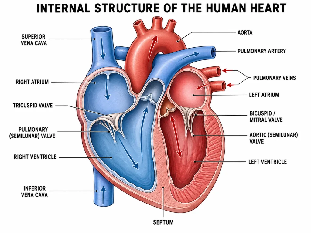
The heart is a muscular pump the size of your fist, located in the chest, slightly tilted to the left.
Four chambers (memorise!)
The heart is divided into FOUR chambers by walls and valves:
Upper chambers (smaller) — Atria (singular: atrium)
- Right atrium — receives deoxygenated blood from the body.
- Left atrium — receives oxygenated blood from the lungs.
Lower chambers (larger) — Ventricles
- Right ventricle — pumps deoxygenated blood to the lungs.
- Left ventricle — pumps oxygenated blood to the entire body.
Atria RECEIVE; ventricles PUMP. Remember this rule.
The septum — why blood doesn't mix
A muscular wall called the septum divides the heart into LEFT half and RIGHT half. This is critical — the septum prevents oxygenated blood (left side) from mixing with deoxygenated blood (right side).
If the septum has a hole (rare birth defect), blood mixes — the person becomes 'blue' from low oxygen.
Four valves — one-way doors
Valves prevent blood from flowing backward.
| Valve | Location | Function |
|---|---|---|
| Tricuspid valve | Between right atrium and right ventricle | Prevents backflow into right atrium |
| Bicuspid (Mitral) valve | Between left atrium and left ventricle | Prevents backflow into left atrium |
| Pulmonary semilunar valve | Exit of right ventricle (into pulmonary artery) | Prevents backflow into right ventricle |
| Aortic semilunar valve | Exit of left ventricle (into aorta) | Prevents backflow into left ventricle |
Result: Blood flows in ONE direction only — atria → ventricles → arteries → never backward.
Why the left ventricle has the thickest wall
Look at the heart carefully — the left ventricle has a much thicker muscular wall than the right ventricle.
Why? Because the left ventricle has to pump blood to the entire body (through aorta) — every organ, every limb, every cell. This requires high pressure.
The right ventricle only has to pump blood to the lungs (a short distance away) — needs much less pressure.
Hence: left ventricle = thicker, more muscular.
The cardiac cycle (briefly)
One complete heartbeat = one cardiac cycle. It has three phases:
- Atrial systole — both atria contract, push blood into ventricles.
- Ventricular systole — both ventricles contract, push blood out to lungs (right) and body (left).
- Diastole — heart relaxes, blood flows in from veins to atria. Cycle repeats.
Normal heart rate: ~70-75 beats/minute at rest. The 'lub-dub' sound you hear is the valves closing.
[Board Important] "Why does the left ventricle have a thicker wall than the right ventricle?" — common 2-mark question. Always link to pumping pressure required for systemic circulation.
Double Circulation
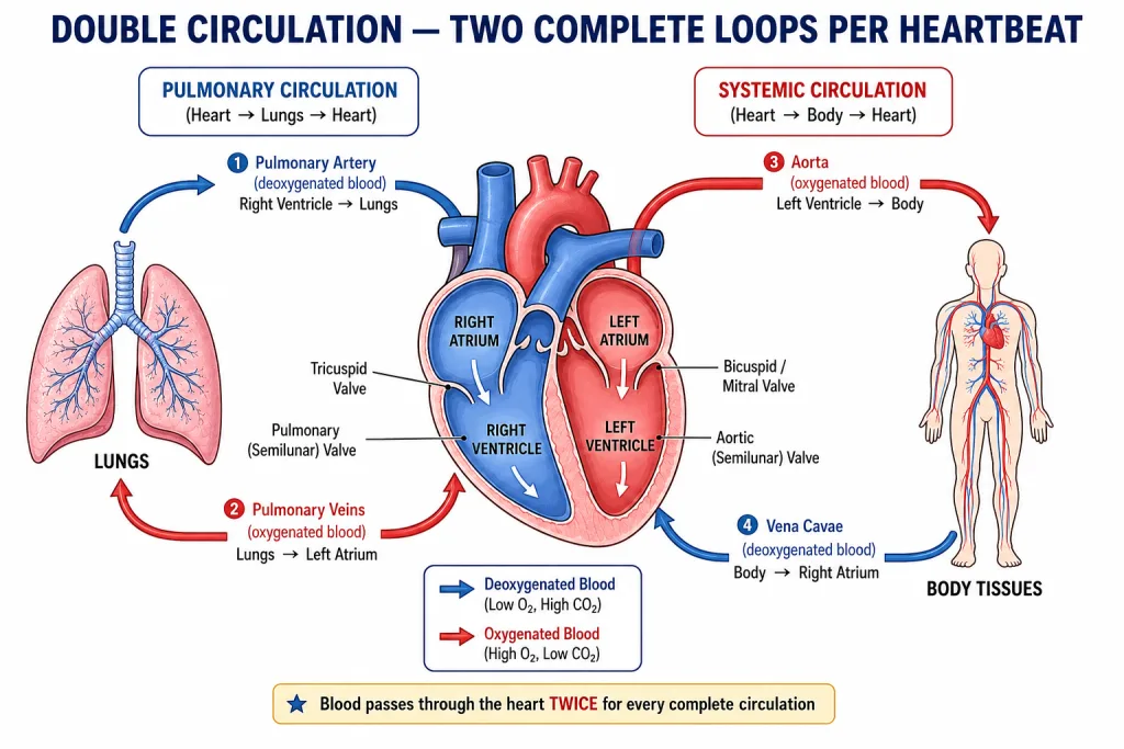
Humans (and all birds and mammals) have double circulation — blood passes through the heart TWICE in one complete journey around the body.
The two circuits
1. Pulmonary circulation (Heart ↔ Lungs) — the shorter loop:
Purpose: pick up O₂, drop off CO₂.
2. Systemic circulation (Heart ↔ Body) — the longer loop:
Purpose: deliver O₂ + nutrients to cells, pick up CO₂ + waste.
Why TWO loops?
Because oxygenated and deoxygenated blood must stay separate.
If only one loop existed, oxygenated and deoxygenated blood would mix. The body's cells would receive partially-oxygenated blood — less efficient. Active animals like birds and mammals (with high metabolic needs) can't afford this mixing.
Fish have single circulation (blood passes through heart only once per cycle, with 2 chambers). They can get away with this because they're cold-blooded and have lower energy demands.
The pulmonary exception (a key NCERT trap!)
Normally:
- Arteries carry OXYGENATED blood (away from heart).
- Veins carry DEOXYGENATED blood (toward heart).
Exception — pulmonary circulation:
- Pulmonary artery carries DEOXYGENATED blood (heart to lungs).
- Pulmonary veins carry OXYGENATED blood (lungs to heart).
This is a frequent NEET MCQ trap! The pulmonary arrangement is opposite. Why? Because the definition of artery is by direction (away from heart), not by the blood content.
Why this design is important
Maintaining separation between O₂-rich and O₂-poor blood means every cell in the body gets the most oxygen-rich blood available. This allows high metabolic rates — which is why mammals/birds can:
- Maintain constant body temperature (warm-blooded).
- Stay active in cold environments.
- Have high energy demands (large brains, active lifestyles).
Summary
| Pulmonary circulation | Systemic circulation | |
|---|---|---|
| Loop length | Short | Long |
| Starts at | Right ventricle | Left ventricle |
| Ends at | Left atrium | Right atrium |
| Blood goes to | Lungs | All body tissues |
| Picks up | O₂ | CO₂ + wastes |
| Drops off | CO₂ | O₂ + nutrients |
[NEET-foundation] Why is pulmonary artery an exception? Because 'artery' means 'vessel going away from the heart', NOT 'vessel carrying oxygenated blood'. Same for pulmonary vein.
Blood Vessels — Arteries, Veins, Capillaries
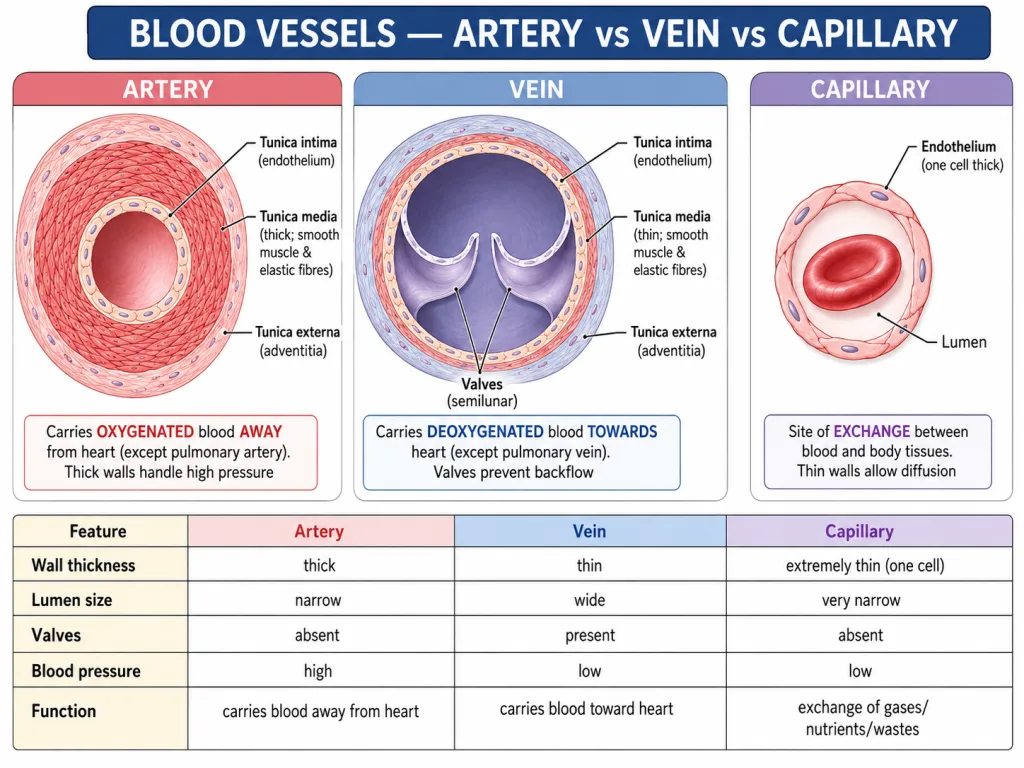
The blood vessels are the body's piping network — and they come in three types, each suited for a specific job.
1. Arteries — carry blood AWAY from heart
Structure:
- Thick, muscular, elastic walls — to withstand high pressure from the pumping heart.
- Narrow lumen (the central hollow).
- No valves (not needed — pressure is high and one-directional).
- Deep inside body — well-protected.
Function: Carry blood from heart to body parts under HIGH PRESSURE.
Blood inside: Usually oxygenated (red). Exception: pulmonary artery carries deoxygenated blood.
Example: Aorta (biggest artery), carotid (to head), femoral (to leg).
Why thick walls? The blood pumped out of the left ventricle is at high pressure (~120 mm Hg systolic). The artery walls must stretch and recoil to maintain this pressure as blood flows.
2. Veins — carry blood TOWARDS heart
Structure:
- Thin, less muscular walls — pressure is low.
- Wider lumen (more blood can flow).
- Contain valves (especially in limbs) — prevent backflow.
- Closer to skin — you can see veins in your wrist!
Function: Carry blood from body parts BACK to heart under LOW PRESSURE.
Blood inside: Usually deoxygenated (darker, purplish). Exception: pulmonary veins carry oxygenated blood.
Example: Vena cavae (biggest veins, returning blood to heart), jugular (from head), femoral vein (from leg).
Why valves? Without the pressure of the heart pushing blood, veins rely on:
- Skeletal muscle contractions squeezing them (especially in legs when walking).
- Valves preventing the blood from flowing backward in between squeezes.
This is why people who stand for long hours sometimes develop varicose veins — when valves fail, blood pools and stretches the veins.
3. Capillaries — the exchange sites
Structure:
- Just ONE cell thick — only the endothelial layer.
- Microscopic (5-10 μm wide).
- Network throughout tissues — close to every cell.
- Connect arteries to veins.
Function: Exchange materials between blood and body cells.
- O₂ and nutrients diffuse OUT of capillary into cells.
- CO₂ and wastes diffuse INTO capillary from cells.
Why so thin? Because exchange happens by diffusion — fast across thin barriers. The single-cell capillary wall is just enough to contain the blood, no more.
Capillaries are so narrow that red blood cells often have to squeeze through one at a time.
Comparison table
| Feature | Artery | Vein | Capillary |
|---|---|---|---|
| Wall | Thick, muscular, elastic | Thinner, less muscular | Single-cell layer |
| Lumen | Narrow | Wider | Very narrow |
| Valves | No | YES (prevent backflow) | No |
| Pressure | High | Low | Very low |
| Blood direction | AWAY from heart | TOWARDS heart | Connects them |
| Blood content | Oxygenated (mostly) | Deoxygenated (mostly) | Mixed |
| Location | Deep | Closer to skin | Throughout tissues |
From artery to vein — a hierarchy
Vessels get progressively smaller toward capillaries, then progressively larger toward veins.
[Board Important] "Differentiate between arteries and veins." Standard 3-mark question. Must include: wall thickness, lumen size, presence of valves, blood direction, pressure.
Components of Blood
Blood is not just "red liquid" — it is a complex tissue with four major components, each with specific functions.
Composition of blood
| Component | % by volume | Role |
|---|---|---|
| Plasma | ~55% | Liquid medium |
| Red Blood Cells (RBCs) | ~45% | Carry O₂ |
| White Blood Cells (WBCs) | <1% | Immune defence |
| Platelets | <1% | Blood clotting |
1. Plasma — the liquid medium
Plasma is ~90% water, with dissolved:
- Nutrients — glucose, amino acids, fatty acids (from intestine).
- Wastes — urea, CO₂ (as bicarbonate).
- Proteins — albumin (maintains osmotic pressure), fibrinogen (clotting), antibodies.
- Hormones — chemical messengers.
- Salts — Na⁺, K⁺, Cl⁻, Ca²⁺, etc.
- Heat — distributed around body.
Plasma is yellowish in colour.
2. Red Blood Cells (RBCs / Erythrocytes)
Features:
- Most numerous (~5 million per mm³ of blood).
- Biconcave disc shape — gives more surface area for O₂ uptake.
- No nucleus (in mature mammalian RBCs) — leaves more room for haemoglobin.
- Lifespan: ~120 days (then broken down in liver/spleen).
- Produced in bone marrow.
- Contain haemoglobin — the iron-containing red pigment that binds O₂.
Function: Transport O₂ from lungs to body cells (and some CO₂ back).
RBCs give blood its red colour.
3. White Blood Cells (WBCs / Leucocytes)
Features:
- Fewer in number (~7,000 per mm³).
- Have a nucleus (unlike RBCs).
- Larger than RBCs.
- Can change shape to squeeze through capillary walls.
Function: Defence against pathogens (bacteria, viruses, etc.).
- Phagocytes — engulf and digest invaders.
- Lymphocytes — produce antibodies.
WBCs are part of the immune system. During an infection, their count rises dramatically.
4. Platelets (Thrombocytes)
Features:
- Tiny fragments (not whole cells) of larger marrow cells.
- No nucleus.
- ~250,000 per mm³.
- Lifespan: ~7-10 days.
Function: Blood clotting — when you cut yourself, platelets stick to the wound and release chemicals that form a fibrin mesh, stopping the bleeding.
Without platelets, a small cut would never stop bleeding — life-threatening.
Why is blood called a "connective tissue"?
Many students get confused — blood is liquid, not solid. But in tissue biology, blood is classified as connective tissue because:
- It has cells (RBCs, WBCs, platelets) suspended in…
- An intercellular matrix (plasma).
Connective tissues are defined by this cells-in-matrix pattern — and blood fits, even though its matrix happens to be liquid.
Blood groups (brief)
Humans have different blood groups based on antigens on RBC surface:
- ABO system: A, B, AB, O (depending on which antigens are present).
- Rh system: Rh+ or Rh- (based on Rh antigen).
Correct blood matching is critical for transfusions. Class 10 just mentions this; details come later.
Functions of blood — summary
Blood does transport, but also more:
| Function | How |
|---|---|
| Transport of O₂ | RBCs (haemoglobin) |
| Transport of CO₂ | Mostly plasma (as bicarbonate), some on Hb |
| Transport of nutrients | Plasma (glucose, amino acids dissolved) |
| Transport of wastes | Plasma (urea to kidneys) |
| Transport of hormones | Plasma |
| Defence against infections | WBCs |
| Clotting | Platelets + fibrinogen |
| Maintaining body temperature | Distributing heat via plasma |
| Maintaining pH | Buffering by plasma proteins |
Blood does a LOT more than just "move stuff around".
[Board Important] "What are the components of blood and their functions?" — 3-mark Board question. Always list all 4: plasma, RBC, WBC, platelets, with function of each.
The Lymphatic System — A Parallel Highway
Most of this section has been about the blood circulatory system — but there's a second, often-overlooked transport network: the lymphatic system.
What is lymph?
When blood flows through capillaries, some of the plasma leaks out into the tissues (because of capillary pressure). This leaked fluid bathes the cells and is now called tissue fluid (or interstitial fluid).
This tissue fluid is mostly returned to the blood — but not all of it. The remaining tissue fluid enters tiny vessels called lymph capillaries and is now called lymph.
Composition of lymph
Lymph is similar to plasma but:
- Less protein (large proteins can't easily leak out of blood capillaries).
- More lymphocytes (a type of WBC).
- Carries fats (absorbed from intestinal lacteals).
Lymph circulation
Lymph flows in one direction — from tissues back to blood:
Unlike blood, lymph is not pumped by the heart. It moves due to:
- Skeletal muscle contractions (when you walk).
- Valves in lymph vessels (preventing backflow).
- Pressure differences.
Lymph nodes
Along lymph vessels are small bean-shaped structures called lymph nodes. They:
- Filter lymph — trap bacteria, viruses, cancer cells.
- House lymphocytes — WBCs that fight infection.
When you get an infection, lymph nodes near the infection site swell up — like swollen "glands" in your neck during a sore throat.
Functions of the lymphatic system
- Returning tissue fluid to blood — preventing tissue swelling (edema).
- Transport of fats absorbed from intestine (via lacteals).
- Defence — lymph nodes filter pathogens; lymphocytes attack invaders.
- Maintaining body fluid balance.
Connection to digestion (recall Section 4)
In Section 4, we saw that fatty acids and glycerol absorbed in the small intestine enter lacteals (not blood capillaries). The lacteals are part of the lymphatic system! Fats first travel in lymph, then eventually merge into blood — explaining why dietary fat takes a different route from glucose/amino acids.
Why is this less famous?
The lymphatic system has no pump (no "heart of its own"), so it doesn't get the dramatic attention of the blood circulation. But it is essential — major lymphatic problems lead to swelling (lymphedema) and immune deficiencies.
[Board-important] "What is lymph? Mention its three functions." Standard 2-3 mark question. Always include: source (leaked tissue fluid), composition (less protein, more lymphocytes), functions (return fluid, transport fats, defence).
Memory Capsule — Section 6
A compact recap before moving to Section 7 (Transportation in Plants).
Three components of the circulatory system
- Heart — the pump.
- Blood vessels — arteries, veins, capillaries.
- Blood — the fluid.
Plus the parallel lymphatic system.
The four-chambered heart
| Chamber | What it does |
|---|---|
| Right atrium | Receives deoxygenated blood from BODY |
| Right ventricle | Pumps deoxygenated blood to LUNGS |
| Left atrium | Receives oxygenated blood from LUNGS |
| Left ventricle | Pumps oxygenated blood to BODY |
Atria receive; ventricles pump.
Left ventricle has the thickest wall (high pressure needed to reach all body parts).
Four valves
- Tricuspid — right atrium ↔ right ventricle.
- Bicuspid (Mitral) — left atrium ↔ left ventricle.
- Pulmonary semilunar — exit of right ventricle.
- Aortic semilunar — exit of left ventricle.
All prevent backflow.
Double circulation
Pulmonary loop: Right ventricle → Pulmonary artery → Lungs → Pulmonary veins → Left atrium. Purpose: oxygenate blood.
Systemic loop: Left ventricle → Aorta → Body tissues → Vena cavae → Right atrium. Purpose: deliver O₂ + nutrients to cells.
Blood passes through heart TWICE per cycle. Hence DOUBLE circulation.
Why double circulation is important
Keeps oxygenated and deoxygenated blood separate. This is essential for warm-blooded, active animals like humans.
Fish have single circulation (only 2-chambered heart) — less efficient, but adequate for their cold-blooded lifestyle.
Blood vessels comparison
| Feature | Artery | Vein | Capillary |
|---|---|---|---|
| Direction | AWAY from heart | TOWARDS heart | Connects |
| Wall | Thick, muscular | Thinner | 1 cell thick |
| Lumen | Narrow | Wider | Very narrow |
| Valves | No | Yes | No |
| Pressure | High | Low | Very low |
| Blood (usual) | Oxygenated | Deoxygenated | Mixed |
Exception: pulmonary artery carries DEOXYGENATED blood; pulmonary vein carries OXYGENATED blood.
Blood components
| Component | Function |
|---|---|
| Plasma (~55%) | Liquid medium; carries dissolved substances |
| RBC (~5 million/mm³) | Carries O₂ via haemoglobin |
| WBC (~7000/mm³) | Defence against pathogens |
| Platelets (~250,000/mm³) | Blood clotting |
RBC special features
- Biconcave disc (more surface area).
- No nucleus in mature mammalian RBCs (more room for Hb).
- Produced in bone marrow.
- Lifespan ~120 days.
Lymph (parallel system)
- Source: leaked tissue fluid from blood capillaries.
- Composition: less protein, more lymphocytes, fats from intestine.
- Flow: tissues → lymph capillaries → lymph nodes → large ducts → veins.
- Functions: return tissue fluid, transport fats (from intestine), defence (via lymph nodes).
Adult human blood
- ~5 litres total.
- Normal heart rate: 70-75 beats/min.
- 'Lub-dub' sound = valves closing.
NCERT-canonical phrases
- "Mammals and birds have four-chambered hearts with complete separation of left and right sides."
- "This separation allows for double circulation, with no mixing of oxygenated and deoxygenated blood."
- "Blood is the fluid which flows in the blood vessels."
One-line takeaway
The human circulatory system is a four-chambered muscular pump (heart) connected to a network of pipes (arteries, veins, capillaries) carrying ~5 litres of blood — with double circulation keeping oxygenated and deoxygenated blood separate for maximum efficiency.
Solved Examples
Example 1: Why is the left ventricle thicker than the right ventricle?
Explain the structural difference between the two ventricles in terms of their functions.
Solution:
The observation
When you cut open a heart, you can clearly see: the left ventricle has a much thicker, more muscular wall than the right ventricle. The septum that divides them is also thick on the left side.
The reason — pumping pressure
The two ventricles do similar jobs (both pump blood out of the heart) but to very different destinations:
Right ventricle:
- Pumps blood to the lungs (a short distance away).
- Pulmonary circulation is low pressure (~25 mm Hg systolic).
- Less force needed → less muscle needed.
Left ventricle:
- Pumps blood to the entire body — head to toes.
- Systemic circulation needs to push blood through kilometres of vessels against gravity (especially up to the brain).
- This is high pressure (~120 mm Hg systolic — what doctors measure as 'blood pressure').
- Needs much more force → needs much more muscle.
Like a water pump
Imagine two pumps:
- One pump that just lifts water to the room next door — needs a small motor.
- One pump that has to push water up a 5-storey building, across a network of pipes, to taps everywhere — needs a much bigger motor.
The left ventricle is the bigger pump.
Numerical comparison
In an adult human:
- Right ventricular wall thickness: ~3-5 mm.
- Left ventricular wall thickness: ~10-15 mm (about 3× thicker).
The left ventricle does roughly the same volume of work, but with much higher pressure — hence its size.
When this design fails
- If the left ventricle weakens (heart failure), blood can't be pumped to the body efficiently — leads to fatigue, shortness of breath, fluid retention.
- If a person has chronic high blood pressure (hypertension), the left ventricle works harder, becomes even thicker (left ventricular hypertrophy), and eventually fails.
A neat fact
The thicker left ventricle is why hearts "point" slightly to the left. The bulk of muscle is on the left side, shifting the heart's apex left.
Answer: The left ventricle has a thicker wall than the right ventricle because it pumps oxygenated blood to the entire body (systemic circulation), which requires high pressure to overcome resistance and reach every cell. The right ventricle only pumps blood to the lungs (pulmonary circulation), a short distance and low pressure. Hence the left ventricle needs more muscle to generate the higher force.
[Board Important] Classic 2-mark question. Always link thickness to pumping pressure for systemic circulation.
Example 2: Why is the pulmonary artery an exception?
Usually arteries carry oxygenated blood and veins carry deoxygenated blood. Explain the exception in pulmonary circulation.
Solution:
This is a classic 'definition trap' question — designed to test whether you really understand what defines an artery vs. a vein.
The standard rule
Most people learn:
- Arteries = oxygenated blood.
- Veins = deoxygenated blood.
But this is a generalisation, not the actual definition.
The real definition
- Artery = a blood vessel that carries blood AWAY FROM the heart.
- Vein = a blood vessel that carries blood TOWARDS the heart.
The definition is based on direction, not on blood content.
Why does the general rule usually work?
In most cases:
- Arteries leave the left ventricle (carrying oxygenated blood from the lungs to body) → they have oxygenated blood.
- Veins return to the right atrium (carrying deoxygenated blood from body back to heart) → they have deoxygenated blood.
So for ~99% of blood vessels in your body, the rule holds.
The pulmonary exception
But pulmonary circulation is different:
Pulmonary artery:
- Leaves the right ventricle (so it's an artery — going AWAY from heart).
- Carries deoxygenated blood (from body → about to go to lungs for oxygenation).
- Result: it's an artery, but it carries deoxygenated blood.
Pulmonary vein:
- Returns to the left atrium (so it's a vein — going TOWARDS the heart).
- Carries oxygenated blood (from lungs → about to be distributed to body).
- Result: it's a vein, but it carries oxygenated blood.
Why the design makes sense
The heart needs both:
To receive deoxygenated blood from body (via vena cavae).
To send that deoxygenated blood to lungs (via pulmonary artery) — needs an outflow vessel = an artery.
To receive the now-oxygenated blood back from lungs (via pulmonary vein) — needs an inflow vessel = a vein.
To send the oxygenated blood to the body (via aorta).
This is exactly what double circulation requires — and the naming follows the direction rule, not the content rule.
How to remember
For any blood vessel, ask: "Is it going away from or returning to the heart?"
- Away → artery.
- Returning → vein.
Don't think about blood colour. Think about direction.
NEET trap warning
NEET questions often phrase this as: "Identify the blood vessel that carries deoxygenated blood but is structurally an artery."
Answer: Pulmonary artery.
This is one of the most-tested 'gotcha' questions in NEET-foundation Class 10.
Answer: The general rule that 'arteries carry oxygenated blood and veins carry deoxygenated blood' is a consequence of typical circulation patterns, not the actual definition. The real definition is by direction: arteries carry blood AWAY from the heart, veins TOWARDS it. In pulmonary circulation, the artery (pulmonary artery, from right ventricle to lungs) carries deoxygenated blood, and the vein (pulmonary vein, from lungs to left atrium) carries oxygenated blood — opposite of the usual rule, but still arteries and veins by the direction definition.
[NEET-foundation] Classic NEET MCQ trap. Memorise: 'artery' is about direction, not content.
Example 3: Trace the path of a drop of blood from a leg muscle to the lungs and back
Follow a drop of deoxygenated blood from a leg muscle through the heart to the lungs (where it gets oxygenated) and back to the leg muscle. Name every chamber, vessel, and major organ.
Solution:
This is a 'follow the molecule' question — tests your understanding of double circulation.
Phase 1: Deoxygenated blood travels FROM leg muscle TO right side of heart
- Leg muscle cell — CO₂ has just been released; deoxygenated blood collected by tiny capillaries.
- Capillary → Venule → Vein in leg.
- Inferior vena cava (the large vein that collects blood from the lower body).
- Right atrium — blood enters here.
- Through the tricuspid valve.
- Right ventricle.
Phase 2: Pulmonary circulation (right ventricle → lungs → left atrium)
- Through the pulmonary semilunar valve.
- Pulmonary artery (note: artery carrying DEOXYGENATED blood — the exception).
- Lungs — gas exchange happens at alveoli.
- CO₂ exits blood (into alveoli, exhaled).
- O₂ enters blood (binds to haemoglobin).
- Blood is now OXYGENATED.
- Pulmonary vein (note: vein carrying OXYGENATED blood — the exception).
- Left atrium — blood enters here.
- Through the bicuspid (mitral) valve.
- Left ventricle.
Phase 3: Systemic circulation — oxygenated blood travels back to leg muscle
- Through the aortic semilunar valve.
- Aorta (the body's biggest artery).
- Down through branching arteries.
- Femoral artery (the main artery to the leg).
- Smaller arteries → arterioles.
- Capillary in leg muscle — gas exchange here.
- O₂ exits blood (into muscle cells).
- CO₂ enters blood (waste from cells).
- Blood is now DEOXYGENATED again.
- Back at the leg muscle — ready to repeat the journey.
Visualizing the loop
Leg muscle (deox.)
    ↓
Vein → Inferior vena cava
    ↓
Right atrium → Right ventricle
    ↓
Pulmonary artery → LUNGS (now oxygenated!) → Pulmonary vein
    ↓
Left atrium → Left ventricle
    ↓
Aorta → Femoral artery → ...
    ↓
Back to leg muscle (oxygenated)
Time and numbers
At rest, this whole loop takes about 1 minute. During exercise it can be as fast as 20 seconds because the heart pumps faster.
Each heart beat (~75 per minute) pushes ~70 mL of blood out — about 5 litres/minute of cardiac output at rest.
Two key points
- Blood passes through the heart TWICE in one complete loop (once on the right side, once on the left).
- Pulmonary artery and pulmonary vein are exceptions to the usual artery-vein blood content rule.
Answer: Leg muscle → capillary → venule → vein → inferior vena cava → right atrium → (tricuspid valve) → right ventricle → (pulmonary semilunar valve) → pulmonary artery → LUNGS (gas exchange) → pulmonary vein → left atrium → (bicuspid valve) → left ventricle → (aortic semilunar valve) → aorta → femoral artery → arterioles → capillary in leg muscle → muscle cell.
[NEET-foundation] Common 'trace the path' question. Practice naming each valve and each major vessel.
Example 4: What are the 4 components of blood and their functions?
List and describe the four components of blood.
Solution:
Composition overview
Blood is roughly:
- 55% plasma (the liquid).
- 45% cells (mostly RBCs).
- Total: ~5 litres in an adult.
Component 1: Plasma
Plasma is the liquid medium of blood — about 90% water, 10% dissolved substances.
What's in plasma:
- Water (90%).
- Proteins: albumin, globulins, fibrinogen.
- Nutrients: glucose, amino acids, lipids.
- Wastes: urea, CO₂ (as bicarbonate).
- Hormones.
- Salts: Na⁺, K⁺, Cl⁻, Ca²⁺.
Functions of plasma:
- Carries dissolved substances around the body.
- Maintains blood volume and pressure.
- Distributes heat throughout the body.
- Carries CO₂ (mainly as bicarbonate ions).
- Plasma proteins help in clotting (fibrinogen) and immunity (antibodies).
Plasma is yellowish in colour.
Component 2: Red Blood Cells (RBCs / Erythrocytes)
Features:
- Most numerous cells: ~5 million per mm³ of blood.
- Biconcave disc shape (looks like a flattened donut without a hole).
- No nucleus (in mature mammalian RBCs).
- Lifespan: ~120 days.
- Produced in bone marrow; destroyed in liver and spleen.
- Packed with haemoglobin (iron-containing protein).
Function: Transport O₂ from lungs to body cells.
- Each haemoglobin binds 4 O₂ molecules.
- Each RBC contains ~250 million Hb molecules → 1 billion O₂ binding sites.
Why biconcave?
- More surface area for gas exchange.
- Flexibility to squeeze through capillaries.
Why no nucleus?
- More room for haemoglobin.
- (Trade-off: RBCs can't repair themselves or divide → short lifespan.)
RBCs give blood its red colour.
Component 3: White Blood Cells (WBCs / Leucocytes)
Features:
- Fewer than RBCs: ~7,000 per mm³.
- Have a nucleus.
- Larger than RBCs.
- Can change shape to squeeze through capillary walls and reach infection sites.
- Lifespan: hours to years (varies by type).
Function: Defence against pathogens (bacteria, viruses, parasites).
Types of WBCs:
- Neutrophils & Macrophages (phagocytes) — eat invaders by phagocytosis.
- Lymphocytes — produce antibodies that target specific pathogens.
- Eosinophils & Basophils — fight parasites and allergic reactions.
During an infection, WBC count rises dramatically — doctors check this in blood tests.
Component 4: Platelets (Thrombocytes)
Features:
- Not whole cells — they are tiny fragments of larger bone-marrow cells called megakaryocytes.
- No nucleus.
- ~250,000 per mm³.
- Lifespan: ~7-10 days.
Function: Blood clotting (stops bleeding from cuts).
How clotting works:
- Cut occurs → blood vessel is damaged.
- Platelets stick to the damaged area.
- Platelets release chemicals that activate fibrinogen (a plasma protein).
- Fibrinogen → fibrin (a sticky mesh of threads).
- The fibrin mesh traps RBCs → forms a clot that plugs the wound.
- The clot hardens to a scab, allowing the wound to heal underneath.
Without platelets, even a small cut would bleed continuously.
Summary table
| Component | % of blood | Has nucleus? | Function |
|---|---|---|---|
| Plasma | ~55% | (it's liquid) | Liquid medium, dissolved substances, heat distribution |
| RBCs | ~45% | NO (mature) | Carry O₂ |
| WBCs | <1% | YES | Defence (immunity) |
| Platelets | <1% | NO (fragments) | Clotting |
Numerical comparison
| Cell type | Count (per mm³ of blood) |
|---|---|
| RBC | ~5,000,000 |
| Platelets | ~250,000 |
| WBC | ~7,000 |
Ratio: RBC : Platelets : WBC ≈ 700 : 30 : 1. RBCs are by far the most numerous.
Answer: Four components of blood: Plasma (liquid medium, 55%, carries dissolved substances and heat); RBCs (carry O₂ via haemoglobin, biconcave, no nucleus, 5 million/mm³); WBCs (defence against pathogens, have nucleus, 7,000/mm³); Platelets (blood clotting via fibrinogen, fragments not whole cells, 250,000/mm³).
[Board Important] Standard 3-mark Board question. Always list all 4 + one function each.
Example 5: Why is double circulation important?
What is double circulation? Why is it necessary in humans but not in fish?
Solution:
What is double circulation?
Double circulation = a blood circulation system where blood passes through the heart TWICE in one complete cycle around the body.
In humans (and all mammals and birds), this is the case.
The two passes through the heart:
Pass 1 — Pulmonary circuit: Heart (right side) → Lungs → Heart (left side). Purpose: pick up O₂, drop off CO₂.
Pass 2 — Systemic circuit: Heart (left side) → Body → Heart (right side). Purpose: deliver O₂ and nutrients to cells, pick up wastes.
At each pass, the right and left sides of the heart are kept completely separate by the septum. Deoxygenated and oxygenated blood never mix.
Why is it necessary in humans?
Humans are warm-blooded animals with high metabolic rates. We need:
- Constant body temperature (37°C) maintained by metabolic heat.
- High O₂ supply to muscles, brain, organs for rapid metabolism.
- Quick removal of CO₂ and other wastes.
This requires the highest-quality oxygenated blood to reach every cell. If oxygenated and deoxygenated blood were to mix in the heart, every cell would receive blood that's only ~50% oxygenated — half-strength. Not enough to fuel a hot, active body.
So double circulation = pure oxygenated blood delivered to body. Maximum efficiency.
Why don't fish need double circulation?
Fish have:
- Cold-blooded metabolism (slower, less energy demand).
- Lower body temperature (matches surrounding water).
- Lower activity levels (most fish don't sustain high-energy activities).
Their circulatory system is single circulation:
- 2-chambered heart (1 atrium + 1 ventricle).
- Blood: heart → gills (oxygenated) → body → back to heart.
- Blood passes through heart only ONCE per cycle.
This is less efficient — the oxygenated blood leaving the gills loses pressure before reaching distant body parts. But for fish, it works because their oxygen demand is modest.
Comparison
| Feature | Fish (single circulation) | Humans (double circulation) |
|---|---|---|
| Heart chambers | 2 (1 atrium + 1 ventricle) | 4 (2 atria + 2 ventricles) |
| Times blood passes heart | 1 | 2 |
| O₂/deox blood mixing | Some mixing | No mixing |
| Efficiency | Lower | Higher |
| Metabolism | Slow, cold-blooded | Fast, warm-blooded |
Where the design came from
Evolutionary path:
- Fish: 2-chambered heart, single circulation.
- Amphibians (frogs): 3-chambered heart (1 ventricle, 2 atria) — partial double circulation, some mixing of blood.
- Reptiles: 3-chambered, with incomplete septum (in most) — better separation but still some mixing.
- Birds and mammals: 4-chambered, complete separation, full double circulation.
Evolution refined the design as animals became more active and metabolically demanding.
Why this is important to understand
The four-chambered heart is one of evolution's most successful innovations. It supports:
- Endothermy (warm-bloodedness).
- High brain activity (rich O₂ supply).
- Sustained physical activity.
Without double circulation, we couldn't be the active, intelligent creatures we are.
Answer: Double circulation means blood passes through the heart TWICE in one cycle — once via the pulmonary circuit (heart → lungs → heart) and once via the systemic circuit (heart → body → heart). It is necessary in humans because we are warm-blooded with high metabolic demands; mixing oxygenated and deoxygenated blood would deliver inefficient half-oxygenated blood to cells, unable to sustain our activity. Fish, being cold-blooded with slower metabolism, can manage with single circulation through a 2-chambered heart.
[Board Important] 5-mark Board question. Must include: definition of double circulation, two circuits, why warm-blooded animals need it, fish comparison.
Example 6: Differentiate arteries and veins (5-mark style)
Differentiate between arteries and veins on the basis of any 5 features.
Solution:
Five differences in tabular form
| # | Feature | Arteries | Veins |
|---|---|---|---|
| 1 | Direction of blood flow | Carry blood AWAY from heart | Carry blood TOWARDS heart |
| 2 | Wall thickness | Thick, muscular, elastic walls | Thinner walls, less muscular |
| 3 | Lumen (internal diameter) | Narrow | Wider |
| 4 | Presence of valves | NO valves | YES — semilunar valves at intervals (prevent backflow) |
| 5 | Blood pressure inside | HIGH pressure (due to heart's pumping) | LOW pressure |
| 6 (bonus) | Blood content (usually) | Oxygenated blood (red) — exception: pulmonary artery | Deoxygenated blood (darker) — exception: pulmonary vein |
| 7 (bonus) | Location in body | Deep, well protected | Closer to skin surface (visible in wrist) |
Explanations for each difference
1. Direction: The defining feature. Artery comes from the same root as "art" — going forth. Veins return.
2. Wall thickness — why? Arteries handle high pressure from the heart pumping. Their thick muscular walls:
- Withstand the pressure surge.
- Expand and recoil with each heartbeat, helping push blood forward.
Veins have low pressure (the pump's effect is much diminished by the time blood gets back). Their walls don't need to be thick.
3. Lumen size — why? Arteries: narrow lumen, thick wall — like a narrow but strong pipe. Veins: wider lumen, thinner wall — more blood can pool in them. (About 60% of the body's blood is in veins at any given moment!)
4. Valves — why? In arteries: blood is pushed forward by high pressure from the heart. No need for valves. In veins: blood is returning to the heart against gravity (in legs, against a metre of height!). Without valves, blood would slip back when not being actively pushed. Valves prevent backflow — they only allow blood to flow toward the heart.
If you've ever felt your veins after a long walk, you can feel pulsing — that's the muscles pumping and the valves working.
5. Pressure — why?
- Just after leaving heart: ~120 mm Hg systolic in arteries.
- By the time blood reaches capillaries: pressure drops to ~30 mm Hg.
- In veins: ~5-15 mm Hg.
- In vena cavae returning to heart: nearly 0 mm Hg.
The pressure decreases steadily as blood moves further from the heart.
6. Blood content (with exceptions): In the systemic circulation (the usual case): arteries carry oxygenated blood, veins carry deoxygenated. But in the pulmonary circulation, it's reversed (pulmonary artery → deoxygenated; pulmonary vein → oxygenated).
Always remember: the definitions are by direction, not by oxygen content.
Real-world consequences
- Bleeding from an artery vs vein: Arterial bleeding is bright red, pulses out forcefully (high pressure). Venous bleeding is darker red, flows steadily (low pressure).
- Injections: Most injections go into veins (intravenous, or 'IV') — because veins are accessible (near skin) and have low pressure.
- Varicose veins: When venous valves fail (often in legs of people who stand long hours), blood pools and stretches the vein — visible as bulging blue veins.
Summary diagram (in your mind)
Artery cross-section:    Vein cross-section:
   ___________               ___________
  /    ____   \             /             \
 |   |    |   |             |              |
 |   |____|   |             |  ____________ |
 |   thick    |             |  thin wall   |
  \___________/             \______________/
  Small lumen,                Large lumen,
  thick wall                  thin wall, valves
Answer: As tabulated above — at minimum mention 5 differences: (1) direction, (2) wall thickness, (3) lumen size, (4) presence of valves, (5) internal pressure. Always note that the blood-content rule has exceptions in pulmonary circulation.
[Board Important] Classic 3-5 mark Board question. Tabular format preferred. Always include 4-5 well-explained differences.