
What is Respiration?
In Sections 2-4 you learnt how food enters the body (nutrition + digestion). Now we ask: what happens to that food once it's inside the cells? The answer is respiration — the process that turns food into usable energy.
A common confusion
When most people say "respiration", they mean breathing. But biology uses the word more precisely. Watch this carefully:
- Breathing = the physical process of taking air in (inhaling) and pushing it out (exhaling). It is just one part of respiration.
- Cellular respiration = the chemical process inside cells that breaks down glucose to release energy.
Both meanings exist in NCERT, but cellular respiration is the biologically important one.
NCERT-canonical definition
"The process in which glucose is broken down to release energy is called respiration."
More precisely: cellular respiration is the breakdown of glucose (and other food molecules) inside cells to release energy stored in their chemical bonds. The energy is captured as ATP (adenosine triphosphate) — the universal energy currency of the cell.
Why do cells need this energy?
Every activity in your body needs ATP:
- Muscle contraction (every move you make).
- Building proteins, DNA, membranes.
- Active transport across cell membranes.
- Maintaining body temperature.
- Sending nerve signals.
- Powering the millions of "molecular movements" we discussed in Section 1.
Without respiration, no ATP. Without ATP, no life.
The simple equation
For glucose + oxygen:
Notice: this is the exact reverse of photosynthesis (Section 2). Plants build glucose using CO₂, H₂O, and light. Cells (including plant cells) break it back down to release the stored energy.
[Board Important] "Write the equation for aerobic respiration." — a 1-mark Board question. Always include ATP/energy on the product side.
Two Types of Respiration — Aerobic vs Anaerobic
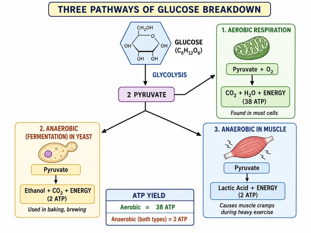
Glucose can be broken down in two main ways, depending on whether oxygen is available.
Step 1 (common to both): Glycolysis
In the cytoplasm of the cell, every glucose molecule is first broken into 2 molecules of pyruvate (also called pyruvic acid).
This step does NOT need oxygen and is the same for everyone — plants, animals, bacteria, yeast. After this, the two paths diverge.
Aerobic Respiration (with O₂)
'Aerobic' = with air (oxygen).
- Pyruvate enters mitochondria.
- In presence of O₂, pyruvate is completely broken down to CO₂ + H₂O + 38 ATP.
Used by: humans, all animals, plants (yes, plants respire too!), most bacteria.
Why mitochondria? Because mitochondria are the only organelle with the enzyme machinery for the Krebs cycle and electron transport chain — the energy-extracting reactions. That's why mitochondria are called the "powerhouses" of the cell.
Anaerobic Respiration (without O₂)
'Anaerobic' = without air (oxygen).
When O₂ is not available, pyruvate cannot enter mitochondria or get completely broken down. Instead, it follows one of two pathways:
Path 1 — In yeast (fermentation):
This is how alcoholic beverages and bread are made. The CO₂ makes bread rise; the ethanol becomes beer/wine.
Path 2 — In our muscle cells during heavy exercise:
Why this happens: When you sprint or lift heavy weights, your muscles use ATP faster than your lungs and heart can supply oxygen. Cells switch to anaerobic respiration as a backup. The lactic acid that builds up causes muscle cramps — that burning sensation after intense exercise.
Why so much less ATP from anaerobic respiration?
Aerobic respiration: 38 ATP per glucose. Anaerobic respiration: 2 ATP per glucose.
That's 19 times less energy!
Why? Because anaerobic respiration doesn't fully break down glucose. The end products (ethanol or lactic acid) still contain a lot of unused chemical energy. Aerobic respiration extracts everything — leaves nothing but CO₂ and water.
Comparison table
| Feature | Aerobic | Anaerobic |
|---|---|---|
| O₂ needed? | Yes | No |
| Location in cell | Cytoplasm + Mitochondria | Cytoplasm only |
| End products | CO₂ + H₂O | Ethanol + CO₂ (yeast) OR Lactic acid (muscle) |
| ATP yield (per glucose) | 38 | 2 |
| Efficiency | High | Low |
| Glucose fully broken? | Yes | No |
| Examples | Most cells of plants, animals, humans | Yeast (always), muscle (during heavy exercise), some bacteria |
A subtle but important point
Anaerobic respiration is NOT a separate thing happening in some special organisms. It is what every cell does in part of glycolysis — and what cells do completely when O₂ runs out.
[Board Important] Standard 3-mark Board question: "Differentiate between aerobic and anaerobic respiration." Always include: (a) O₂ requirement, (b) location, (c) end products, (d) ATP yield.
The Human Respiratory System
For humans to do aerobic respiration, we need a constant supply of O₂. Our respiratory system is the body's air-handling machinery that brings O₂ in and takes CO₂ out.
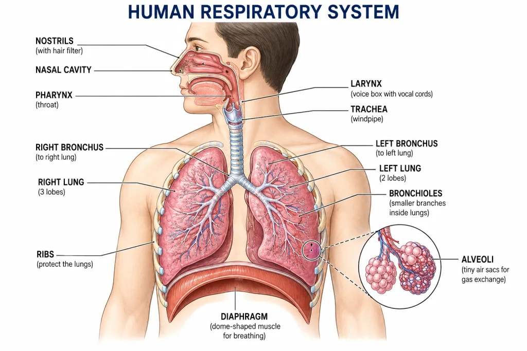
The air pathway (memorise this!)
Let's walk through each part.
1. Nostrils + Nasal Cavity
Two openings to the nose. The nasal cavity:
- Filters dust — fine hairs and mucus catch particles.
- Warms the air — body-warm blood vessels under the lining heat the cold air.
- Moistens the air — mucus adds moisture so the lungs aren't dried out.
This is why breathing through your nose is healthier than through your mouth — your nose is a free air conditioner.
2. Pharynx
The throat. Common passage for both food (going to oesophagus) and air (going to larynx).
At the top of the larynx is a flap called the epiglottis — when you swallow, it covers the windpipe to prevent food going down it. (Sometimes it fails — and food "goes the wrong way". You cough to clear it.)
3. Larynx — the voice box
Contains vocal cords. Air passing through them vibrates them, producing sound. The larynx also bridges the pharynx to the trachea.
4. Trachea — the windpipe
A 10-12 cm tube running down the front of the neck. Key features:
- C-shaped cartilage rings keep it open at all times (so it doesn't collapse when you breathe in).
- Ciliated lining + mucus traps any dust that escaped the nose; cilia wave the dust upward to be swallowed.
5. Two Bronchi
The trachea splits into two bronchi (one to each lung). They are like the trunks of two branching trees.
6. Bronchioles
Each bronchus branches into thousands of smaller tubes called bronchioles. Each bronchiole gets thinner and thinner, until…
7. Alveoli — the gas-exchange surface
'Alveoli' (singular: alveolus) = tiny balloon-like air sacs at the end of each bronchiole.
This is where the magic happens. Each lung has ~300 million alveoli. If you spread them all out flat, they'd cover ~100 m² — about half a tennis court. That's the surface area where O₂ enters your blood and CO₂ leaves.
Alveoli are:
- Thin-walled (1 cell thick) — easy for gases to diffuse.
- Richly supplied with blood capillaries — quick removal of O₂, quick supply of CO₂ for exhalation.
- Moist inside — gases dissolve in the moisture before diffusing.
Two lungs
The lungs are two pink, spongy organs in the chest cavity. The right lung has 3 lobes; the left lung has 2 lobes (smaller, to leave room for the heart).
Protective bones — Ribs
The lungs sit inside the rib cage — a protective bony cage formed by the ribs (12 pairs) and the sternum (breastbone). The rib cage protects the lungs and helps in breathing (via intercostal muscles between the ribs).
The diaphragm
A large dome-shaped muscle below the lungs, separating the chest cavity from the abdomen. The diaphragm is the main muscle of breathing — when it contracts, the lungs expand. More on this below.
[Board Important] "List the parts of the respiratory system in order, starting from the nostril." — 2-mark Board question. Memorise the pathway exactly: nostrils → nasal cavity → pharynx → larynx → trachea → bronchi → bronchioles → alveoli.
Gas Exchange at the Alveolus
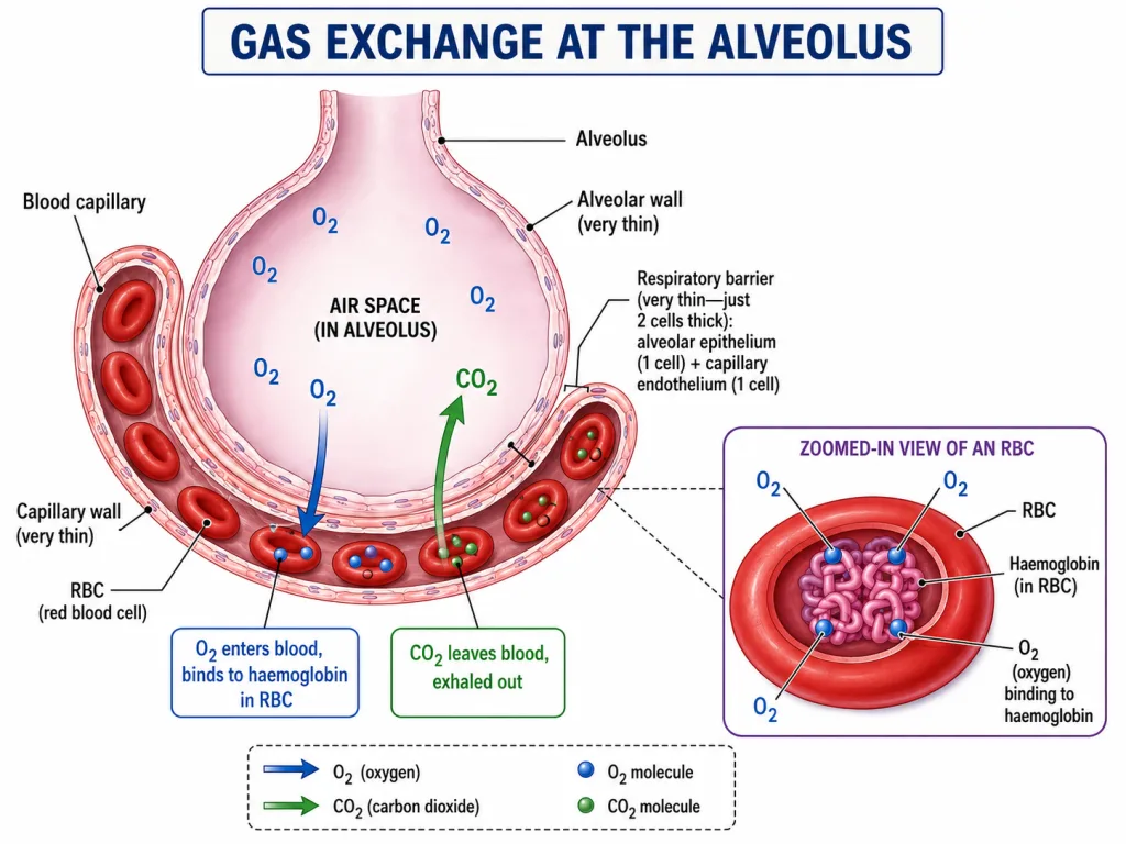
This is THE most important diagram of respiration — Board exam favourite.
The setup
Each alveolus is wrapped tightly by a network of blood capillaries. The alveolar wall + capillary wall together form a very thin barrier (~0.5 μm — about 100× thinner than a sheet of paper). This is the respiratory membrane.
What happens at the membrane
Two gases move in opposite directions, driven by diffusion (high to low concentration):
O₂ goes from alveolus → blood:
- Alveolus has high O₂ (you just inhaled air).
- Capillary blood has low O₂ (used up by body cells).
- O₂ diffuses INTO the blood.
- It then binds to haemoglobin inside red blood cells (RBCs).
CO₂ goes from blood → alveolus:
- Capillary blood has high CO₂ (waste from cells).
- Alveolus has low CO₂ (fresh air).
- CO₂ diffuses OUT into the alveolus.
- It is then exhaled out.
The role of haemoglobin
Haemoglobin is a red protein inside red blood cells. Each haemoglobin molecule has 4 iron-containing heme groups, and each heme can bind one O₂ molecule. So one haemoglobin can carry 4 O₂ molecules.
Blood with oxyhaemoglobin is bright red (arterial blood, leaving lungs). Blood that has dropped off O₂ is darker red, almost purplish (venous blood, returning to lungs).
Why haemoglobin?
O₂ doesn't dissolve well in water/plasma. Without haemoglobin, blood could carry only tiny amounts of O₂ — not enough to keep us alive. Haemoglobin multiplies oxygen-carrying capacity by ~70 times.
This is why anaemic patients (low haemoglobin) feel tired easily — their blood can't carry enough O₂.
What about CO₂ transport?
CO₂ is mostly carried in the blood as bicarbonate ions (HCO₃⁻) dissolved in the plasma. A small amount is carried bound to haemoglobin (as carbaminohaemoglobin). It is released at the alveoli for exhalation.
The trip in numbers
Normal breathing:
- 12-20 breaths per minute (at rest).
- ~500 mL air per breath (tidal volume).
- Total ~6-8 litres of air per minute exchanged.
- During exercise: up to 100 litres/minute!
Why this design is brilliant
The respiratory system maximises three things:
- Surface area — alveoli (300 million) provide ~100 m² of exchange surface.
- Thinness — the membrane is just 0.5 μm thin, so diffusion is fast.
- Continuous fresh supply — breathing brings new air; the heart pumps blood through capillaries continuously.
This is the same design principle as villi in the small intestine — increase surface area, minimise thickness, maintain a constant gradient.
[Board Important] Standard 3-5 mark Board question: "How does gas exchange take place at the alveoli?" Must include: thin walls, blood capillaries, diffusion of O₂ in, CO₂ out, role of haemoglobin.
How Do We Actually Breathe?
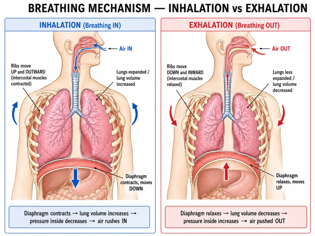
Breathing (the physical act) is purely mechanical — it's driven by changing the volume of your chest cavity, which changes the air pressure inside the lungs.
The principle
Air moves from high pressure to low pressure.
So to breathe IN, you create low pressure inside the lungs (relative to outside). To breathe OUT, you create high pressure inside the lungs.
How do you change the pressure inside the lungs? By changing the volume of the chest cavity.
Two muscles do all the work
1. Diaphragm — a dome-shaped muscle below the lungs. 2. Intercostal muscles — muscles between the ribs.
Inhalation (breathing IN)
- Diaphragm contracts → it FLATTENS (moves down).
- Intercostal muscles contract → ribs move UP and OUTWARD.
- Combined: chest cavity volume increases.
- By Boyle's Law (volume up → pressure down): pressure inside lungs drops below atmospheric pressure.
- Air rushes IN from the atmosphere through the nose → trachea → lungs.
Exhalation (breathing OUT)
- Diaphragm relaxes → returns to its dome shape (moves up).
- Intercostal muscles relax → ribs move DOWN and INWARD.
- Combined: chest cavity volume decreases.
- Pressure inside lungs rises above atmospheric.
- Air is pushed OUT.
Comparison table
| Feature | Inhalation | Exhalation |
|---|---|---|
| Diaphragm | Contracts (flattens, moves DOWN) | Relaxes (dome-shaped, moves UP) |
| Ribs | Lift UP and OUTWARD | Move DOWN and INWARD |
| Intercostal muscles | Contract | Relax |
| Chest cavity volume | Increases | Decreases |
| Pressure in lungs | Decreases (below atm) | Increases (above atm) |
| Air flow | IN | OUT |
A simple test
Put your hand on your belly and breathe deeply. When you breathe in, your belly should EXPAND (because the diaphragm is moving down and pushing your abdominal organs down). When you breathe out, your belly should RETURN to normal.
Most people don't breathe deeply enough — they only use their chest muscles, not the diaphragm. Practising belly breathing (diaphragmatic breathing) is recommended in yoga and pranayama for better oxygen supply.
Special breathing situations
- Forced breathing (during exercise, or playing wind instruments) — abdominal muscles also help push air out forcefully.
- Coughing — sudden forceful exhalation to clear airways.
- Sneezing — like coughing, but through the nose.
- Yawning — deep involuntary inhalation, possibly to wake up alertness or cool the brain.
- Hiccups — spasms of the diaphragm.
Why the cartilage rings in the trachea?
The trachea has C-shaped cartilage rings keeping it open. Without these, the windpipe would collapse during inhalation (when pressure inside drops). The cartilage provides structural support, like the rings of a vacuum cleaner hose.
Why C-shaped, not full circles? The open side faces the back, allowing the oesophagus behind to expand when food passes — without crushing the trachea.
[Board Important] "Describe the mechanism of inhalation and exhalation." — 3-5 mark Board favourite. Always mention: (1) diaphragm + intercostal muscles, (2) volume change, (3) pressure change, (4) air movement.
Respiration in Other Organisms — A Quick Tour
We've covered humans in detail. NCERT also touches on how other organisms respire — these are common 2-mark Board questions.
Plants
Plants respire too! Many students mistakenly think plants "breathe in CO₂ and breathe out O₂" — that's photosynthesis (Section 2), only during the day. Plants also need O₂ for respiration:
- During the day: photosynthesis (CO₂ in, O₂ out) > respiration (O₂ in, CO₂ out). Net: O₂ released, CO₂ absorbed.
- At night: photosynthesis stops, only respiration happens. Net: O₂ absorbed, CO₂ released.
This is why old wisdom said don't sleep under a tree at night — they release CO₂.
Where do plants exchange gases?
- Stomata in leaves (you met them in Section 2).
- Lenticels in stems — tiny pores in bark.
- Roots — exchange gases with air in soil.
No specialised respiratory organs — diffusion is sufficient because plants are not very metabolically active.
Fish — Gills
Fish live in water, where O₂ is much scarcer than in air (water holds about 30× less O₂ per volume than air). They have evolved gills to extract this O₂ efficiently.
- Gills are richly supplied with blood capillaries.
- Water flows over them, and O₂ diffuses from water into blood.
- CO₂ moves the other way.
Why gills don't work in air: Out of water, gill filaments stick together, drastically reducing surface area. Plus they dry out quickly. This is why a fish out of water suffocates even though there's plenty of O₂ around.
Insects — Tracheal tubes
Insects don't have lungs OR gills. They have a network of tiny tubes called trachea (different from the human trachea — same name, different structure!) that extend into the body, opening to the outside through pores called spiracles.
- Air enters spiracles → travels through trachea → reaches every cell directly.
- No blood transport of O₂ needed!
Why this works only for small animals: Tracheal diffusion is slow over long distances. That's why insects are small — large bodies can't be supplied with O₂ through tracheal tubes alone.
Amphibians — Multiple methods
Frogs respire in three ways:
- Through the skin (cutaneous respiration) — must be moist.
- Through the buccal cavity (mouth lining).
- Through lungs — like simpler versions of human lungs.
This is why frogs need wet environments. If their skin dries out, they can't breathe properly.
A summary table
| Organism | Respiratory organ | O₂ source |
|---|---|---|
| Humans | Lungs | Air |
| Plants | Stomata, lenticels | Air |
| Fish | Gills | Water |
| Insects | Tracheal tubes + spiracles | Air |
| Frogs | Skin + lungs + buccal | Air + water |
| Amoeba | Whole cell membrane | Water (dissolved O₂) |
NCERT-canonical phrasing
"Terrestrial organisms use the oxygen in the atmosphere for respiration. This oxygen is taken up by different organs specialised for this function. The lungs in humans are the organs of respiration."
[NEET-foundation] This 'comparative respiration' theme deepens in Class 11. Master the basics now.
Memory Capsule — Section 5
A compact recap before moving to Section 6 (Transportation).
The aerobic respiration equation
Exactly the reverse of photosynthesis.
Glycolysis (common step)
After pyruvate — three paths
| Path | Location | Conditions | End products | ATP |
|---|---|---|---|---|
| Aerobic | Mitochondria | With O₂ | CO₂ + H₂O | 38 |
| Anaerobic (yeast) | Cytoplasm | No O₂, fermentation | Ethanol + CO₂ | 2 |
| Anaerobic (muscle) | Cytoplasm | No O₂, heavy exercise | Lactic acid | 2 |
Lactic acid = muscle cramps.
Human respiratory pathway (memorise!)
Nostrils → Nasal cavity → Pharynx → Larynx → Trachea → Bronchi → Bronchioles → Alveoli
Key facts about each organ
- Nasal cavity — filters, warms, moistens air.
- Larynx — voice box (vocal cords).
- Trachea — C-shaped cartilage rings keep it open.
- Bronchi — 2, one per lung.
- Bronchioles — branching network.
- Alveoli — 300 million; ~100 m² surface area; gas exchange site.
Right lung vs left lung
- Right lung: 3 lobes.
- Left lung: 2 lobes (less space; heart is there).
Mechanism of breathing
| Inhalation | Exhalation | |
|---|---|---|
| Diaphragm | Contracts, flattens, moves DOWN | Relaxes, dome-shaped, moves UP |
| Ribs | Up and OUT | Down and IN |
| Lung volume | UP | DOWN |
| Lung pressure | DOWN | UP |
| Air | IN | OUT |
Gas exchange at alveoli (key facts)
- Thin walls — 1 cell thick.
- Wrapped in capillaries.
- O₂ diffuses IN to blood, binds to haemoglobin in RBCs.
- CO₂ diffuses OUT, carried mostly as bicarbonate in plasma.
Haemoglobin
- Red iron-containing protein in RBCs.
- Each haemoglobin carries 4 O₂ molecules.
- Without it, blood couldn't carry enough O₂.
- Low Hb = anaemia.
Other organisms
- Plants: stomata + lenticels.
- Fish: gills (in water).
- Insects: tracheal tubes + spiracles.
- Frogs: skin + lungs + buccal cavity.
NCERT-canonical phrases
- "The breakdown of glucose to release energy is called respiration."
- "In humans, gaseous exchange takes place at the alveoli of the lungs."
- "Sometimes, when there is a lack of oxygen in our muscle cells, another pathway for the breakdown of pyruvate is taken… Lactic acid is produced."
One-line takeaway
Respiration is the cellular process that releases energy (ATP) from food (glucose) — aerobically with O₂ (38 ATP per glucose) or anaerobically without (only 2 ATP). Humans bring O₂ via lungs and alveoli, where it diffuses into blood and is carried by haemoglobin to every cell.
Solved Examples
Example 1: Differentiate aerobic and anaerobic respiration with examples
Describe at least 4 differences between aerobic and anaerobic respiration. Give one organism/situation example of each.
Solution:
The four key differences
| Feature | Aerobic respiration | Anaerobic respiration |
|---|---|---|
| Oxygen | Requires O₂ | Occurs WITHOUT O₂ |
| Location in cell | Cytoplasm + mitochondria | Cytoplasm only |
| End products | CO₂ + H₂O | Ethanol + CO₂ (yeast) OR Lactic acid (muscle) |
| ATP yield (per glucose) | 38 ATP | 2 ATP |
| Glucose broken | Completely | Partially |
Examples
Aerobic example: Most cells of plants and animals, including human cells. When you sit reading this, your muscle and brain cells are doing aerobic respiration — using O₂ from your lungs.
Anaerobic examples:
Yeast (fermentation): Yeast cells in dough or in a fermenting tank carry out anaerobic respiration: This is how bread rises (CO₂ inflates the dough) and alcohol is made.
Muscle cells during heavy exercise: When you sprint or lift heavy weights, your lungs and heart can't supply O₂ fast enough. Muscle cells switch to anaerobic respiration: The lactic acid causes the muscle cramps and burning sensation you feel.
Why such a huge ATP difference?
38 vs 2 — that's 19 times more energy from aerobic respiration.
Reason: anaerobic respiration leaves a lot of energy locked up in the end products (ethanol or lactic acid still have chemical energy). Aerobic respiration completely breaks glucose down to CO₂ and water — extracting maximum energy.
The shared first step — glycolysis
Both aerobic and anaerobic respiration share the first step: glycolysis, where glucose (C₆) is broken into 2 pyruvate molecules (C₃ each), yielding 2 ATP. After this:
- With O₂: pyruvate enters mitochondria → 36 more ATP.
- Without O₂: pyruvate stays in cytoplasm → becomes ethanol or lactic acid → no more ATP.
Answer: Aerobic respiration uses O₂, occurs in mitochondria, produces CO₂ + H₂O + 38 ATP per glucose. Anaerobic respiration occurs without O₂, only in cytoplasm, produces ethanol + CO₂ (yeast) or lactic acid (muscle), yielding only 2 ATP. Aerobic example: human body cells. Anaerobic examples: yeast (fermentation), human muscle during heavy exercise.
[Board Important] Classic 3-mark Board question. Always include the 4 key differences AND examples.
Example 2: Why do we get muscle cramps after heavy exercise?
Explain in terms of cellular respiration.
Solution:
What happens during heavy exercise
Step 1: Demand for energy spikes. When you sprint or lift weights, your muscles need lots of ATP very fast — for muscle contraction.
Step 2: Aerobic respiration tries to keep up. Normally, your muscle cells do aerobic respiration: For this, they need a constant supply of O₂ from your lungs (via blood).
Step 3: O₂ supply can't match the demand. During intense exercise, even though your heart rate and breathing rate increase, your lungs cannot deliver O₂ fast enough to match the rapidly-firing muscle cells. Oxygen becomes the limiting factor.
Step 4: Cells switch to anaerobic respiration. With insufficient O₂, the muscle cells switch to anaerobic respiration: This gives quick energy but only 2 ATP per glucose (vs 38 in aerobic). Also, it produces lactic acid as an end product.
Step 5: Lactic acid accumulates in muscles. The lactic acid builds up in the muscle cells faster than blood can carry it away. High lactic acid concentration causes:
- Burning sensation in muscles.
- Pain.
- Muscle cramps.
Step 6: Recovery. After you stop exercising, oxygen-rich breathing resumes normally. The accumulated lactic acid is gradually transported to the liver and converted back to glucose, or completely broken down. This is called "repaying the oxygen debt." This is why you keep breathing heavily for a few minutes even after stopping — your body is paying back its oxygen debt.
Why doesn't this happen during gentle exercise?
During walking or light jogging, your lungs CAN supply enough O₂ for full aerobic respiration. No lactic acid build-up. That's why you can walk for hours without cramps but only sprint for seconds.
A real-world example
Usain Bolt's 9.58-second 100m sprint — his muscles are doing almost entirely anaerobic respiration. That's why no one can sprint at full speed for more than a few seconds.
The takeaway
Muscle cramps during/after heavy exercise are caused by lactic acid accumulation due to anaerobic respiration in muscle cells when O₂ supply is insufficient.
Answer: During heavy exercise, muscles need more ATP than the lungs can supply oxygen for. Cells switch to anaerobic respiration: glucose → lactic acid + 2 ATP. The accumulating lactic acid causes muscle cramps and burning sensation. After exercise, breathing remains heavy to supply extra O₂ that breaks down the lactic acid — repaying the 'oxygen debt'.
[Board Important] 3-mark NCERT question. Always link to: anaerobic respiration in muscles, lactic acid build-up, oxygen debt.
Example 3: How does gas exchange take place at the alveolus?
Describe the structure of an alveolus and the process of gas exchange there.
Solution:
What is an alveolus?
An alveolus (plural: alveoli) is a tiny, balloon-shaped air sac at the end of each bronchiole in the lungs. It is the actual site of gas exchange.
Anatomy of an alveolus
- Walls are very thin (just 1 cell thick).
- Surrounded by a dense network of blood capillaries — also 1 cell thick.
- Inner surface is moist — gases dissolve here before diffusing.
- About 300 million alveoli per lung — total surface area ~100 m².
The respiratory membrane
The respiratory membrane = alveolar wall + capillary wall. Together it's only ~0.5 μm thick — incredibly thin, allowing rapid gas diffusion.
Gas exchange — two gases, two directions
The key principle: diffusion from high to low concentration.
Oxygen (O₂):
- Alveolus: HIGH O₂ (you just inhaled).
- Capillary blood: LOW O₂ (used up by cells).
- Direction: O₂ diffuses INTO blood, across the thin membrane.
- Fate: O₂ binds to haemoglobin in red blood cells, forming oxyhaemoglobin.
Carbon dioxide (CO₂):
- Capillary blood: HIGH CO₂ (waste from cells).
- Alveolus: LOW CO₂ (fresh air).
- Direction: CO₂ diffuses OUT of blood into alveolus.
- Fate: CO₂ is exhaled out via bronchioles → bronchi → trachea → nose.
Why this is so efficient
Three design features:
- Massive surface area. 300 million alveoli = ~100 m² (half a tennis court) in your chest.
- Minimum thickness. 0.5 μm = molecules diffuse across in microseconds.
- Continuous gradient.
- Breathing brings fresh O₂ to alveoli (refreshes one side).
- Heart pumps blood through capillaries (refreshes other side).
- So the concentration gradient never gets exhausted.
The role of haemoglobin
Haemoglobin (Hb) is a red iron-containing protein inside RBCs. It binds 4 O₂ molecules per Hb:
Without Hb, blood plasma alone could carry only ~1.5% as much O₂ — far too little. Hb multiplies oxygen-carrying capacity by ~70x.
CO₂ transport
Unlike O₂, CO₂ is transported mostly dissolved in plasma as bicarbonate ions (HCO₃⁻), plus a small amount bound to haemoglobin and dissolved as CO₂.
NCERT-canonical phrase
"The exchange of gases takes place between the alveolar air and the blood. Gas exchange is by diffusion."
Diagram description (for Board answers)
Draw a balloon-shaped alveolus with thin walls. Wrap it with a coiled blood capillary. Draw two arrows:
- One arrow showing O₂ moving from alveolus → blood (label "O₂ + Hb → oxyhaemoglobin").
- One arrow showing CO₂ moving from blood → alveolus (label "CO₂ exhaled").
Answer summary
An alveolus is a thin-walled air sac wrapped by blood capillaries — the site of gas exchange in lungs. By diffusion: O₂ moves from alveolus (high) → blood (low), binding to haemoglobin in RBCs; CO₂ moves from blood (high) → alveolus (low), to be exhaled. The very thin respiratory membrane (~0.5 μm), enormous total surface area (~100 m²), and continuous fresh supply on both sides make this exchange extremely efficient.
[Board Important] 5-mark Board question. Must include: alveolus structure, thin walls, capillaries, diffusion direction for each gas, haemoglobin role.
Example 4: Trace the path of an oxygen molecule from the air to a body cell
From a breath of air outside your body to a muscle cell deep in your leg — describe the journey of an O₂ molecule. Name every structure it passes through.
Solution:
This is a 'follow the molecule' question — a classic NEET-style problem. Let's trace step by step.
Phase 1: External respiration (air → blood)
Atmosphere — O₂ is in the air around you.
Nostrils — O₂ enters during inhalation.
Nasal cavity — filtered, warmed, moistened.
Pharynx — passes through throat.
Larynx — past the voice box.
Trachea — down the windpipe (kept open by C-rings).
Bronchi — left or right branch into lung.
Bronchioles — through smaller and smaller branches.
Alveolus — finally reaches an alveolar air sac.
Alveolar wall + capillary wall (respiratory membrane) — O₂ diffuses across the thin barrier from alveolar air into blood capillary.
Red blood cell (RBC) — O₂ binds to haemoglobin inside an RBC.
Phase 2: Transport (blood → tissue)
- Pulmonary vein — oxygenated blood travels from lungs back to the heart.
- Heart — left atrium — blood enters here.
- Heart — left ventricle — pumped out with high pressure.
- Aorta — main artery, carrying oxygenated blood.
- Arteries — branching network throughout the body.
- Arterioles — smaller arteries.
- Capillaries in leg muscle — finest blood vessels, in direct contact with muscle cells.
Phase 3: Internal respiration (blood → cell)
- Hb releases O₂ — at the muscle cell capillary, where O₂ is low, haemoglobin lets go.
- Tissue fluid — O₂ diffuses out into the fluid surrounding cells.
- Muscle cell membrane — O₂ diffuses across.
- Cytoplasm — O₂ inside the cell.
- Mitochondria — O₂ finally enters mitochondria.
- Aerobic respiration — O₂ is used in the electron transport chain to break down glucose → CO₂ + H₂O + ATP.
A simplified summary
Reverse journey of CO₂
The CO₂ produced in mitochondria travels the reverse route out of the body:
Mitochondria → cytoplasm → tissue fluid → blood (as bicarbonate) → heart → lungs → alveolus → exhaled out through trachea → nose → atmosphere.
The same path, in reverse, for a different gas.
Why is this question important?
It links:
- Respiratory system (Section 5).
- Circulatory system (Section 6 — coming next).
- Cellular respiration (the biochemistry).
A single O₂ molecule connects everything. If any step fails, the cell dies.
Answer: Air → nostrils → nasal cavity → pharynx → larynx → trachea → bronchi → bronchioles → alveolus → through respiratory membrane → blood capillary → binds to haemoglobin in RBC → pulmonary vein → left atrium → left ventricle → aorta → arteries → arterioles → leg muscle capillary → tissue fluid → muscle cell → cytoplasm → mitochondria → used in aerobic respiration.
[NEET-foundation] 'Trace the path' is a frequent NEET MCQ pattern. Memorise the order — that's what they test.
Example 5: NCERT in-text question — Mechanism of breathing
Describe the mechanism of breathing in humans (inhalation and exhalation), with reference to the diaphragm and ribs.
Solution:
Breathing is mechanical — it's about changing the volume of the chest cavity, which changes air pressure inside the lungs, causing air to move in or out.
Boyle's Law (the underlying physics)
For a gas at constant temperature: Volume × Pressure = constant.
That is: when volume increases, pressure decreases (and vice versa).
In breathing:
- Increase chest volume → pressure inside lungs drops → air rushes IN.
- Decrease chest volume → pressure inside lungs rises → air pushed OUT.
Inhalation (breathing IN) — Step by step
Step 1: The diaphragm (dome-shaped muscle below the lungs) contracts. As it contracts, it FLATTENS — pulling DOWNWARD.
Step 2: The intercostal muscles (muscles between ribs) contract simultaneously. They pull the ribs UPWARD and OUTWARD.
Step 3: Both these movements together increase the volume of the chest cavity (thoracic cavity). The lungs, which are inside, expand along with it.
Step 4: As the lung volume increases, the air pressure inside the lungs drops below atmospheric pressure.
Step 5: Atmospheric air, being at higher pressure, rushes INTO the lungs through the nose, trachea, bronchi, bronchioles, and finally into the alveoli.
Exhalation (breathing OUT) — Step by step
Step 1: The diaphragm relaxes and returns to its dome shape — moves UPWARD.
Step 2: The intercostal muscles relax, allowing the ribs to drop DOWN and INWARD.
Step 3: The chest cavity volume decreases. The lungs deflate slightly.
Step 4: Air pressure inside the lungs rises above atmospheric pressure.
Step 5: Air is pushed OUT through the bronchioles, bronchi, trachea, and nose.
A simple analogy
Think of a syringe.
- Pull the plunger BACK → volume inside increases → pressure drops → air gets sucked IN.
- Push the plunger FORWARD → volume decreases → pressure rises → air pushed OUT.
Your chest cavity is the syringe; the diaphragm + ribs are the plunger.
Comparison table
| Inhalation (IN) | Exhalation (OUT) | |
|---|---|---|
| Diaphragm | Contracts (flattens, moves DOWN) | Relaxes (dome, moves UP) |
| Intercostal muscles | Contract | Relax |
| Ribs | Up and OUTWARD | Down and INWARD |
| Chest volume | Increases | Decreases |
| Pressure inside lungs | Decreases (below atm) | Increases (above atm) |
| Air flow | IN | OUT |
Why two muscles, not one?
The diaphragm alone could move air, but the intercostal muscles add extra capacity. Together, they handle both rest (mostly diaphragm) and exertion (both fully active).
How fast?
- At rest: ~12-20 breaths/minute.
- During exercise: up to 40-50 breaths/minute.
- In sleep: 12-15 breaths/minute.
- Each normal breath: ~500 mL air (tidal volume).
Forced breathing
During intense exercise or wind instrument playing:
- Inhalation is helped by additional neck and chest muscles.
- Exhalation is helped by abdominal muscles pushing the diaphragm UP forcefully.
Normally exhalation is mostly passive (just muscle relaxation + lung elastic recoil). Forced exhalation is active.
Answer: Inhalation: diaphragm contracts and flattens, intercostal muscles contract lifting ribs UP and OUT. Chest cavity volume increases, lung pressure drops below atmospheric, air rushes IN. Exhalation: diaphragm and intercostal muscles relax, diaphragm domes UP, ribs move DOWN and IN. Chest cavity volume decreases, lung pressure rises above atmospheric, air is pushed OUT. The underlying principle is Boyle's law — volume and pressure are inversely related.
[Board Important] 5-mark Board favourite. Must include: (1) diaphragm contracting/relaxing, (2) intercostal muscles + rib movement, (3) volume change, (4) pressure change, (5) air movement.
Example 6: Why are alveoli a perfect example of "form follows function"?
List all the structural features of the alveoli and explain how each is specifically adapted for efficient gas exchange.
Solution:
The alveoli are a beautiful example of biological design — every structural feature exists for a functional reason. Let's break it down.
Feature 1: Thin walls (1 cell thick)
Why: Gases (O₂, CO₂) must DIFFUSE across the wall. Diffusion is faster across thinner barriers. The respiratory membrane (alveolar wall + capillary wall) is only ~0.5 μm — about 100× thinner than a sheet of paper.
Function served: Rapid gas exchange.
Feature 2: Huge total surface area
The numbers: Each lung has ~300 million alveoli (so 600 million total). When you spread out all the alveolar surfaces, you get ~100 m² of surface area — about half a tennis court crammed into your chest.
Why: More surface area = more places for gases to diffuse simultaneously. If lungs were a single big bag, the surface area would be only ~0.01 m² — far too little.
Function served: Maximum gas exchange in minimum volume.
Feature 3: Wrapped in blood capillaries
Why: O₂ that diffuses out of the alveolus needs to be quickly carried away — and CO₂ needs to be quickly brought in. A dense capillary network ensures every alveolus has blood flowing past at all times.
Function served: Maintains concentration gradient (driving force for diffusion).
Feature 4: Moist inner surface
Why: Gases must dissolve in a liquid before they can cross a cell membrane. The thin layer of moisture on the alveolar inner wall ensures O₂ and CO₂ first dissolve, then diffuse.
Function served: Enables gas dissolution → diffusion.
Feature 5: Elastic walls
Why: Alveoli need to expand during inhalation and recoil during exhalation. Elastic fibres in the alveolar wall allow this.
Function served: Helps in passive exhalation (lungs spring back).
Feature 6: Located deep in lungs, not at body surface
Why: Gas exchange surfaces need to be protected. Putting alveoli deep inside protects them from drying out, dust, pathogens, and physical damage. The trachea + bronchi serve as protective pipes leading to them.
Function served: Protection.
Feature 7: Branching tree structure (bronchi → bronchioles → alveoli)
Why: A single tube couldn't reach 300 million alveoli efficiently. The branching tree (called the bronchial tree) brings air close to every alveolus quickly.
Function served: Efficient distribution of air.
Feature 8: Surfactant on the inner surface
Why: Soap-like molecules called surfactants are secreted into the alveoli. They reduce surface tension, preventing the moist alveoli from collapsing during exhalation.
Function served: Keeps alveoli inflated and functional.
Summary table — feature → function
| Feature | Function served |
|---|---|
| Thin walls (1 cell thick) | Fast diffusion |
| Huge surface area (100 m²) | Lots of exchange surface |
| Wrapped in capillaries | Maintains gradient |
| Moist inner lining | Gases dissolve first |
| Elastic walls | Expansion + recoil |
| Deep inside lungs | Protection |
| Branching airways | Air reaches all alveoli |
| Surfactant | Prevents collapse |
Compare with intestinal villi (Section 4)
This design principle — thin walls + huge surface area + close to transport network — also appears in:
- Villi of the small intestine (digestion + absorption).
- Capillaries themselves (gas + nutrient + waste exchange).
- Nephrons in kidneys (filtration — Section 8).
Evolution has rediscovered this design pattern again and again — whenever a body needs to exchange materials with a fluid medium.
Answer: Alveoli are perfectly adapted for gas exchange. Key features: (1) thin walls (1 cell thick) for fast diffusion; (2) huge surface area (~100 m² total) for maximum exchange; (3) wrapped in blood capillaries to maintain gradient; (4) moist inner lining so gases dissolve before diffusing; (5) elastic walls for breathing; (6) deep in lungs for protection; (7) branching airways for efficient air supply; (8) surfactant to prevent collapse. Each feature exists because it serves a specific functional need — a perfect example of 'form follows function'.
[Board Important] Standard 5-mark question. List features AND link each to a function. The 'form follows function' phrasing is bonus marks-worthy.