
The Complete Human Digestive Tract
In Section 3 we saw how Amoeba digests food inside a single cell. Now we step up to a multicellular giant — the human digestive system, a ~9-metre-long tube running from mouth to anus, with specialised organs at every stage.
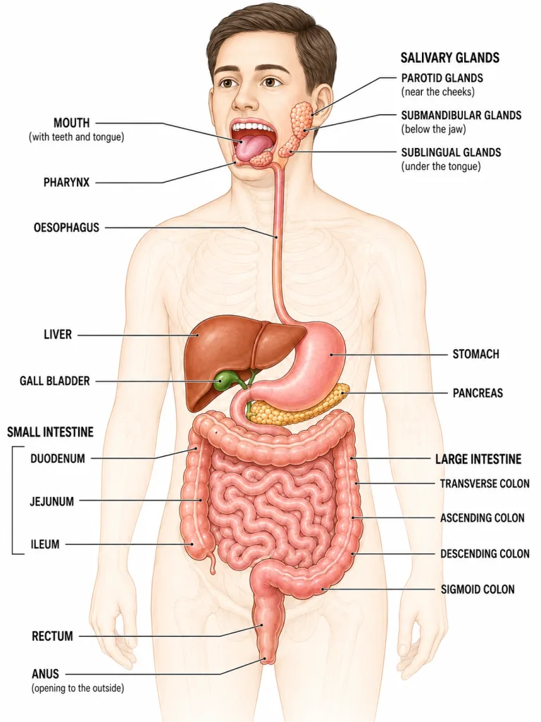
The two parts of the system
1. Alimentary canal — the long continuous tube from mouth to anus:
- Mouth → Pharynx → Oesophagus → Stomach → Small intestine → Large intestine → Rectum → Anus.
2. Accessory (digestive) glands — glands that secrete juices into the canal but are NOT part of the canal itself:
- Salivary glands (3 pairs, around the mouth).
- Liver (largest gland in body, produces bile).
- Pancreas (produces pancreatic juice).
The journey of food in one sentence
Food enters the mouth, is chewed and mixed with saliva (carbohydrate digestion begins), is swallowed into the stomach (protein digestion begins in acid), enters the small intestine (full digestion finishes; absorption happens at villi), passes into the large intestine (water absorbed; waste forms), and exits via the rectum and anus as faeces.
The next several screens unpack each organ in detail.
NCERT-canonical phrase: "The food we eat has to be broken down so that it can be absorbed and used by our cells." This is the one-line raison d'être of digestion.
Stop 1 — The Mouth (Buccal Cavity)
The mouth is the point of ingestion and also the first chemical attack site on food.
Three things happen in the mouth
1. Mechanical digestion (chewing) — teeth do this.
Humans are heterodont — different teeth do different jobs:
| Tooth | Function | Number (adult, total) |
|---|---|---|
| Incisors | Cutting (front teeth) | 8 |
| Canines | Tearing (pointed) | 4 |
| Premolars | Grinding (broader) | 8 |
| Molars | Crushing (back) | 12 |
| Total | 32 |
The chewing action turns large food chunks into small particles — increasing surface area for enzymes.
Dental formula (adult human): 2-1-2-3 / 2-1-2-3 (one side, top/bottom). Memorise this — frequent NEET MCQ.
Two sets of teeth:
- Milk teeth (deciduous) — 20 teeth, appear in childhood.
- Permanent teeth — 32 teeth, replace milk teeth.
2. Chemical digestion — saliva does this
Three pairs of salivary glands open into the mouth:
- Parotid, sub-mandibular, sub-lingual.
- They secrete saliva (~1 litre/day).
Saliva contains:
- Water + mucus — moistens food, makes it slippery.
- Salivary amylase (Ptyalin) — an enzyme that digests starch.
Carbohydrate digestion begins in the mouth itself. Try chewing a piece of bread for a minute — it starts to taste sweet. That's amylase converting starch to maltose.
3. Tongue
- Moves food around in mouth (mixing with saliva).
- Contains taste buds (sweet, sour, salty, bitter, umami).
- Helps in swallowing.
- Helps in speech.
From mouth to stomach
The chewed-and-amylase-mixed food is called a bolus. It is pushed into the pharynx and then down the oesophagus.
Oesophagus = a ~25 cm long muscular tube that uses peristalsis (waves of muscle contraction) to push food down to the stomach.
[Board Important] A 2-mark Board favourite: "What is the function of saliva?" Answer must mention (a) moistening food, (b) starch digestion via amylase, (c) mucus for swallowing.
Stop 2 — The Stomach

The stomach is a J-shaped muscular bag — your body's chemical-attack chamber.
What happens in the stomach
1. Storage — food is held here for ~3-4 hours. 2. Mechanical churning — stomach walls contract, mixing food into a soupy paste (chyme). 3. Chemical digestion — gastric juice acts on proteins.
Gastric juice — three components, three jobs
The stomach's inner wall has gastric glands that secrete gastric juice (~2-3 litres/day). It has three main components, each with a separate role.
| Component | Source | Function |
|---|---|---|
| HCl (Hydrochloric acid) | Parietal cells | Kills bacteria; creates acidic medium (pH ~1.5-2) for pepsin to work |
| Pepsin | Chief cells (as pepsinogen, activated by HCl) | Digests proteins → peptides |
| Mucus | Goblet cells | Protects stomach lining from HCl + pepsin (self-protection!) |
Why these three together?
This is one of the most elegant designs in biology:
- HCl alone would burn the stomach.
- Pepsin alone wouldn't work (needs acidic medium).
- Mucus alone wouldn't digest anything.
Together: HCl creates the right environment, pepsin does the digestion, mucus protects the stomach from itself.
A key NCERT fact
Pepsin is secreted as an inactive precursor called pepsinogen by chief cells. HCl converts pepsinogen → active pepsin only INSIDE the stomach lumen. This way, chief cells don't accidentally digest their own proteins.
Protein digestion equation
Note: pepsin only partially digests proteins. Full breakdown to amino acids happens in the small intestine.
What does NOT happen in the stomach
- No carbohydrate digestion — salivary amylase is inactivated by HCl.
- No fat digestion — fats stay intact (will be digested later in small intestine by bile + lipase).
- Very little absorption — only small molecules like alcohol and some drugs are absorbed here. Real absorption happens in the small intestine.
The pyloric sphincter
At the exit of the stomach is a valve called the pyloric sphincter. It opens periodically to let small amounts of chyme into the small intestine. This slow release ensures the small intestine isn't overwhelmed at once.
Common conditions
- Acidity / heartburn — too much HCl, or HCl in the oesophagus.
- Ulcer — damage to stomach wall (often by H. pylori bacterium).
- Why do we feel "hungry"? — empty stomach signals brain via hormones.
[Board Important] Standard 3-mark Board question: "What are the three components of gastric juice? State the function of each." Memorise the table above. Always mention that mucus protects the stomach from self-digestion.
Stop 3 — The Small Intestine (Where Real Digestion Finishes)
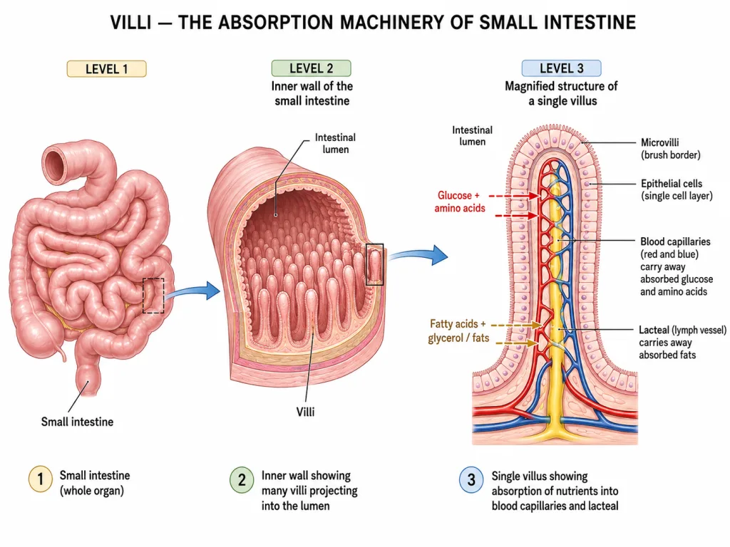
The small intestine is the most important part of the digestive system. Why? Because:
- Most digestion happens here — proteins, fats, and carbs all finished.
- Almost all absorption happens here — nutrients enter the blood.
- It is the longest part — ~6 metres in adults (3x your height!).
Three sections
- Duodenum — first ~25 cm. Receives pancreatic juice + bile.
- Jejunum — middle section (~2.5 metres).
- Ileum — last section (~3.5 metres). Main absorption site.
Three digestive juices act here
(a) Bile — from the liver, stored in gall bladder.
- NOT an enzyme. Bile contains bile salts.
- Function: emulsification of fats — breaks large fat globules into tiny droplets so lipase can act on them.
- Think of bile as soap for fats.
(b) Pancreatic juice — from the pancreas.
Contains THREE major enzymes:
| Enzyme | Acts on | Product |
|---|---|---|
| Trypsin | Proteins → peptides | Smaller peptides + amino acids |
| Pancreatic Lipase | Emulsified fats | Fatty acids + glycerol |
| Pancreatic Amylase | Starch (any remaining) | Maltose |
(c) Intestinal juice (Succus entericus) — from intestinal walls.
| Enzyme | Acts on | Product |
|---|---|---|
| Maltase | Maltose | Glucose + Glucose |
| Lactase | Lactose (milk sugar) | Glucose + Galactose |
| Sucrase | Sucrose (table sugar) | Glucose + Fructose |
| Erepsin (peptidase) | Peptides | Amino acids |
By the time food leaves the small intestine, all complex molecules have been reduced to their simplest absorbable forms.
The absorption miracle — VILLI
The inner wall of the small intestine is covered with millions of finger-like projections called villi (singular: villus). Each villus is also covered with even smaller projections called microvilli.
Why this elaborate structure? To maximise surface area for absorption.
If you flattened out the entire small intestine's villi, the surface area would be about 250 m² — equivalent to a tennis court!
Inside a villus
Each villus has:
- Single layer of epithelial cells on the outside — for easy diffusion.
- Network of blood capillaries inside — carry away absorbed glucose, amino acids, water-soluble vitamins.
- A central lacteal (lymph vessel) — carries away absorbed fats (fatty acids + glycerol go into lymph, not blood directly).
Adaptations for absorption
- Large surface area — villi + microvilli.
- Thin walls — only one cell thick.
- Rich blood supply — every villus is packed with capillaries.
- Lacteal — separate channel for fats.
- Length — 6 metres = lots of time for absorption.
NCERT-canonical phrase
"The small intestine is the site of complete digestion of carbohydrates, proteins and fats." — verbatim NCERT, memorise.
What is absorbed and where it goes
| Nutrient | Absorbed into | Final destination |
|---|---|---|
| Glucose | Blood capillaries → liver → cells | Energy (respiration) |
| Amino acids | Blood capillaries → cells | Build proteins |
| Fatty acids + Glycerol | Lacteal (lymph) | Body fat / energy |
| Vitamins, minerals, water | Blood/lymph | Various uses |
[Board Important] "Why is the small intestine the longest part of the alimentary canal?" — Because complete digestion + absorption of all nutrients happens here. Length and villi give maximum time + surface area.
Stop 4 — The Large Intestine (Water Recovery)
After the small intestine has absorbed all the useful nutrients, the leftover material — mostly water + undigested fibre + dead bacteria — enters the large intestine.
Anatomy
The large intestine is shorter (~1.5 m) but wider than the small intestine. Its parts:
- Caecum (with the appendix attached — a vestigial organ in humans)
- Ascending colon — goes up on right side.
- Transverse colon — goes across.
- Descending colon — goes down on left side.
- Sigmoid colon — S-shaped.
- Rectum — storage area.
- Anus — exit.
What happens in the large intestine
1. Water absorption — the main job! Up to 1.5 litres of water are reabsorbed daily. This is critical — without it, you'd lose dangerous amounts of water in faeces (which is why diarrhoea causes dehydration — water isn't reabsorbed).
2. Some salts and vitamins are also absorbed. Bacteria living in the colon make some Vitamin K and Vitamin B12 — small amounts absorbed.
3. Formation of faeces. As water is absorbed, the contents thicken into solid waste — faeces. They are stored in the rectum until egestion.
4. Egestion. Faeces are expelled through the anus via the act of defecation.
What does NOT happen in the large intestine
- No digestion — there are no digestive enzymes here.
- No absorption of food nutrients — those were already absorbed in the small intestine.
Why we need fibre in our diet
Dietary fibre (cellulose from vegetables) cannot be digested by human enzymes. But it serves a crucial role:
- Adds bulk to the contents of the large intestine.
- This bulk stimulates peristalsis — keeps food moving along.
- Prevents constipation.
Hence the medical advice: eat plenty of vegetables and whole grains for fibre.
A simple summary of the whole tract
| Organ | Main role | Time |
|---|---|---|
| Mouth | Mechanical + start of starch digestion | seconds |
| Oesophagus | Transport (peristalsis) | 5-10 sec |
| Stomach | Protein digestion (HCl + pepsin) | 3-4 hours |
| Small intestine | Complete digestion + absorption | 4-6 hours |
| Large intestine | Water absorption + faeces formation | 12-24 hours |
| Rectum + anus | Storage + egestion | variable |
Total transit time: roughly 24-72 hours from mouth to anus.
[Board Important] Standard question: "State the role of large intestine in human digestive system." — Mention: water absorption, vitamin/salt absorption, faeces formation, egestion. NOT digestion.
Memory Capsule — Section 4
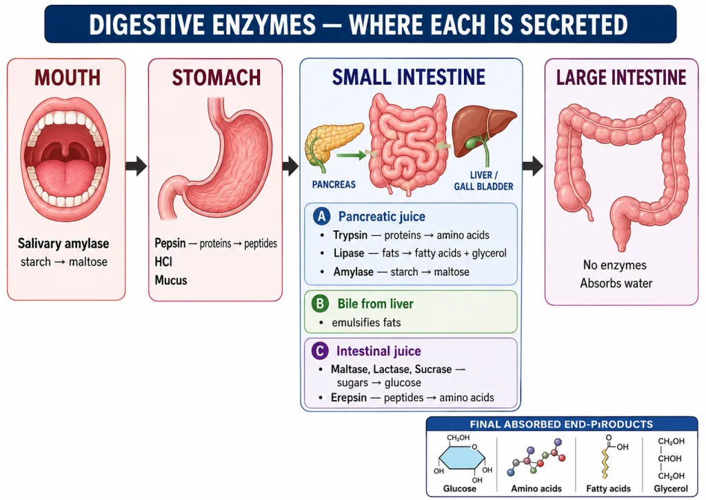
A compact recap before moving to Section 5 (Respiration).
The full alimentary canal (in order)
Mouth → Pharynx → Oesophagus → Stomach → Small Intestine → Large Intestine → Rectum → Anus
Three accessory glands
- Salivary glands → saliva (amylase).
- Liver → bile (emulsifies fats; no enzymes).
- Pancreas → pancreatic juice (trypsin, lipase, amylase).
Master enzyme table (memorise!)
| Location | Enzyme | Substrate | Product |
|---|---|---|---|
| Mouth | Salivary amylase (ptyalin) | Starch | Maltose |
| Stomach | Pepsin | Proteins | Peptides |
| Small intestine — Pancreatic juice | Trypsin | Proteins/peptides | Amino acids |
| Pancreatic amylase | Starch | Maltose | |
| Pancreatic lipase | Fats (after bile) | Fatty acids + glycerol | |
| Small intestine — Intestinal juice | Maltase | Maltose | Glucose |
| Lactase | Lactose | Glucose + galactose | |
| Sucrase | Sucrose | Glucose + fructose | |
| Erepsin (peptidase) | Peptides | Amino acids |
Bile is special
- From liver, stored in gall bladder.
- NOT an enzyme.
- Function: emulsifies fats (breaks large drops to tiny ones).
- Makes the small intestine medium alkaline (neutralises acid from stomach).
Stomach — three secretions
| Secretion | Function |
|---|---|
| HCl | Acidic medium + kills bacteria |
| Pepsin | Protein digestion |
| Mucus | Protects stomach wall |
Small intestine — site of:
- Complete digestion (carbs, proteins, fats).
- Almost all absorption (via villi).
- Glucose + amino acids → blood capillaries.
- Fatty acids + glycerol → lacteals (lymph).
Large intestine — only:
- Water absorption.
- Faeces formation.
- No digestion, no nutrient absorption.
Teeth (dental formula)
Adult: 2-1-2-3 / 2-1-2-3 → Total 32 teeth. Milk teeth: 20.
NCERT-canonical phrases
- "Small intestine is the site of complete digestion of carbohydrates, proteins and fats."
- "Inner lining of small intestine has numerous finger-like projections called villi which increase the surface area for absorption."
- "Bile salts emulsify fats and break them into smaller globules."
One-line takeaway
The human digestive tract is a 9-metre tube where food is mechanically broken in the mouth, chemically attacked in the stomach (proteins) and small intestine (everything), absorbed at villi into the blood, and the dry waste is finally expelled — a four-stage assembly line with specialised enzymes at every step.
Solved Examples
Example 1: Trace a piece of paneer through the human digestive tract
Paneer is a milk product rich in protein and fat. Trace its complete digestion from mouth to absorption.
Solution:
We trace each macronutrient separately.
The protein in paneer
Mouth: No protein digestion here — salivary amylase acts only on carbohydrates. So the protein passes through untouched.
Stomach: This is where protein digestion begins. HCl creates acidic medium; pepsin acts on proteins, breaking them into smaller peptides.
Small intestine: Pancreatic juice's trypsin continues breaking peptides → amino acids. Intestinal juice's erepsin (peptidase) finishes the job.
Absorption: Amino acids enter the blood capillaries of villi.
The fat in paneer
Mouth, stomach: No fat digestion here. Fats remain intact.
Small intestine: This is where ALL fat digestion happens.
- First, bile from the liver/gall bladder emulsifies large fat globules into tiny droplets. (Bile is not an enzyme — it's a detergent!)
- Then pancreatic lipase acts on emulsified fats.
Absorption: Fatty acids + glycerol enter the lacteal (lymph vessel) in each villus — NOT directly into blood.
Summary table
| Component | Mouth | Stomach | Small intestine | Absorbed into |
|---|---|---|---|---|
| Protein | No action | Pepsin → peptides | Trypsin + Erepsin → amino acids | Blood |
| Fat | No action | No action | Bile emulsifies; Lipase → fatty acids + glycerol | Lacteal (lymph) |
Answer: As shown. Protein digestion starts in stomach (pepsin) and finishes in small intestine (trypsin, erepsin). Fat digestion happens entirely in the small intestine (bile emulsifies → lipase digests). Amino acids enter blood; fatty acids enter the lacteal.
[Board Important] Frequent 5-mark Board question. Show the path enzyme-by-enzyme. Mention bile's special role (emulsifies, not digests).
Example 2: The three components of gastric juice — function of each
Name the three main components of gastric juice and state the function of each in 2-3 sentences.
Solution:
Gastric juice is secreted by glands in the stomach wall. It has three components — each plays a separate role.
1. Hydrochloric acid (HCl)
Source: Parietal cells (oxyntic cells) of gastric glands.
Functions:
- Creates a highly acidic medium (pH ~1.5-2) in the stomach. This is the optimal pH for pepsin to work.
- Kills bacteria that enter with food — a major immune defence.
- Activates pepsinogen → pepsin.
Without HCl, pepsin wouldn't work, and the food would have many live bacteria entering deeper into the gut.
2. Pepsin (an enzyme)
Source: Chief cells (peptic cells) of gastric glands. Secreted as the inactive precursor pepsinogen; HCl converts it to active pepsin.
Function:
- Begins protein digestion — breaks large proteins into smaller fragments called peptides.
3. Mucus
Source: Goblet cells / mucus cells of the stomach lining.
Function:
- Forms a thick protective layer over the inner stomach wall.
- Prevents HCl and pepsin from digesting the stomach itself (self-digestion).
- Without mucus, the stomach would form ulcers.
Why all three together?
This is one of NCERT's elegant teaching points. Each component is useless or dangerous alone:
- HCl alone — burns the stomach.
- Pepsin alone — can't work without acid.
- Mucus alone — does nothing to food.
Together: HCl creates the condition + pepsin does the work + mucus protects the stomach. Perfect teamwork.
Answer:
- HCl — creates acidic medium for pepsin; kills bacteria.
- Pepsin — digests proteins into peptides.
- Mucus — protects stomach wall from HCl and pepsin.
[Board Important] A classic 3-mark Board question. Always mention the protective role of mucus — that's the part most students forget.
Example 3: Why is the small intestine the longest part of the alimentary canal?
Give at least three reasons.
Solution:
The small intestine is ~6 metres long — far longer than any other part of the digestive tract (stomach: 30 cm; large intestine: 1.5 m). Why?
Reason 1: It is the site of complete digestion
By the time food reaches the small intestine, it has been partially digested in the mouth (starch → maltose) and stomach (protein → peptides). But fats are still completely undigested, and carbs and proteins are not yet fully broken down.
The small intestine must finish digesting all three macronutrients:
- Carbohydrates → glucose.
- Proteins → amino acids.
- Fats → fatty acids + glycerol.
This requires multiple enzymes acting in sequence — and takes time. A long intestine gives the enzymes more contact time.
Reason 2: It is the main absorption site
Almost ALL nutrients are absorbed in the small intestine (only water absorption happens in large intestine).
Absorption requires:
- Surface area — solved by villi and microvilli (250 m² equivalent area).
- Time — solved by length of the intestine (6 metres = lots of time as food moves through).
If the small intestine were short, food would race past the villi too quickly for absorption.
Reason 3: It receives juices from multiple sources
Three juices meet in the small intestine:
- Bile (from liver) — emulsifies fats.
- Pancreatic juice (from pancreas) — trypsin, lipase, amylase.
- Intestinal juice (from intestinal wall) — maltase, lactase, sucrase, erepsin.
Each juice acts at slightly different points along the intestine. A long tube allows space for each to act in sequence.
Quantitative perspective
- Small intestine length: ~6 metres in adults.
- This is 3x the height of an average person — folded up into the abdomen.
- Time for food to traverse: ~4-6 hours.
- Surface area (with villi): ~250 m².
No other organ in the body has this combination of length, surface area, and enzymatic activity. That's why it's so long.
Answer: The small intestine is the longest part of the alimentary canal because (1) complete digestion of all three macronutrients happens here — requires sequential action of multiple enzymes; (2) absorption of all digested nutrients into blood/lymph occurs here — needs maximum surface area and time; (3) it receives three different digestive juices (bile, pancreatic juice, intestinal juice) which need space and time to act. Hence its length is essential for both digestion and absorption.
[Board Important] A 3-mark NCERT-style question. Always link length to (a) complete digestion, (b) full absorption, (c) multiple digestive juices.
Example 4: What is the role of bile? Why is it called an emulsifier, not an enzyme?
Bile is secreted by the liver and stored in the gall bladder. It enters the small intestine via the bile duct.
Solution:
What bile contains
Bile is a yellow-greenish fluid containing:
- Bile salts — the main functional component.
- Bile pigments — bilirubin, biliverdin (waste products of haemoglobin breakdown).
- Water, cholesterol, lecithin.
- NO enzymes. This is the key point.
What bile does (its three functions)
1. Emulsification of fats (main function)
Fats in food enter the small intestine as large globules (think of oil floating on water — large fat blobs). These are very hard for lipase to digest, because lipase can only act on the surface, and large globules have low surface area per volume.
Bile salts work like a detergent: they break the large fat globules into tiny droplets.
Now these tiny droplets have HUGE total surface area, and lipase can attack them efficiently:
2. Neutralisation of acid
Food entering the small intestine from the stomach is very acidic (pH ~2 due to HCl). Bile is alkaline (basic). It neutralises the acid, making the intestinal contents slightly alkaline — the right pH for pancreatic and intestinal enzymes (which work best at pH 7-8, not acidic).
3. Excretion of waste
Bile pigments (bilirubin) are waste products of dead RBC breakdown. The body excretes them via bile, into the intestine, and finally out in faeces (this is what gives faeces their brown colour).
Why bile is NOT called an enzyme
Enzymes are proteins that catalyse chemical reactions. They have a specific shape, an active site, and they actually break or form chemical bonds.
Bile salts do NOT catalyse a chemical reaction. They don't break any bond. They simply physically disrupt the fat globules by acting as soap-like molecules. The chemical digestion of fats is done by lipase, not bile.
Analogy: Bile is like a chef who chops vegetables. Lipase is like the fire that cooks them. Bile prepares the substrate; lipase does the digestion.
Summary
| Property | Bile | Enzymes (e.g., lipase) |
|---|---|---|
| Type of molecule | Bile salts (steroid-derived) | Proteins |
| Role | Emulsifies fats + neutralises acid | Catalyses chemical reactions |
| Source | Liver | Various glands |
| Acts on | Large fat globules (physically) | Specific substrates (chemically) |
Answer: Bile is a yellow-greenish fluid from the liver that emulsifies large fat globules into tiny droplets, vastly increasing surface area for lipase to act on. It also neutralises stomach acid in the small intestine. Bile is NOT an enzyme because it does not catalyse any chemical reaction — it acts physically like a detergent. The actual chemical digestion of fats is done by lipase.
[Board Important] Frequent 3-mark question. Always mention: (a) emulsification, (b) bile is NOT an enzyme, (c) lipase does the actual digestion.
Example 5: Describe the structure of a villus and explain how it is adapted for absorption.
Draw a labelled diagram of a single villus and describe its adaptations.
Solution:
What is a villus?
A villus (plural: villi) is a finger-like projection lining the inner wall of the small intestine. Each villus is about 1 mm tall. The small intestine has millions of villi — they look like a thick velvety carpet under a microscope.
Structure of a single villus (labelled parts)
Outside (surrounding the villus):
- A single layer of epithelial cells covers the entire surface — thin walls = easy diffusion of nutrients.
- The epithelial cells themselves have microvilli on their surface (even smaller projections — a "brush border") that further multiply surface area.
Inside the villus:
- A dense network of blood capillaries — receives glucose, amino acids, water-soluble vitamins, and minerals.
- A central lacteal (a lymph vessel) — receives fatty acids and glycerol (fat-digestion products).
- Smooth muscle cells — to keep the villus moving slightly (mixes intestinal contents, exposes more surface).
Adaptations for absorption — five key features
1. Enormous surface area. Numbers: Small intestine length: 6 m. Villi: millions. Microvilli: each cell has 1000+. Net result: ~250 m² of absorbing surface. This is why a 6 m tube can absorb a day's worth of nutrients.
2. Thin walls (just one cell thick). Why it matters: Diffusion is faster across a thinner barrier. Glucose and amino acids can quickly pass from the gut lumen → epithelial cell → blood capillary.
3. Rich blood supply. Why it matters: Each villus is packed with capillaries. As blood continuously moves nutrients away, the concentration gradient is maintained — nutrients keep diffusing in.
4. Central lacteal (lymph vessel). Why it matters: Fatty acids and glycerol (which can't dissolve in water-based blood directly) enter the lacteal — a parallel lymph transport. This is a separate pathway for fats.
5. Length and shape — increase contact time. Why it matters: Food moves slowly through the 6-metre tube, giving each section time to absorb nutrients.
Putting it together — how absorption happens
- Digested nutrients (glucose, amino acids, etc.) sit in the gut lumen.
- They contact the villus surface (microvilli).
- They cross the single-layer epithelial cells (by diffusion or active transport).
- They enter either:
- Blood capillaries (glucose, amino acids, water-soluble vitamins).
- Lacteal (fatty acids, glycerol, fat-soluble vitamins).
- Blood/lymph carries them away to the rest of the body.
Why this is brilliant
The small intestine doesn't just have a long flat surface — that would be inefficient. By using villi + microvilli, evolution has multiplied the surface area 600 times over a flat tube. It's like comparing a folded fan to a flat piece of paper.
NCERT-canonical phrasing
"The inner lining of the small intestine has numerous finger-like projections called villi, which increase the surface area for absorption."
Answer: A villus is a 1-mm finger-like projection from the inner wall of the small intestine, covered by a single layer of epithelial cells (with microvilli on top). Inside it has a network of blood capillaries and a central lacteal. Adaptations for absorption: (1) huge surface area (villi + microvilli = 250 m² total); (2) thin walls for easy diffusion; (3) rich blood supply maintains concentration gradient; (4) central lacteal for fat absorption; (5) length of intestine ensures contact time.
[Board Important] 5-mark question. Must include labelled diagram + list of adaptations. NCERT phrase about "finger-like projections to increase surface area" is a mark-winner.
Example 6: Trace the digestion of a chapati (mostly starch)
A chapati is mainly carbohydrate (starch from wheat flour) with a bit of protein (gluten). Describe its complete digestion through the alimentary canal.
Solution:
Let's trace it organ by organ.
Mouth
The chapati is chewed by the teeth (mechanical digestion) and mixed with saliva.
- Salivary amylase (ptyalin) in saliva starts digesting starch.
Note: Salivary amylase only partially digests starch in the mouth — there isn't enough time for complete digestion in the brief stay there.
Oesophagus
No digestion. Just transport via peristalsis to the stomach. (5-10 seconds.)
Stomach
The stomach is acidic, but salivary amylase needs an alkaline/neutral pH. So:
- Salivary amylase is inactivated by stomach HCl. Starch digestion temporarily PAUSES here.
- However, the little bit of protein (gluten) in the chapati starts being digested by pepsin.
Small intestine — where starch digestion really finishes
Now comes the main act. When food enters the small intestine, pancreatic juice + bile + intestinal juice all enter.
Step 1: Pancreatic amylase finishes starch → maltose conversion.
Step 2: Intestinal juice's maltase breaks maltose → glucose.
Step 3 (for the protein bit): Trypsin and erepsin finish peptides → amino acids.
Absorption (small intestine)
Glucose and amino acids enter the blood capillaries of villi.
Large intestine
No digestion here. Water from the leftover material is absorbed. (The undigested fibre in the chapati provides bulk.)
Egestion
Undigested cellulose fibre exits through the anus as part of faeces.
Summary table
| Location | What happens to starch | What happens to gluten |
|---|---|---|
| Mouth | Salivary amylase → some maltose | No digestion |
| Stomach | Amylase inactivated; pauses | Pepsin → peptides |
| Small intestine | Pancreatic amylase → maltose; Maltase → glucose | Trypsin/Erepsin → amino acids |
| Absorption | Glucose into blood capillaries | Amino acids into blood capillaries |
| Large intestine | Fibre/cellulose unchanged | nothing |
| Egestion | Fibre out as faeces | nothing |
Final fate of glucose
Once absorbed:
- Blood carries glucose to all body cells.
- Cells use it in respiration to make ATP (Section 5).
- Excess is stored as glycogen in the liver/muscles.
And that's how a chapati powers your day!
Answer: Starch in a chapati is digested in stages: mouth (salivary amylase → some maltose), then paused in the stomach (amylase inactivated by acid), then completed in the small intestine (pancreatic amylase → maltose; maltase → glucose). Glucose is absorbed into blood at the villi. Gluten is digested in stomach (pepsin → peptides) and small intestine (trypsin + erepsin → amino acids). Undigested cellulose fibre exits as faeces.
[NEET-foundation] 'Trace the path' questions are common. The trick is: every macronutrient (starch, protein, fat) starts being digested somewhere, gets paused/continued somewhere, and finishes in the small intestine.