
The Neuron — Basic Unit of Nervous System
The nervous system is built from cells called neurons. These are the 'wires' that carry electrical signals throughout your body.
Definition
'Neuron' (or nerve cell) = the structural and functional unit of the nervous system.
Numbers in the Body
Total neurons in human body: ~100 billion (10¹¹). All in your brain alone: ~86 billion! Each neuron can connect to up to 10,000 others.
That's an unimaginable web of connections — your brain.
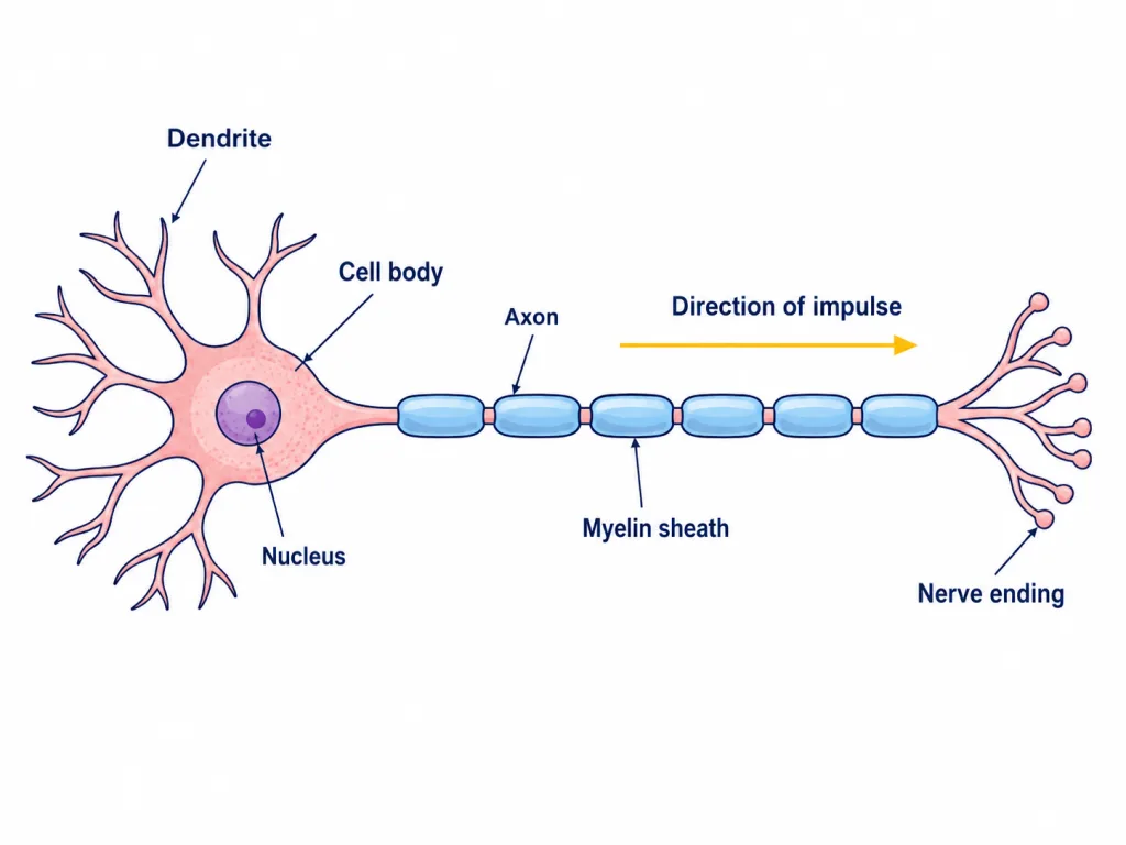
Structure of a Neuron
A typical neuron has 4 main parts:
1. Cell Body (Cyton): Contains:
- Nucleus.
- Cytoplasm with all standard cell organelles. Function: maintains cell life, makes proteins.
2. Dendrites: Short, branched extensions. Function: receive signals from other neurons. Like 'antennas' picking up information.
3. Axon: Long, single extension (can be 1 m long in legs). Function: carries signal away from cell body. Often covered with myelin sheath — fatty insulating layer.
4. Axon Terminals: End branches of axon. Function: transmit signal to next neuron or muscle. Form synapses with target cells.
Diagram (Verbal)
  Dendrites (input)
      |
  Cell Body (with nucleus)
      |
      |  ← Axon (long fiber)
      |  ← Myelin sheath (insulation)
      |
  Axon Terminals (output)
Neuron looks like a tree with roots (dendrites) and a long trunk (axon).
Myelin Sheath
Fatty layer wrapping the axon. Function: speeds up signal transmission (~100 m/s). Without it: signals would be slower (~1 m/s).
In multiple sclerosis (a disease), myelin is damaged → signals slow → motor problems.
[NCERT — diagram important]
Types of Neurons
Three main types based on function:
1. Sensory Neurons (Afferent)
Carry signals from sense organs (skin, eyes, ears) to brain/spinal cord.
Direction: receptor → CNS.
Examples:
- Touch a hot pan → skin receptor → sensory neuron → spinal cord.
- See light → eye → sensory neuron → brain.
2. Motor Neurons (Efferent)
Carry signals from brain/spinal cord to muscles or glands.
Direction: CNS → effector.
Examples:
- Brain decides to move arm → motor neuron → arm muscle.
- Brain says 'salivate' → motor neuron → salivary glands.
3. Relay/Interneurons
Found inside CNS (brain, spinal cord). Connect sensory neurons to motor neurons. Process and decide.
Most numerous type — billions in brain.
Quick Summary Table
| Type | Direction | Function |
|---|---|---|
| Sensory | Receptor → CNS | Carry sensation in |
| Motor | CNS → Effector | Carry response out |
| Relay | Within CNS | Connect/process |
How a Signal Travels (Example)
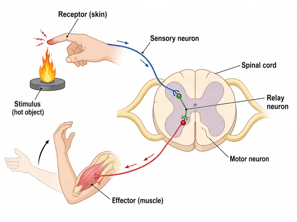
Touching hot pan: 1. Receptor (skin) → detects heat. 2. Sensory neuron → carries signal to spinal cord. 3. Relay neuron in spinal cord → processes (decides 'pull back!'). 4. Motor neuron → carries signal to arm muscle. 5. Effector (muscle) → contracts → hand pulls back.
All in 0.1 seconds!
[NCERT — important]
Synapse — The Junction
Neurons don't actually touch each other — there's a tiny gap between them. This gap is the synapse.
Definition
'Synapse' = junction (gap) between two neurons or between neuron and muscle/gland. Width: ~20-40 nanometers.
How Signals Cross the Synapse
The signal in a neuron is electrical, but at the synapse it becomes chemical for crossing.
Steps: 1. Electrical signal reaches axon terminal. 2. Triggers release of neurotransmitters (chemical messengers). 3. Neurotransmitters cross the synapse gap. 4. They bind to receptors on next neuron. 5. New electrical signal starts in next neuron.
Common Neurotransmitters
1. Acetylcholine — at neuromuscular junctions. 2. Dopamine — pleasure, movement (Parkinson's = dopamine deficiency). 3. Serotonin — mood, sleep (low = depression). 4. GABA — inhibitory (calms nervous system). 5. Glutamate — excitatory.
Why Synapses Matter
1. Allow processing: signal can be modified, blocked, or amplified. 2. Enable learning: strong synapses form with practice. 3. Memory storage: memories are patterns of synaptic connections. 4. Drugs target synapses: painkillers, antidepressants, etc.
Number of Synapses
Brain has ~100 trillion synapses! Each thought, memory, action involves many synapses firing.
Diagram (Verbal)
    Axon Terminal (Neuron 1)
         |
         | ← Synaptic vesicles (with neurotransmitters)
         |
   ----------- Synapse (gap)
         |
         | ← Receptors on Neuron 2
         |
      Dendrite (Neuron 2)
Synapse = bottleneck where signals are processed.
[NCERT — important concept]
Reflex Action — Quick Automatic Response
When you accidentally touch a hot pan, your hand pulls back BEFORE you 'feel' the pain. This automatic, fast response is a reflex action.
Definition
'Reflex action' = an immediate, involuntary, automatic response to a stimulus.
Speed: ~0.1 seconds. Bypasses the conscious brain — handled by spinal cord.
Why Reflex Actions are Important
1. Saves time — no need to think. 2. Protects body — pulls hand from hot pan before burn worsens. 3. Survival — instant response in dangerous situations. 4. Saves brain capacity — frees brain for higher functions.
Reflex Arc — The Pathway
The path that signal takes during reflex.
5 components of reflex arc:
1. Receptor — detects stimulus (e.g., skin). 2. Sensory neuron — carries signal to spinal cord. 3. Spinal cord (with relay neuron) — processes signal. 4. Motor neuron — carries response back. 5. Effector — performs action (e.g., muscle).
Diagram (Verbal)
Stimulus (hot pan)
     ↓
  Receptor (skin)
     ↓
Sensory Neuron
     ↓
Spinal Cord (Relay neuron)
     ↓
Motor Neuron
     ↓
  Effector (muscle)
     ↓
Response (hand pulled back)
Note: brain is informed AFTER the reflex. That's why you 'feel' pain after the action.
Examples of Reflex Actions
1. Knee-jerk reflex: tap below knee → leg kicks out. 2. Pupillary reflex: bright light → pupils contract. 3. Sneezing: dust irritates nose → involuntary sneeze. 4. Coughing: food enters wrong pipe → cough to clear. 5. Blinking: object near eye → eyes close instantly. 6. Swallowing reflex: food in throat → automatic swallow. 7. Salivation: seeing/smelling food.
Why Through Spinal Cord, Not Brain?
Brain is far from many body parts. Spinal cord is closer → signal reaches faster.
Example: hand to spinal cord = 0.5 m. Hand to brain = 1 m. Reflex via spinal cord saves precious time during emergencies.
Brain is informed afterwards (when you 'feel' the pain).
[NCERT — Board favourite]
Voluntary vs Involuntary Actions
Voluntary Actions
'Voluntary' = under conscious control. You decide to do them. Controlled by cerebrum (front of brain).
Examples: 1. Walking, running. 2. Writing, typing. 3. Talking. 4. Eating. 5. Reading this!
Pathway: brain → motor neuron → muscles.
Involuntary Actions
'Involuntary' = NOT under conscious control. Happen automatically. Controlled by medulla, hypothalamus, autonomic nervous system.
Examples: 1. Heartbeat. 2. Breathing (mostly). 3. Digestion. 4. Sweating. 5. Reflexes (pulling hand from hot). 6. Pupil dilation. 7. Saliva secretion.
Often controlled in background — you can't 'tell' your heart to beat slower (well, with practice you can — meditation!).
Comparison Table
| Feature | Voluntary | Involuntary |
|---|---|---|
| Control | Conscious | Automatic |
| Brain part | Cerebrum | Medulla, hypothalamus |
| Speed | Variable | Often fast (reflexes) |
| Examples | Walking, talking | Heartbeat, reflexes |
| Can you stop it? | Yes | No (mostly) |
Reflex Action — A Special Type
Reflex action is involuntary. But unique because it bypasses brain. Handled by spinal cord directly.
Other involuntary actions (heartbeat) are controlled by medulla in brain.
Comparison: Reflex vs Voluntary
| Aspect | Reflex | Voluntary |
|---|---|---|
| Speed | Very fast (~0.1 s) | Slower (decision time) |
| Pathway | Spinal cord | Brain |
| Control | Involuntary | Conscious |
| Examples | Pulling hand from hot | Picking up cup |
Practical Examples
1. Driving a Car: Voluntary at first (consciously turning steering). Becomes 'automatic' with practice (semi-reflex). Habits form through repeated synaptic connections.
2. Doctor's Knee-Jerk Test: Doctor taps below knee → reflex kick. Tests if reflex arc is working. Damaged spinal cord → no reflex.
3. Sneezing in Class: Involuntary, can't control. Cover mouth (voluntary action) to be polite!
[NCERT — important]
Memory Capsule — Section 2
Quick revision card for neurons and reflex action.
Neuron — Basic Unit
4 Parts: 1. Cell body (cyton) — nucleus, cytoplasm. 2. Dendrites — receive signals. 3. Axon — carries signals away. 4. Axon terminals — transmit to next.
Myelin sheath = fatty insulation, speeds signal.
3 Types of Neurons
1. Sensory — receptor → CNS. 2. Motor — CNS → effector. 3. Relay/Interneurons — within CNS.
Synapse
Junction between two neurons. Signal: electrical → chemical (neurotransmitters) → electrical. Common neurotransmitters: acetylcholine, dopamine, serotonin.
Reflex Action
Definition: immediate, involuntary, automatic response. Speed: ~0.1 seconds. Pathway: skips brain, goes through spinal cord.
Reflex Arc — 5 Components
1. Receptor. 2. Sensory neuron. 3. Spinal cord (relay). 4. Motor neuron. 5. Effector.
Voluntary vs Involuntary
| Voluntary | Involuntary | |
|---|---|---|
| Brain part | Cerebrum | Medulla, etc. |
| Control | Conscious | Automatic |
| Examples | Walking | Heartbeat, reflexes |
Examples of Reflexes
1. Pulling hand from hot pan. 2. Knee-jerk. 3. Pupillary reflex (bright light). 4. Sneezing. 5. Blinking.
Why Reflexes Matter
1. Save time (~0.1 s vs 0.5 s for thinking). 2. Protect body from injury. 3. Survival in emergencies.
Numbers
- Total neurons: ~100 billion.
- In brain: ~86 billion.
- Synapses in brain: ~100 trillion.
- Signal speed (myelinated): ~100 m/s.
One-Liner Insights
1. Neuron = nerve cell, 4 parts. 2. Sensory carries IN, motor carries OUT. 3. Synapse = junction; uses neurotransmitters. 4. Reflex bypasses brain (uses spinal cord). 5. Voluntary = conscious; involuntary = automatic.
[Quick reference for last-minute revision!]
Example 1: NCERT — Neuron Structure
Draw a labelled diagram of a neuron. State the function of each part.
Solution:
Neuron Structure (Verbal Diagram)
  Dendrites (input)
      ↓
  Cell Body (nucleus + cytoplasm)
      ↓
   ━━━━━━━ ← Axon (with myelin sheath)
      ↓
  Axon Terminals (output)
4 Parts and Functions
1. Cell Body (Cyton): Contains nucleus, cytoplasm, organelles. Function: maintains cell life, makes proteins, metabolises.
2. Dendrites: Short, branched extensions of cell body. Function: RECEIVE signals from other neurons. Like 'antennas' or 'ears'.
3. Axon: Long, single extension (can be 1 m in long nerves). Function: CARRY signal AWAY from cell body. Often covered with myelin sheath (insulation). Speed: up to 100 m/s with myelin.
4. Axon Terminals: End branches of axon. Function: TRANSMIT signal to next neuron, muscle, or gland. Form synapses with target cells.
Direction of Signal
Dendrites → Cell body → Axon → Axon terminals. One way only.
Myelin Sheath
Fatty insulating layer around axon. Speeds up signal: 100 m/s (with myelin) vs 1 m/s (without). Made by glial cells (Schwann cells). Damage causes diseases like multiple sclerosis.
[NCERT — must-know diagram]
Example 2: NCERT — Types of Neurons
Name the 3 types of neurons. State the function of each with an example.
Solution:
3 Types of Neurons
1. Sensory Neurons (Afferent): Carry signals TO the brain/spinal cord (CNS). From: receptors (sense organs). Direction: in.
Example: when you touch a hot pan: Skin receptor → sensory neuron → spinal cord.
2. Motor Neurons (Efferent): Carry signals FROM the brain/spinal cord (CNS). To: effectors (muscles, glands). Direction: out.
Example: when you decide to lift hand: Brain → motor neuron → arm muscle.
3. Relay/Interneurons: Found WITHIN the CNS. Connect sensory and motor neurons. Process information.
Example: in reflex action: Sensory neuron → relay neuron in spinal cord → motor neuron.
Comparison Table
| Type | Direction | Found Where | Function |
|---|---|---|---|
| Sensory | To CNS | Body to CNS | Carry sensation |
| Motor | From CNS | CNS to body | Carry response |
| Relay | Within CNS | Brain, spinal cord | Process and connect |
Example: Reflex Action Path
Touch hot pan (using all 3 types):
1. Skin (receptor) detects heat. 2. Sensory neuron carries signal to spinal cord. 3. Relay neuron processes — decides 'pull back!'. 4. Motor neuron carries signal to arm muscle. 5. Arm muscle (effector) contracts → hand pulls back.
All 3 types work together!
Numerical Insight
Brain has billions of relay/interneurons. This vast network is what makes 'thinking' possible.
[NCERT — important]
Example 3: NCERT — Synapse
What is a synapse? How does a signal cross it?
Solution:
Synapse
'Synapse' = the junction (small gap) between two neurons, or between a neuron and an effector (muscle, gland).
Width: ~20-40 nanometers (very tiny!).
Why is There a Gap?
Direct touch between neurons would mean signals could flow randomly. Gap allows: 1. Control of signal flow. 2. Processing/modulation. 3. Decision-making. 4. Learning.
How Signal Crosses (5 Steps)
Step 1: Electrical signal arrives Signal travels along axon as electrical impulse. Reaches axon terminal.
Step 2: Triggers chemical release Synaptic vesicles in axon terminal release neurotransmitters (chemical messengers).
Step 3: Diffusion across gap Neurotransmitters diffuse across the synapse. Takes ~0.5 milliseconds.
Step 4: Binding to receptors On the other side, receptors on the next neuron's dendrite catch the neurotransmitters.
Step 5: New electrical signal Binding triggers a new electrical impulse in the next neuron. Signal continues!
So:
Signal converts: electrical → chemical → electrical. Synapse is a bottleneck where conversion happens.
Common Neurotransmitters
| Neurotransmitter | Function |
|---|---|
| Acetylcholine | Muscle contraction, learning |
| Dopamine | Pleasure, reward, movement |
| Serotonin | Mood, sleep, appetite |
| GABA | Inhibitory (calms) |
| Glutamate | Excitatory |
Why is the Synapse Important?
1. Direction of signal: Synapse forces signal to go one way only (cannot go backwards).
2. Modulation: Signal can be amplified, blocked, or changed at synapse.
3. Learning: Repeated signals strengthen synaptic connections. This is how we learn and remember.
4. Drug targets: Most psychiatric drugs work at synapses (antidepressants, anxiolytics).
A Mind-Boggling Number
Your brain has ~100 trillion synapses. Each thought, memory, decision involves countless synapses firing in patterns. This is what makes us 'us'!
[NCERT — important + medical relevance]
Example 4: NCERT — Reflex Action and Reflex Arc
Describe reflex action with an example. Draw and label a reflex arc.
Solution:
Reflex Action
'Reflex action' = an immediate, involuntary, automatic response to a stimulus.
Example: Withdrawing Hand from Hot Pan
Steps: 1. Hand touches hot pan (stimulus). 2. Heat receptors in skin detect. 3. Sensory neuron carries signal. 4. Spinal cord processes (relay neuron). 5. Motor neuron carries signal back. 6. Arm muscle contracts. 7. Hand pulls back!
Total time: ~0.1 seconds. You feel pain only AFTER hand is safe.
Reflex Arc — Path of Signal
STIMULUS (hot pan)
     ↓
  RECEPTOR (skin)
     ↓
  SENSORY NEURON ──→ SPINAL CORD
                       ↓
                     RELAY NEURON
                       ↓
                     MOTOR NEURON
     ↓             ↓
  EFFECTOR (arm muscle)
     ↓
  RESPONSE (hand pulls back)
5 Components of Reflex Arc
1. Receptor — detects stimulus (e.g., skin). 2. Sensory neuron — carries signal to CNS. 3. Spinal cord (with relay neuron) — processes signal. 4. Motor neuron — carries response away from CNS. 5. Effector — performs action (e.g., muscle).
Why Through Spinal Cord, Not Brain?
Brain is far → would take ~0.5 seconds. Spinal cord is closer → ~0.1 seconds. In emergency, every fraction of second matters!
Note: brain is informed AFTER the reflex (that's why we feel pain after).
More Examples of Reflex Actions
1. Knee-jerk reflex: doctor taps below knee → leg kicks. 2. Pupillary reflex: bright light → pupils contract. 3. Blinking: object near eye → eyes close. 4. Sneezing: dust irritates nose → sneeze. 5. Salivation: smelling food → saliva. 6. Coughing: food in airway → cough. 7. Swallowing reflex: food in throat → swallow.
Reflexes Test Nervous System
Doctors test reflexes (e.g., knee-jerk) to check:
- Spinal cord function.
- Nerve damage.
- Disease.
Slow reflexes can indicate: nerve damage, drugs, alcohol, age, disease.
Significance
1. Survival: instant response saves life. 2. Protection: prevents injury (hot, sharp, light). 3. Internal regulation: breathing, swallowing. 4. Diagnostic: doctors use reflexes for diagnosis.
[NCERT — Board favourite, every year]
Example 5: Voluntary vs Involuntary Actions
Differentiate between voluntary and involuntary actions. Give 5 examples of each.
Solution:
Comparison Table
| Feature | Voluntary | Involuntary |
|---|---|---|
| Control | Conscious | Automatic |
| Decision | You decide | Body decides |
| Brain part | Cerebrum | Medulla, hypothalamus |
| Speed | Variable (decision time) | Often fast |
| Can stop? | Yes | Usually no |
Voluntary Actions (5 Examples)
Under conscious control. You think, then do.
1. Walking — you decide direction and speed. 2. Writing — choose words, move pen. 3. Talking — choose words, control voice. 4. Eating — pick up food, chew, swallow. 5. Reading — focus eyes, process text.
Pathway: cerebrum → motor neurons → skeletal muscles.
Involuntary Actions (5 Examples)
Happen automatically without conscious thought.
1. Heartbeat — heart pumps continuously. 2. Breathing — usually automatic (can also be voluntary). 3. Digestion — food moves through gut. 4. Sweating — when hot. 5. Pupil dilation — in dim light.
Plus all reflexes: - Pulling hand from hot. - Sneezing, coughing. - Blinking. - Salivation.
Pathway: medulla, hypothalamus, autonomic nervous system → smooth/cardiac muscles.
Mixed Cases
Breathing: Usually automatic (involuntary). Can be controlled (voluntary) — e.g., holding breath, deep breathing.
Heart Rate: Mostly automatic. Can influence with meditation, exercise.
Walking: Voluntary at first (when you learned). Becomes automatic with practice.
Why This Distinction Matters
1. Survival: Important functions (heart, breathing) can't depend on conscious thought — they happen no matter what.
2. Free will: Voluntary actions allow us to choose, plan, decide.
3. Health: Disorders affecting voluntary actions: paralysis, ataxia. Disorders affecting involuntary: heart problems, autonomic dysfunction.
Brain Areas Involved
Voluntary actions: Cerebrum (especially motor cortex) — top of brain. Decisions made here, signals sent to muscles.
Involuntary actions: Medulla — at base of brain — controls heart, breathing. Hypothalamus — temperature, hormones. Autonomic nervous system — gut, glands.
Brain has division of labour.
[NCERT — important comparison]
Example 6: Application — Why Reflex Action is Faster Than Conscious Action
Why does pulling hand from hot pan happen faster than picking up a cup?
Solution:
Time Comparison
Pulling hand from hot pan: ~0.1 second. Picking up cup: ~0.5-1 second.
Reflex is 5-10× faster!
Why? Different Pathways
Reflex (Hot Pan):
Skin → Sensory neuron → SPINAL CORD → Motor neuron → Muscle
Bypasses brain. Distance: short (skin to spinal cord, then back). Time: ~0.1 seconds.
Conscious (Picking Up Cup):
Eyes → Sensory neuron → BRAIN (decides!) → Motor neuron → Muscle
Brain has to: 1. Recognise object (cup). 2. Calculate distance. 3. Plan movement. 4. Send precise signals. Distance: longer. Time: ~0.5-1 second.
Why Spinal Cord for Reflex?
1. Closer to most body parts. 2. Doesn't need 'thinking'. 3. Pre-programmed responses. 4. Saves brain's processing power.
Why Brain for Voluntary?
1. Complex decision-making needed. 2. Requires planning. 3. Adapts to context (light, distance, weight). 4. Allows choice.
Real-Life Implications
1. Driver Reaction Time: Voluntary: 1-1.5 seconds (recognise danger, decide, act). Trained reflex: faster (drivers train for emergencies). Why young drivers crash more: less practice = slower reflexes.
2. Sports: Trained athletes have faster 'reflexes' (actually trained motor patterns). Cricket batsman dodges bouncer in milliseconds. Boxer dodges punch.
3. Medical Diagnosis: Doctors test reflexes (knee-jerk, etc.) for nerve damage. Slow reflex = possible problem.
Brain vs Spinal Cord Roles
| Brain | Spinal Cord | |
|---|---|---|
| Function | Thinking, decisions | Quick reflexes, transmission |
| Speed | Slower (decisions take time) | Fast (pre-programmed) |
| Examples | Choosing food, talking | Pulling from hot, knee-jerk |
Final Insight
Body has two response systems: 1. Fast reflex (spinal cord) — for emergencies. 2. Considered action (brain) — for decisions.
Both are essential. Together, they allow us to react quickly AND think carefully.
Evolution gave us this dual system.
[Application — Real life]
Example 7: Synthesis Question
(a) What happens at a synapse when a signal arrives? (b) Why are reflex actions important for survival? (c) Trace the pathway of signal in a reflex when you accidentally step on a thorn.
Solution:
(a) At the Synapse
Signal arriving at synapse:
Step 1: Electrical signal reaches axon terminal. Step 2: Triggers vesicles to release neurotransmitters (e.g., acetylcholine). Step 3: Neurotransmitters diffuse across the synaptic gap (~20-40 nm). Step 4: Bind to receptors on next neuron's dendrites. Step 5: New electrical impulse generated. Step 6: Signal continues onwards.
Conversion: Electrical → Chemical → Electrical.
This conversion at synapse: 1. Allows control of signal flow. 2. Enables modulation (amplification/blocking). 3. Enables learning (repeated signals strengthen synapses). 4. Storage of memories (synapse patterns).
(b) Why Reflexes are Important
1. Survival in emergencies: Pulling hand from hot pan instantly prevents burn. Without reflex, conscious thought would take too long.
2. Speed: 0.1 second vs 0.5 second — 5× difference!
3. Protects body: Eye blink protects eye. Coughing clears airway. Sneezing expels irritants.
4. Saves brain capacity: Routine tasks handled by spinal cord. Brain free for higher functions.
5. Diagnostic value: Doctors check reflexes to assess nerve health.
(c) Stepping on Thorn — Reflex Pathway
Stimulus: Thorn pricks foot
    ↓
Receptor: pain receptors in foot skin
    ↓
Sensory neuron carries signal up the leg
    ↓
Spinal cord receives signal
    ↓
Relay neuron processes (decides: pull foot back!)
    ↓
Motor neuron carries signal down to leg
    ↓
Effector: leg muscles contract
    ↓
Response: foot lifted off thorn
    ↓
(Brain informed afterwards → 'OUCH!')
Time Sequence
1. Thorn touch: 0 ms. 2. Skin receptor activated: 1 ms. 3. Signal reaches spinal cord: ~30 ms. 4. Signal sent back to leg: ~30 ms. 5. Foot lifted: ~80 ms total. 6. Pain felt: ~150 ms (after foot is already up).
Body acts before you 'feel' pain. Amazing!
Why This is Brilliant
Without reflex: 1. You'd push thorn deeper into foot. 2. More damage. 3. More blood loss.
With reflex: 1. Foot lifted instantly. 2. Less damage. 3. Better survival.
Hence, reflexes have evolved over millions of years to keep us safe!
Comparison with Conscious Action
If you were consciously aware of stepping on thorn: 1. Sensory signal → brain (longer path). 2. Brain processes (decision time). 3. Brain → motor signal → muscles. 4. Time: ~0.5 seconds. Damage worse, more pain.
Hence: reflex is much better for protection.
Final Word
The nervous system uses multiple pathways for different needs: 1. Reflex arc (spinal cord) for emergencies. 2. Conscious processing (brain) for decisions. 3. Synapses everywhere for processing/control.
All work together brilliantly.
[Board: 5-mark synthesis]