
The Dorsiventral (Dicotyledonous) Leaf
The vertical section of a dorsiventral leaf through the lamina shows three main parts: epidermis, mesophyll and vascular system.
Epidermis. It covers both the upper surface, the adaxial epidermis, and the lower surface, the abaxial epidermis, and it has a conspicuous cuticle. The abaxial epidermis generally bears more stomata than the adaxial epidermis, and the adaxial may even lack stomata altogether.
Mesophyll. The tissue between the upper and the lower epidermis. It possesses chloroplasts and carries out photosynthesis, and it is made up of parenchyma. It has two types of cells:
- Palisade parenchyma - adaxially placed, made up of elongated cells arranged vertically and parallel to each other.
- Spongy parenchyma - oval or round and loosely arranged, situated below the palisade cells and extending to the lower epidermis, with numerous large spaces and air cavities between the cells.
Vascular system. It includes the vascular bundles, which can be seen in the veins and the midrib. The size of the vascular bundles depends on the size of the veins, and the veins vary in thickness in the reticulate venation of dicot leaves. The vascular bundles are surrounded by a layer of thick-walled bundle sheath cells.
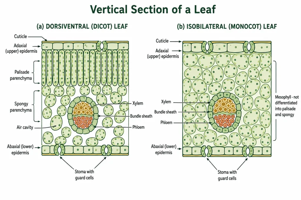
[NEET Important] Two things are asked from this block more than anything else. The palisade is adaxial, elongated and vertical; the spongy is below it, loosely arranged, with large air spaces - and the reason for the split is worth knowing, since the palisade sits where the light is strongest and the spongy layer keeps the air channels open for gas exchange. The other is the stomatal distribution: more on the abaxial surface, sometimes none on the adaxial.
The Isobilateral (Monocotyledonous) Leaf
The anatomy of the isobilateral leaf is similar to that of the dorsiventral leaf in many ways. It shows the following characteristic differences.
- The stomata are present on both the surfaces of the epidermis.
- The mesophyll is not differentiated into palisade and spongy parenchyma.
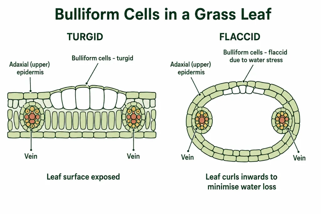
Bulliform cells. In grasses, certain adaxial epidermal cells along the veins modify themselves into large, empty, colourless cells. These are the bulliform cells.
- When the bulliform cells in the leaves have absorbed water and are turgid, the leaf surface is exposed.
- When they are flaccid due to water stress, they make the leaves curl inwards to minimise water loss.
Vascular bundles. The parallel venation in monocot leaves is reflected in the near similar sizes of the vascular bundles, except in the main veins.
| Feature | Dorsiventral (dicot) leaf | Isobilateral (monocot) leaf |
|---|---|---|
| Stomata | More on the abaxial surface; adaxial may lack them | On both surfaces |
| Mesophyll | Differentiated into palisade and spongy parenchyma | Not differentiated |
| Bulliform cells | Absent | Present in grasses, on the adaxial epidermis along the veins |
| Vascular bundle size | Varies with vein thickness, following reticulate venation | Near similar, following parallel venation, except in main veins |
[NEET Important] The bulliform-cell mechanism is a standard three-mark answer and the logic has to be stated both ways: turgid means the leaf surface is exposed, flaccid under water stress means the leaves curl inwards to minimise water loss. Curling is the plant reducing the surface it presents to dry air.
Quick Recap
Dorsiventral (dicot) leaf - three parts: epidermis, mesophyll, vascular system.
- Epidermis on both surfaces - adaxial above, abaxial below - with a conspicuous cuticle; more stomata on the abaxial, and the adaxial may lack them.
- Mesophyll - between the two epidermal layers, parenchyma with chloroplasts, carries out photosynthesis; palisade (adaxial, elongated, vertical, parallel) and spongy (oval or round, loosely arranged, with large air cavities, reaching the lower epidermis).
- Vascular system - bundles in the veins and midrib, size depending on vein size, each surrounded by a layer of thick-walled bundle sheath cells.
Isobilateral (monocot) leaf
- Stomata on both surfaces.
- Mesophyll not differentiated into palisade and spongy.
- Bulliform cells - in grasses, large, empty, colourless adaxial epidermal cells along the veins; turgid, the leaf surface is exposed; flaccid under water stress, the leaves curl inwards to minimise water loss.
- Vascular bundles of near similar size, matching the parallel venation, except in the main veins.
Solved Examples
Question 1
Q. Name the three main parts seen in a vertical section of a dorsiventral leaf.
Answer. The epidermis, the mesophyll and the vascular system.
Question 2
Q. What are the adaxial and abaxial epidermis?
Answer. The adaxial epidermis covers the upper surface of the leaf and the abaxial covers the lower surface.
Question 3
Q. How are the stomata distributed on a dorsiventral leaf?
Answer. The abaxial epidermis generally bears more stomata than the adaxial, and the adaxial may even lack stomata.
Question 4
Q. What is the mesophyll, and what is it for?
Answer. The tissue between the upper and lower epidermis. It is made of parenchyma, it possesses chloroplasts, and it carries out photosynthesis.
Question 5
Q. Describe palisade parenchyma.
Answer. It is adaxially placed and made of elongated cells arranged vertically and parallel to each other.
Question 6
Q. Describe spongy parenchyma.
Answer. Its cells are oval or round and loosely arranged. It sits below the palisade cells and extends to the lower epidermis, and there are numerous large spaces and air cavities between the cells.
Question 7
Q. Describe the internal structure of a dorsiventral leaf.
Answer. A vertical section through the lamina shows three parts.
- Epidermis - covering both the adaxial (upper) and abaxial (lower) surfaces, with a conspicuous cuticle. The abaxial bears more stomata, and the adaxial may lack them.
- Mesophyll - the tissue between the two epidermal layers, made of parenchyma with chloroplasts, carrying out photosynthesis. It has palisade parenchyma adaxially, made of elongated cells arranged vertically and parallel to each other, and spongy parenchyma below it, oval or round, loosely arranged, with numerous large spaces and air cavities, extending to the lower epidermis.
- Vascular system - vascular bundles in the veins and the midrib, their size depending on the size of the veins, each surrounded by a layer of thick-walled bundle sheath cells.
This is one of the chapter-end exercises, and it also asks for a labelled diagram.
Question 8
Q. Give the two characteristic differences of an isobilateral leaf.
Answer. The stomata are present on both surfaces of the epidermis, and the mesophyll is not differentiated into palisade and spongy parenchyma.
Question 9
Q. What are bulliform cells and where are they found?
Answer. In grasses, certain adaxial epidermal cells along the veins modify into large, empty, colourless cells. Those are the bulliform cells.
Question 10
Q. Explain how bulliform cells work.
Answer. They act on their water content. When they have absorbed water and are turgid, the leaf surface is exposed and the leaf lies open. When they are flaccid because of water stress, they make the leaves curl inwards, and the curling minimises water loss by reducing the surface the leaf presents to dry air.
Question 11
Q. Why are the vascular bundles of a monocot leaf all about the same size?
Answer. Because the leaf has parallel venation, so the veins themselves are of near similar thickness, and the size of a vascular bundle depends on the size of its vein. The exception is the main veins, which are larger. In a dicot leaf the venation is reticulate, the veins vary in thickness, and so do the bundles.
Question 12
Q. A leaf section shows stomata on both surfaces and a mesophyll that is uniform throughout. Name the leaf type and the class of plant.
Answer. An isobilateral leaf, so the plant is a monocotyledon. Both features are exactly the two differences that define it.