
Same Syllabus. A Different Exam.
The Important Questions and Answers section worked this chapter the way a textbook does - name the layer, describe the section, compare the two. NEET does not ask you to describe anything. It hands you two or three features, four options and about half a minute, and it wants the right one.
So this section covers the same syllabus with a different question in mind: not "can you describe this section?" but "can you place it from two features and kill the other three options?"
The marking is what forces the change. A correct answer is +4 and a wrong one is -1, so an option you are only half sure of costs you a mark and, worse, costs you the time you spent hesitating over it.
This is the most predictable chapter in the unit. The whole of it is six transverse sections, three tissue systems and about a dozen named structures. Nothing has to be worked out, nothing is derived, and the same handful of facts is recycled year after year. Short, dense and closed - which means it can be finished, and a finished chapter should never cost you more than 25 seconds a question.
Key Point: Every question from this chapter is really one question - where does this structure sit, and what does that make the section? Learn each section as an ordered list of rings from the outside inward and you are answering from a list rather than from a picture you are trying to remember.
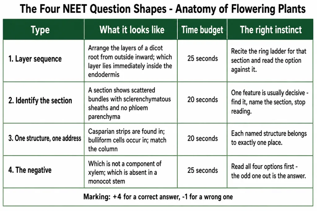
The four shapes, and what each should cost you
| Type | What it looks like | Budget | The right instinct |
|---|---|---|---|
| 1. Layer sequence | "Arrange the layers of a dicot root from outside inward." "Which layer lies immediately inside the endodermis?" | 25 seconds | Recite the ring ladder for that section and read the option against it. Do not picture the diagram. |
| 2. Identify the section | "A section shows scattered bundles with sclerenchymatous sheaths and no phloem parenchyma." | 20 seconds | One feature is usually decisive. Find it, name the section, and do not read the rest. |
| 3. One structure, one address | "Casparian strips are found in…" "Bulliform cells occur in…" Match-the-column items. | 20 seconds | Each named structure belongs to exactly one place. If you have the address, the option is read, not reasoned. |
| 4. The negative | "Which is not a component of xylem?" "Which is absent in a monocot stem?" | 25 seconds | Read the whole option list first. The odd one out is what is being asked for, and rushing here is what loses the mark. |
What this section repeats, and what it does not
It does not re-describe the six sections. It gives you the ring orders as lists, the one address per structure, the component lists the negatives are built from, and the distractor patterns the paper reuses - and then drills them at speed.
Secondary growth is not used here. It sits outside the current syllabus and is kept in this chapter only as clearly flagged bonus material, so nothing in this section depends on it.
The Six Sections, as Ordered Rings
Most questions in this chapter are really testing one thing: can you say what comes next as you move inward? So learn each section as a list, not as a drawing. Recite each one until it runs without a pause.
1. Dicot root
- Epiblema - the outermost layer; many cells protrude as unicellular root hairs.
- Cortex - several layers of thin-walled parenchyma with intercellular spaces.
- Endodermis - the innermost layer of the cortex; a single layer of barrel-shaped cells with no intercellular spaces, their tangential and radial walls carrying casparian strips of suberin.
- Pericycle - a few layers of thick-walled parenchymatous cells; lateral roots and the vascular cambium are initiated here.
- Vascular bundles - two to four xylem and phloem patches, radial, with conjunctive tissue between them.
- Pith - small or inconspicuous.
Stele = everything inside the endodermis - pericycle, vascular bundles and pith.
2. Monocot root
The same six rings, in the same order, with the outermost called the epidermis. Three differences, and they are the whole of what is asked:
- More than six xylem bundles - polyarch.
- Pith large and well developed.
- No secondary growth at all.
3. Dicot stem
- Epidermis - outermost protective layer, with a thin cuticle, trichomes and a few stomata.
- Hypodermis - collenchymatous, giving mechanical strength to the young stem.
- Cortical parenchyma - rounded, thin-walled cells with conspicuous intercellular spaces.
- Endodermis - the innermost cortical layer, rich in starch grains, so called the starch sheath.
- Pericycle - inside the endodermis and above the phloem, as semi-lunar patches of sclerenchyma.
- Medullary rays - radially placed parenchymatous cells between the bundles.
- Vascular bundles - a large number arranged in a ring, each conjoint, open and endarch.
- Pith - rounded parenchymatous cells with large intercellular spaces in the centre.
Rings 2 to 4 together are the cortex, in three sub-zones.
4. Monocot stem
- Epidermis.
- Hypodermis - sclerenchymatous.
- Ground tissue - one large conspicuous parenchymatous mass, not divided into cortex and pith.
- Vascular bundles - scattered through that ground tissue, each conjoint and closed, each surrounded by a sclerenchymatous bundle sheath, with water-containing cavities inside the bundle and no phloem parenchyma. Peripheral bundles are generally smaller than the central ones.
Four rings against the dicot stem's eight. That shortness is itself the identification.
5. Dorsiventral leaf, read from the top down
- Cuticle, conspicuous, over the adaxial (upper) epidermis, which may even lack stomata.
- Palisade parenchyma - adaxially placed, elongated cells arranged vertically and parallel to each other.
- Spongy parenchyma - oval or round, loosely arranged, with numerous large spaces and air cavities, extending down to the lower epidermis.
- Vascular bundles - in the veins and the midrib, their size depending on the size of the vein, each surrounded by a layer of thick-walled bundle sheath cells.
- Abaxial (lower) epidermis, which generally bears more stomata.
Palisade plus spongy = the mesophyll, the ground tissue of the leaf.
6. Isobilateral leaf
The same list with two characteristic differences and one extra structure:
- Stomata are present on both surfaces of the epidermis.
- The mesophyll is not differentiated into palisade and spongy parenchyma.
- In grasses, bulliform cells - large, empty, colourless adaxial epidermal cells along the veins.
- Vascular bundles of near similar size, which follows from parallel venation.
[NEET Important] The commonest sequence distractor swaps endodermis and pericycle. Going inward the endodermis always comes first, because it is the innermost layer of the cortex, and the pericycle is the first ring of the stele.
One Grid, Six Sections
Read it down a column to learn one section and across a row to answer a comparison. A dash means the structure is not described for that section.
| Feature | Dicot root | Monocot root | Dicot stem | Monocot stem | Dorsiventral leaf | Isobilateral leaf |
|---|---|---|---|---|---|---|
| Outermost layer | Epiblema, with unicellular root hairs | Epidermis | Epidermis, thin cuticle, trichomes, a few stomata | Epidermis | Adaxial and abaxial epidermis, conspicuous cuticle | Same |
| Cuticle | Absent | Absent | Thin | - | Conspicuous | Conspicuous |
| Hypodermis | - | - | Collenchymatous | Sclerenchymatous | - | - |
| Endodermis | Casparian strips of suberin | Present | Starch sheath | - | - | - |
| Pericycle | Thick-walled parenchyma; lateral roots and vascular cambium | Present | Semi-lunar sclerenchyma above the phloem | - | - | - |
| Bundle type | Radial | Radial | Conjoint, open, endarch | Conjoint, closed | Conjoint | Conjoint |
| Bundle arrangement | Alternating xylem and phloem patches | Alternating patches | In a ring | Scattered, peripheral ones smaller | In the veins and midrib | In veins, near similar sizes |
| Xylem number | Two to four | More than six - polyarch | - | - | - | - |
| Bundle sheath | - | - | Absent | Sclerenchymatous | Thick-walled | Thick-walled |
| Ground tissue | Cortex + small pith | Cortex + large pith | Cortex, endodermis, pericycle, medullary rays, pith | One large parenchymatous mass | Mesophyll - palisade + spongy | Mesophyll, undifferentiated |
| Stomata | - | - | A few | - | More abaxial; adaxial may lack them | Both surfaces |
| Signature structure | Conjunctive tissue | No secondary growth | Starch sheath, medullary rays | Water-containing cavities, no phloem parenchyma | Palisade layer | Bulliform cells |
How this grid is examined. One cell at a time, and almost always from the rows hypodermis, bundle arrangement, ground tissue and signature structure. If you can fill those four rows from memory you can answer most of what this chapter sets.
[NEET Important] A section is placed by two features, not one. Radial or conjoint tells you root against stem or leaf. Then the number of xylem patches settles a root and ring against scattered settles a stem. Two reads, section named.
One Structure, One Address
Every named structure in this chapter lives in exactly one place. Match-the-column items are built entirely from this table, and so is half of everything else.
| Structure | Its one address | The fact that goes with it |
|---|---|---|
| Casparian strips | Endodermis of the root | Deposits of the water-impermeable waxy material suberin on the tangential and radial walls |
| Starch sheath | Endodermis of the dicot stem | Its cells are rich in starch grains |
| Semi-lunar patches of sclerenchyma | Pericycle of the dicot stem, above the phloem | The pericycle here is for support, not for growth |
| Sclerenchymatous bundle sheath | Round each scattered bundle of the monocot stem | Absent in the dicot stem |
| Thick-walled bundle sheath | Round the vascular bundles of a leaf | In the veins and midrib |
| Bulliform cells | Adaxial epidermis of a grass leaf, along the veins | Large, empty and colourless; turgid exposes the surface, flaccid curls the leaf inwards to minimise water loss |
| Conjunctive tissue | Between the xylem and phloem of a root | Parenchymatous |
| Medullary rays | Between the vascular bundles of a dicot stem | Radially placed parenchymatous cells |
| Epiblema | Outermost layer of the dicot root | Cells protrude as unicellular root hairs |
| Water-containing cavities | Within the vascular bundles of a monocot stem | Go with absent phloem parenchyma |
| Subsidiary cells | Epidermis, next to the guard cells | Epidermal cells specialised in shape and size |
| Palisade parenchyma | Adaxial side of a dorsiventral leaf | Elongated, vertical, parallel |
The three pairs that get swapped
| The pair | The separator |
|---|---|
| Endodermis against pericycle | Endodermis is the innermost layer of the cortex; pericycle is the first layer of the stele, just inside it |
| Root pericycle against stem pericycle | In the root, thick-walled parenchyma where lateral roots start; in the dicot stem, semi-lunar sclerenchyma for support |
| Root hair against trichome | Unicellular, on the root, absorbs; against usually multicellular, on the shoot, prevents water loss |
The Two Axes That Must Not Be Collapsed
Students merge these two and the paper is built to catch it. They are independent, and a bundle carries one label from each.
| Axis | The question it answers | The two values |
|---|---|---|
| Radial or conjoint | Where do the xylem and phloem sit relative to each other? | Radial - on alternate radii, as in roots. Conjoint - along the same radius, phloem usually only on the outer side of the xylem, as in stems and leaves |
| Open or closed | Is a cambium present between the phloem and the xylem? | Open - cambium present, so it can form secondary xylem and phloem, as in dicot stems. Closed - no cambium, so no secondary tissue, as in monocots |
The three labelled types the chapter itself gives are radial, conjoint closed and conjoint open. So the wording to have ready is:
- A dicot stem bundle is conjoint, open and endarch.
- A monocot stem bundle is conjoint and closed.
- A root bundle is radial, and its protoxylem is exarch.
Endarch against exarch is a third, separate axis - protoxylem towards the centre in stems, protoxylem towards the periphery in roots.
[NEET Important] "Radial and open" and "conjoint and radial" are both nonsense, and both appear as options. Check that the option carries one word from each axis before you check anything else.
The Component Lists, and the Negatives
The lists the "which is not a component" question is built from
Learn these as closed sets. The question is always "which one does not belong", so a set with a hole in it is worth nothing.
| Set | Its members |
|---|---|
| Xylem | Tracheids, vessels, xylem fibres, xylem parenchyma |
| Phloem | Sieve tube elements, companion cells, phloem parenchyma, phloem fibres |
| Three tissue systems | Epidermal, ground or fundamental, vascular or conducting - classified on the basis of structure and location |
| Epidermal tissue system | Epidermal cells, stomata (guard cells and subsidiary cells), epidermal appendages - root hairs and trichomes |
| Ground tissue system | All tissues except epidermis and vascular bundles - parenchyma, collenchyma and sclerenchyma in the cortex, pericycle, pith and medullary rays, and the mesophyll of a leaf |
| Stomatal apparatus | Stomatal aperture + guard cells + subsidiary cells |
| Stele | Pericycle + vascular bundles + pith - everything inside the endodermis |
| Cortex of the dicot stem | Hypodermis + cortical parenchyma + endodermis |
| Simple tissues | Parenchyma, collenchyma, sclerenchyma |
| Complex tissues | Xylem, phloem |
| Meristems | Apical, intercalary, lateral |
The three traps inside these lists.
- Companion cells belong to phloem, never to xylem. This is the single most-set "not a component" item in the chapter.
- The endodermis is not part of the stele. The stele begins inside it, at the pericycle.
- Sclerenchyma is a simple tissue, even though it looks specialised. Simple means one cell type, not "simple job".
The negatives - the cheapest questions an examiner can set
An absence is easier to test than a presence, so learn these as facts in their own right.
| The negative | Where it is true | The mistake it catches |
|---|---|---|
| No cuticle | In roots | The cuticle prevents water loss, and a root has to absorb water |
| No phloem parenchyma | Monocot stem | Present in the dicot stem |
| No secondary growth | Monocot roots, and monocot stems | Their bundles are closed, so there is no cambium to form secondary tissue |
| No differentiation of the mesophyll | Isobilateral leaf | No palisade against spongy split at all - the whole mesophyll is uniform |
| No bundle sheath | Dicot stem | The sclerenchymatous sheath is the monocot stem's feature |
| No medullary rays, no separate cortex and pith | Monocot stem | It has one undivided parenchymatous ground tissue |
| No intercellular spaces | Endodermis, and collenchyma | Barrel-shaped endodermal cells sit flush; the cortex around them does have spaces |
| Stomata may be absent | Adaxial epidermis of a dorsiventral leaf | The abaxial surface generally bears more |
| No vessels | Gymnosperm xylem | And gymnosperm phloem has albuminous cells and sieve cells instead of sieve tubes and companion cells |
| Not living | Sclerenchyma, and all of the xylem except the xylem parenchyma | Only xylem parenchyma is living |
[NEET Important] When an option list is four true-looking statements, the answer is decided by one word of place - root against stem, dicot against monocot, adaxial against abaxial. Go to that word first, and ignore the rest of the sentence.
Solved Examples at NEET Pace
Each item names the shortcut it uses, because the shortcut is the point.
Question 1
Q. Arrange the following layers of a dicot root in order from the outside inward: pericycle, epiblema, endodermis, cortex.
Answer. Epiblema, cortex, endodermis, pericycle. Use the ring ladder. The epiblema is the outermost layer, the endodermis is the innermost layer of the cortex so it must follow the cortex, and the pericycle is the first ring of the stele, just inside the endodermis. The one swap to watch is endodermis against pericycle, and the ladder settles it every time.
Question 2
Q. A transverse section shows vascular bundles that are conjoint, scattered and surrounded by sclerenchymatous bundle sheaths, and the phloem parenchyma is absent. What is it?
Answer. A monocot stem. Take the decisive feature first - scattered bundles already rules out the dicot stem, where the ring arrangement is characteristic. The sclerenchymatous bundle sheath and the absent phloem parenchyma are both monocot stem features and only confirm it. Twenty seconds, and you should stop reading after the word "scattered".
Question 3
Q. Which of these is not a component of xylem: tracheids, vessels, companion cells, xylem fibres?
Answer. Companion cells. They belong to phloem, together with sieve tube elements, phloem parenchyma and phloem fibres. Xylem is tracheids, vessels, xylem fibres and xylem parenchyma. The shortcut is to hold the two sets as closed lists of four and check membership, rather than to reason about function.
Question 4
Q. Casparian strips are found in which layer, and what are they made of?
Answer. In the endodermis of the root, and they are made of suberin, a water-impermeable waxy material, laid down on the tangential and radial walls of the barrel-shaped endodermal cells. One structure, one address - do not let the option that puts them in the pericycle survive.
Question 5
Q. An option reads: "The vascular bundle of a dicot stem is radial and open." Accept or reject.
Answer. Reject. The two axes have been collapsed. Radial means the xylem and phloem lie on alternate radii, which happens in roots. A dicot stem bundle is conjoint, open and endarch. Check that an option takes one word from each axis - position first, cambium second - and a wrong pairing shows up before you have read the rest of the sentence.
Question 6
Q. A root section shows eight xylem patches and a large, well developed pith. Identify it, and state one thing it cannot do.
Answer. A monocot root. More than six xylem bundles is polyarch, and the large pith confirms it - a dicot root has two to four patches and a small or inconspicuous pith. It cannot undergo secondary growth; monocot roots have none.
Question 7
Q. Which of the following is absent in a monocot stem: sclerenchymatous hypodermis, scattered bundles, phloem parenchyma, water-containing cavities?
Answer. Phloem parenchyma. The other three are all present. This is the chapter's favourite negative because three of the four options are lifted straight out of the monocot stem description and only one is a stated absence. Read all four options before choosing - the negative question punishes a fast first pick.
A Drill Before You Move On
Decide each one before you read the verdict. Aim for 20 to 25 seconds each.
Question 8
Q. Which structure is not part of the stele: pericycle, endodermis, vascular bundles, pith?
Answer. The endodermis. The stele is everything on the inner side of the endodermis - the pericycle, the vascular bundles and the pith. The endodermis is the boundary, and it belongs to the cortex, being its innermost layer.
Question 9
Q. Bulliform cells - where exactly are they, and what happens when they lose water?
Answer. They are adaxial epidermal cells along the veins of a grass leaf, and they are large, empty and colourless. When they are flaccid due to water stress they make the leaves curl inwards, which minimises water loss. When they are turgid, the leaf surface is exposed. The word to hold on to is adaxial - an option that puts them on the lower surface is wrong on the address alone.
Question 10
Q. Name the layer, in a dicot stem, that lies immediately above the phloem, and say what it is made of.
Answer. The pericycle, present as semi-lunar patches of sclerenchyma. Note what changes between organs: in the root the pericycle is thick-walled parenchyma and is where lateral roots and the vascular cambium start. Same name, different tissue, different job - and the option list usually contains both descriptions.
Question 11
Q. In a stoma, which wall of the guard cell is thickened, and what shape are the guard cells in grasses?
Answer. The inner wall, towards the stomatal pore, is highly thickened; the outer wall, away from the pore, is thin. In grasses the guard cells are dumb-bell shaped, against bean-shaped everywhere else. The direction word - inner against outer - is the whole question.
Question 12
Q. A leaf section shows stomata on both surfaces and a mesophyll that is uniform throughout. Identify the leaf and the class of plant, and name one more structure you would expect nearby.
Answer. An isobilateral leaf, so a monocotyledon. Those are exactly the two characteristic differences from a dorsiventral leaf - stomata on both surfaces and a mesophyll not differentiated into palisade and spongy parenchyma. If it is a grass you would also expect bulliform cells in the adaxial epidermis along the veins.
Question 13
Q. Answer each in under 15 seconds. (i) Where is the cuticle absent? (ii) What lies between the xylem and phloem of a root? (iii) Which layer of a dicot stem is the starch sheath? (iv) In which layer are lateral roots initiated? (v) What are the three parts of the stomatal apparatus?
Answer.
- (i) In roots.
- (ii) Conjunctive tissue, which is parenchymatous.
- (iii) The endodermis, because its cells are rich in starch grains.
- (iv) The pericycle.
- (v) The stomatal aperture, the guard cells and the subsidiary cells.