
Anaphase
Anaphase is the stage at which the two copies of the genome finally part company. Everything before it was preparation; from here the cell is committed to producing two nuclei.
At the onset of anaphase, each chromosome arranged at the metaphase plate is split simultaneously, and the two daughter chromatids, now referred to as daughter chromosomes of the future daughter nuclei, begin their migration towards the two opposite poles.
Notice that the name changes at this exact moment, and the change is examinable. While the two threads were held together at the centromere they were sister chromatids; the instant the centromere splits, each thread becomes a daughter chromosome in its own right, headed for one of the two future daughter nuclei.
As each chromosome moves away from the equatorial plate, the centromere of each chromosome remains directed towards the pole and hence at the leading edge, with the arms of the chromosome trailing behind. In other words the chromosome is drawn centromere first, and the arms simply follow behind it.
Thus the anaphase stage is characterised by the following key events:
- Centromeres split and chromatids separate.
- Chromatids move to opposite poles.
[NEET Important] Anaphase is the answer to two of the most frequently set one-line questions in this chapter: the stage at which the centromere splits and chromatids separate, and the stage at which chromatids move to opposite poles. The favourite distractor is metaphase, because metaphase is when the chromosomes are only lying at the plate - the splitting itself is anaphase. Remember also the direction: centromere at the leading edge, arms trailing, never the other way round.
Telophase
At the beginning of the final stage of karyokinesis, that is telophase, the chromosomes that have reached their respective poles decondense and lose their individuality. The individual chromosomes can no longer be seen, and each set of chromatin material tends to collect at each of the two poles.
The simplest way to hold telophase in your head is to see it as prophase running backwards. In prophase the chromatin condensed and the nuclear envelope, nucleolus, golgi complex and endoplasmic reticulum vanished; in telophase the chromatin decondenses and every one of those structures comes back.
This is the stage which shows the following key events:
- Chromosomes cluster at opposite spindle poles and their identity is lost as discrete elements.
- Nuclear envelope develops around the chromosome clusters at each pole, forming two daughter nuclei.
- Nucleolus, golgi complex and ER reform.
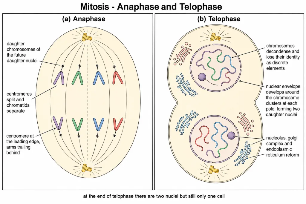
[NEET Important] Telophase is the end of karyokinesis, not the end of cell division. At the close of telophase there are two nuclei but still only one cell - the cell is split only by cytokinesis, which comes next. The three-part key-event list is asked as a set, so learn decondensation, nuclear envelope reformation, and the return of nucleolus, golgi complex and ER together.
Cytokinesis
Mitosis accomplishes not only the segregation of duplicated chromosomes into daughter nuclei, which is karyokinesis, but the cell itself is divided into two daughter cells by the separation of cytoplasm, called cytokinesis, at the end of which cell division gets completed.
The mechanism is not the same in the two kinds of cell, and the reason is structural.
In an animal cell, cytokinesis is achieved by the appearance of a furrow in the plasma membrane. The furrow gradually deepens and ultimately joins in the centre, dividing the cell cytoplasm into two. The cut therefore runs from the outside inward.
Plant cells, however, are enclosed by a relatively inextensible cell wall, and therefore they undergo cytokinesis by a different mechanism. In plant cells, wall formation starts in the centre of the cell and grows outward to meet the existing lateral walls. The formation of the new cell wall begins with the formation of a simple precursor, called the cell-plate, that represents the middle lamella between the walls of two adjacent cells. Here the partition is built from the inside outward.
| Feature | Animal cell | Plant cell |
|---|---|---|
| Reason for the difference | Flexible plasma membrane, no cell wall | Enclosed by a relatively inextensible cell wall |
| Structure formed | A furrow in the plasma membrane | A cell-plate, the precursor of the new cell wall |
| Direction of division | From the periphery inward - the furrow deepens and joins in the centre | From the centre outward - wall formation grows out to meet the existing lateral walls |
| What it becomes | The two separated cytoplasms | The middle lamella between the walls of two adjacent cells |
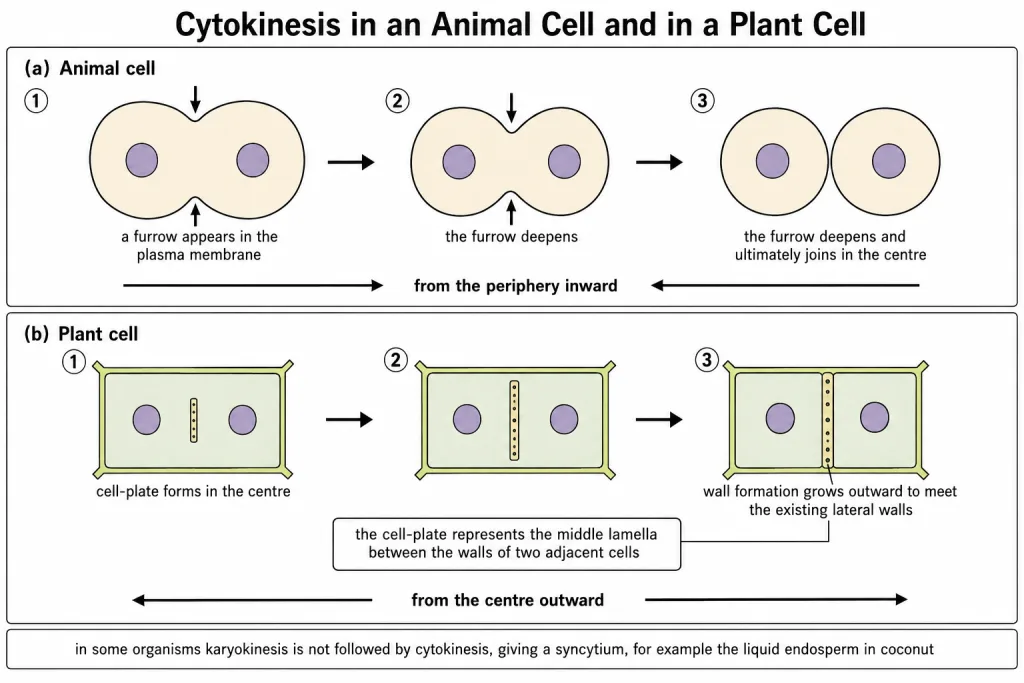
What happens to the organelles. At the time of cytoplasmic division, organelles like mitochondria and plastids get distributed between the two daughter cells.
When cytokinesis does not follow. In some organisms karyokinesis is not followed by cytokinesis, as a result of which a multinucleate condition arises, leading to the formation of a syncytium - for example, the liquid endosperm in coconut.
[NEET Important] The plant-versus-animal contrast is the single most reliable question here, and the way to answer it in one line is by direction: animal cell, furrow, outside inward; plant cell, cell-plate, centre outward. Also fix the identity of the cell-plate - it represents the middle lamella, not the whole wall and not the metaphase plate. And keep the coconut liquid endosperm ready as the example of a syncytium.
Quick Recap
- At the onset of anaphase, each chromosome arranged at the metaphase plate is split simultaneously.
- The two daughter chromatids are now referred to as daughter chromosomes of the future daughter nuclei, and they begin their migration towards the two opposite poles.
- As each chromosome moves away from the equatorial plate, the centromere remains directed towards the pole and hence at the leading edge, with the arms of the chromosome trailing behind.
- Key events of anaphase: centromeres split and chromatids separate; chromatids move to opposite poles.
- Telophase is the final stage of karyokinesis. The chromosomes that have reached their respective poles decondense and lose their individuality.
- The individual chromosomes can no longer be seen, and each set of chromatin material tends to collect at each of the two poles.
- Key events of telophase: chromosomes cluster at opposite spindle poles and their identity is lost as discrete elements; a nuclear envelope develops around the chromosome clusters at each pole forming two daughter nuclei; nucleolus, golgi complex and ER reform.
- Cytokinesis is the division of the cell into two daughter cells by the separation of cytoplasm, and cell division is completed at the end of it.
- In an animal cell, cytokinesis is achieved by the appearance of a furrow in the plasma membrane, which gradually deepens and ultimately joins in the centre, dividing the cell cytoplasm into two.
- Plant cells are enclosed by a relatively inextensible cell wall and therefore undergo cytokinesis by a different mechanism.
- In plant cells, wall formation starts in the centre of the cell and grows outward to meet the existing lateral walls.
- The new cell wall begins with a simple precursor called the cell-plate, which represents the middle lamella between the walls of two adjacent cells.
- At the time of cytoplasmic division, organelles like mitochondria and plastids get distributed between the two daughter cells.
- In some organisms karyokinesis is not followed by cytokinesis, giving a multinucleate condition called a syncytium, for example the liquid endosperm in coconut.
Solved Examples
Question 1
Q. What happens at the onset of anaphase?
Answer. Each chromosome arranged at the metaphase plate is split simultaneously, and the two daughter chromatids begin their migration towards the two opposite poles.
Question 2
Q. Why are the separated chromatids given a new name during anaphase, and what is that name?
Answer. Once the centromere splits, each thread is a complete chromosome on its own, so it is no longer called a chromatid. The two daughter chromatids are now referred to as daughter chromosomes of the future daughter nuclei.
Question 3
Q. During anaphase, which part of the chromosome leads the way towards the pole?
Answer. The centromere. As each chromosome moves away from the equatorial plate, the centromere of each chromosome remains directed towards the pole and hence at the leading edge, with the arms of the chromosome trailing behind.
Question 4
Q. State the two key events of anaphase.
Answer. Centromeres split and chromatids separate, and chromatids move to opposite poles.
Question 5
Q. Name the stage of cell cycle at which the following event occurs: Centromere splits and chromatids separate. This is one of the chapter-end exercises.
Answer. Anaphase, the third stage of karyokinesis in mitosis.
At the onset of anaphase, each chromosome arranged at the metaphase plate is split simultaneously, and the two daughter chromatids, now referred to as daughter chromosomes of the future daughter nuclei, begin their migration towards the two opposite poles.
Be careful not to answer metaphase. Metaphase is only when the chromosomes come to lie at the equator on the metaphase plate, still held together at the centromere. The splitting of the centromere itself is the defining event of anaphase.
Question 6
Q. What happens to the chromosomes at the beginning of telophase?
Answer. The chromosomes that have reached their respective poles decondense and lose their individuality. The individual chromosomes can no longer be seen, and each set of chromatin material tends to collect at each of the two poles.
Question 7
Q. List the key events of telophase.
Answer. There are three.
- Chromosomes cluster at opposite spindle poles and their identity is lost as discrete elements.
- Nuclear envelope develops around the chromosome clusters at each pole, forming two daughter nuclei.
- Nucleolus, golgi complex and ER reform.
Question 8
Q. Which cell structures reappear during telophase?
Answer. The nuclear envelope, which develops around the chromosome clusters at each pole to form two daughter nuclei, and then the nucleolus, the golgi complex and the endoplasmic reticulum, all of which reform. These are exactly the structures that had disappeared by the end of prophase.
Question 9
Q. At the end of telophase, how many nuclei and how many cells are present?
Answer. Two nuclei but still only one cell. Telophase is the end of karyokinesis, the nuclear division. The cell itself is separated into two only during cytokinesis, which follows.
Question 10
Q. What is cytokinesis?
Answer. The division of the cell itself into two daughter cells by the separation of cytoplasm, at the end of which cell division gets completed. Mitosis accomplishes not only the segregation of duplicated chromosomes into daughter nuclei, which is karyokinesis, but this cytoplasmic division as well.
Question 11
Q. How is cytokinesis achieved in an animal cell?
Answer. By the appearance of a furrow in the plasma membrane. The furrow gradually deepens and ultimately joins in the centre, dividing the cell cytoplasm into two.
Question 12
Q. How does cytokinesis in plant cells differ from that in animal cells? This is one of the chapter-end exercises.
Answer. The two differ in why, in what is built, and in which direction the division runs.
The reason for the difference. Plant cells are enclosed by a relatively inextensible cell wall, and therefore they undergo cytokinesis by a different mechanism. An animal cell has only a flexible plasma membrane, so it can simply be pinched in two.
In an animal cell. Cytokinesis is achieved by the appearance of a furrow in the plasma membrane. The furrow gradually deepens and ultimately joins in the centre, dividing the cell cytoplasm into two. The division therefore proceeds from the periphery inward.
In a plant cell. Wall formation starts in the centre of the cell and grows outward to meet the existing lateral walls. The formation of the new cell wall begins with the formation of a simple precursor, called the cell-plate, that represents the middle lamella between the walls of two adjacent cells. The division therefore proceeds from the centre outward.
One-line answer for a short question. An animal cell divides by a cleavage furrow that deepens from outside inward; a plant cell divides by a cell-plate that grows from the centre outward into the new cell wall.
Question 13
Q. What is the cell-plate, and what does it represent?
Answer. It is a simple precursor with which the formation of the new cell wall begins in a dividing plant cell, and it represents the middle lamella between the walls of two adjacent cells.
Question 14
Q. What happens to organelles such as mitochondria and plastids during cytokinesis?
Answer. At the time of cytoplasmic division, organelles like mitochondria and plastids get distributed between the two daughter cells.
Question 15
Q. What is a syncytium, and how does it arise? Give one example.
Answer. In some organisms karyokinesis is not followed by cytokinesis. The nucleus divides again and again while the cytoplasm is never separated, so a multinucleate condition arises, and this is called a syncytium. The standard example is the liquid endosperm in coconut.