
M Phase - the Dramatic Part
This is the most dramatic period of the cell cycle, involving a major reorganisation of virtually all components of the cell.
Since the number of chromosomes in the parent and progeny cells is the same, mitosis is also called equational division.
Though for convenience mitosis has been divided into four stages of nuclear division (karyokinesis), it is very essential to understand that cell division is a progressive process and very clear-cut lines cannot be drawn between the various stages.
Karyokinesis involves the following four stages:
- Prophase
- Metaphase
- Anaphase
- Telophase
[NEET Important] The word equational is a definition question in its own right: mitosis is equational because the parent and progeny cells have the same chromosome number. Contrast it with meiosis, which is reductional. And note the caution the chapter gives - the four stages are a convenience, not four sealed boxes, because division is a progressive process.
Prophase
Prophase, the first stage of karyokinesis of mitosis, follows the S and phases of interphase. In the S and phases, the new DNA molecules formed are not distinct but intertwined.
Prophase is marked by the initiation of condensation of chromosomal material, and the chromosomal material becomes untangled during the process of chromatin condensation.
The completion of prophase is marked by these characteristic events:
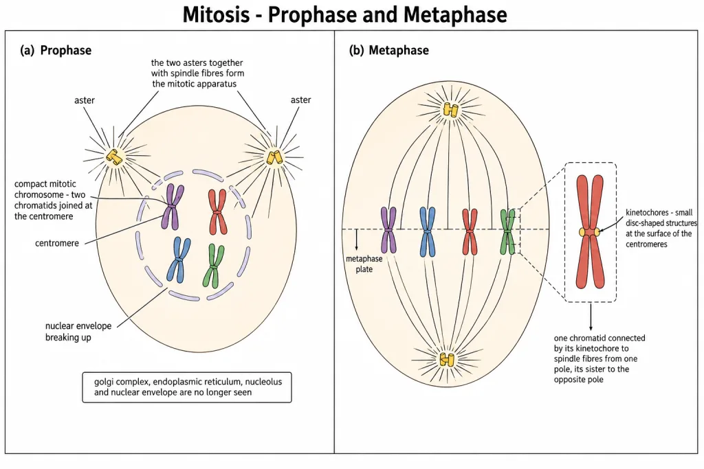
- Chromosomal material condenses to form compact mitotic chromosomes. Chromosomes are seen to be composed of two chromatids attached together at the centromere.
- The centrosome, which had undergone duplication during interphase, begins to move towards opposite poles of the cell. Each centrosome radiates out microtubules called asters. The two asters together with spindle fibres form the mitotic apparatus.
What is missing by the end of prophase. Cells at the end of prophase, when viewed under the microscope, do not show golgi complexes, endoplasmic reticulum, nucleolus and the nuclear envelope.
[NEET Important] That last list is a favourite "which of these is NOT seen at the end of prophase" item, so learn all four together: golgi complex, endoplasmic reticulum, nucleolus, nuclear envelope. Note also that the centrosome duplicated back in S phase, not in prophase - prophase is only when it starts moving to the poles.
Metaphase
The complete disintegration of the nuclear envelope marks the start of the second phase of mitosis, and hence the chromosomes are spread through the cytoplasm of the cell.
By this stage, condensation of chromosomes is completed and they can be observed clearly under the microscope. This is the stage at which the morphology of chromosomes is most easily studied.
At this stage, the metaphase chromosome is made up of two sister chromatids, which are held together by the centromere. Small disc-shaped structures at the surface of the centromeres are called kinetochores, and these structures serve as the sites of attachment of spindle fibres to the chromosomes that are moved into position at the centre of the cell.
Hence metaphase is characterised by all the chromosomes coming to lie at the equator, with one chromatid of each chromosome connected by its kinetochore to spindle fibres from one pole and its sister chromatid connected by its kinetochore to spindle fibres from the opposite pole.
The plane of alignment of the chromosomes at metaphase is referred to as the metaphase plate.
The key features of metaphase are:
- Spindle fibres attach to kinetochores of chromosomes.
- Chromosomes are moved to the spindle equator and get aligned along the metaphase plate through spindle fibres to both poles.
[NEET Important] Metaphase is the stage at which the morphology of chromosomes is most easily studied - that single sentence is asked directly, and it is asked because the answer is not the stage students guess. Also fix the direction of attachment: one chromatid to one pole, its sister to the opposite pole, both through kinetochores, not through the centromere itself.
Quick Recap
- M phase is the most dramatic period of the cell cycle, involving a major reorganisation of virtually all components of the cell.
- Mitosis is called equational division because the number of chromosomes in the parent and progeny cells is the same.
- Division is a progressive process - the four stages are a convenience, and very clear-cut lines cannot be drawn between them.
- Karyokinesis has four stages: prophase, metaphase, anaphase, telophase.
- Prophase follows the S and phases, in which the new DNA molecules are not distinct but intertwined.
- Prophase is marked by the initiation of condensation of chromosomal material; the material becomes untangled during chromatin condensation.
- Chromosomes condense into compact mitotic chromosomes, each of two chromatids attached at the centromere.
- The centrosome, duplicated during interphase, begins to move to opposite poles; each radiates microtubules called asters; the two asters plus spindle fibres form the mitotic apparatus.
- At the end of prophase there is no golgi complex, endoplasmic reticulum, nucleolus or nuclear envelope.
- Metaphase begins with the complete disintegration of the nuclear envelope, so chromosomes spread through the cytoplasm.
- Metaphase is the stage at which chromosome morphology is most easily studied.
- The metaphase chromosome has two sister chromatids held together by the centromere.
- Kinetochores are small disc-shaped structures at the surface of the centromeres and are the sites of attachment of spindle fibres.
- All chromosomes lie at the equator, one chromatid linked to one pole and its sister to the opposite pole.
- The plane of alignment is the metaphase plate.
Solved Examples
Question 1
Q. Why is mitosis called equational division? This is one of the chapter-end exercises.
Answer. Because the number of chromosomes in the parent and progeny cells is the same.
The reason it works out that way is worth stating. Each chromosome entering mitosis is already made of two sister chromatids, copied during S phase, when the DNA content doubled from to while the chromosome number stayed at . At anaphase the centromere splits and one chromatid goes to each pole, so each daughter cell receives one full set - chromosomes, of DNA, exactly what the parent had at .
Contrast this with meiosis, which is called reductional division because it halves the chromosome number.
Question 2
Q. Name the four stages of karyokinesis in order.
Answer. Prophase, metaphase, anaphase, telophase.
Question 3
Q. Why does the chapter warn that the four stages are only a convenience?
Answer. Because cell division is a progressive process and very clear-cut lines cannot be drawn between the various stages. The stages are named to make description possible, not because the cell stops between them.
Question 4
Q. Which phases of interphase does prophase follow, and what is the state of the DNA at that point?
Answer. It follows the S and phases. In the S and phases, the new DNA molecules formed are not distinct but intertwined.
Question 5
Q. What marks the beginning of prophase?
Answer. The initiation of condensation of chromosomal material. The chromosomal material becomes untangled during the process of chromatin condensation.
Question 6
Q. Describe the chromosome as it appears at the end of prophase.
Answer. Chromosomal material has condensed into a compact mitotic chromosome, and each one is composed of two chromatids attached together at the centromere.
Question 7
Q. What is the mitotic apparatus, and how is it formed?
Answer. The centrosome, which had undergone duplication during interphase, begins to move towards opposite poles of the cell. Each centrosome radiates out microtubules called asters. The two asters together with the spindle fibres form the mitotic apparatus.
Question 8
Q. Which four structures are not seen in a cell at the end of prophase?
Answer. Golgi complexes, endoplasmic reticulum, nucleolus and the nuclear envelope.
Question 9
Q. What event marks the start of metaphase?
Answer. The complete disintegration of the nuclear envelope, after which the chromosomes are spread through the cytoplasm of the cell.
Question 10
Q. At which stage of mitosis is the morphology of chromosomes most easily studied, and why?
Answer. At metaphase. By this stage condensation of chromosomes is completed and they can be observed clearly under the microscope.
Question 11
Q. What are kinetochores and what do they do?
Answer. They are small disc-shaped structures at the surface of the centromeres. They serve as the sites of attachment of spindle fibres to the chromosomes.
Question 12
Q. Describe how a metaphase chromosome is attached to the spindle.
Answer. One chromatid of each chromosome is connected by its kinetochore to spindle fibres from one pole, and its sister chromatid is connected by its kinetochore to spindle fibres from the opposite pole.
Question 13
Q. What is the metaphase plate?
Answer. The plane of alignment of the chromosomes at metaphase, at the equator of the cell.
Question 14
Q. Name the stage of the cell cycle at which chromosomes are moved to the spindle equator. This is one of the chapter-end exercises.
Answer. Metaphase. Metaphase is characterised by all the chromosomes coming to lie at the equator, aligned along the metaphase plate through spindle fibres running to both poles.