
From Functional Megaspore to Embryo Sac — A Quick Recap
In Section 4 we ended with the functional megaspore — one surviving haploid cell out of the original four. That single cell now becomes the starting material for one of the most elegant developmental sequences in plant biology: the formation of the 7-celled, 8-nucleate embryo sac.
This section deserves your highest attention. Why? Because:
- The embryo sac is THE most heavily-tested structure in this entire chapter.
- Every NEET paper has at least 1-2 MCQs on its cell count, nucleus count, ploidy, or function.
- The CBSE Board's 5-mark diagram question on "the embryo sac" appears frequently.
- The structure is also the entry point for Section 8 (Double Fertilisation).
By the end of this section, you should be able to draw the mature embryo sac from memory, label every cell, state every ploidy, and explain the function of each component.
NCERT-canonical phrase: "This method of embryo sac formation from a single megaspore is termed monosporic development."
The Polygonum-Type Development — 3 Mitoses, 8 Nuclei
The dominant pattern of embryo sac formation in angiosperms is called the Polygonum-type (named after the genus where it was first described). It involves a specific developmental choreography.
Step-by-step development
\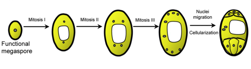
Stage 1: Functional megaspore (1 nucleus, n)
The functional megaspore — the chalazal one of the linear tetrad — remains inside the nucellus. It has a single haploid nucleus.
Stage 2: First mitosis (2 nuclei, free)
The nucleus divides mitotically — without cell division (no cytokinesis). Result: 2 nuclei in the same cell. The two nuclei migrate to opposite poles (one to micropyle, one to chalaza), with a large central vacuole forming between them.
Stage 3: Second mitosis (4 nuclei, free)
Each of the 2 nuclei divides again — still no cytokinesis. Now: 4 nuclei total, with 2 at each pole. Still one big cell, still a large vacuole.
Stage 4: Third mitosis (8 nuclei, free)
Each of the 4 nuclei divides one more time — still no cytokinesis. Result: 8 free nuclei (4 at each pole).
Stage 5: Cellularisation (final 7-celled, 8-nucleate embryo sac)
Now the magic happens. Cell walls form around the nuclei in a precise pattern:
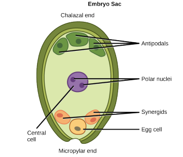
- At the micropylar pole (3 cells): 1 egg cell + 2 synergids = the egg apparatus.
- At the centre: 1 large central cell with 2 polar nuclei (which migrate inward from each pole) — this is the central cell.
- At the chalazal pole (3 cells): 3 antipodal cells.
So we end up with 7 cells but 8 nuclei (because the central cell has 2 nuclei).
The defining numbers
This is the line you must memorise for every NEET attempt:
3 mitotic divisions → 8 nuclei → 7 cells → at the micropylar end there are 3 cells (the egg apparatus), at the chalazal end there are 3 cells (the antipodals), and the central cell has 2 polar nuclei.
Why this development is "monosporic"
Because the entire embryo sac develops from a single megaspore (just 1 of the 4 produced). Contrast with bisporic (2 megaspores contribute) or tetrasporic (all 4 contribute) types found in other species — but Polygonum-type (monosporic) is the dominant pattern in flowering plants.
The Egg Apparatus — Micropylar End
The three cells at the micropylar end form the egg apparatus. This is the receiving end of the embryo sac — where the pollen tube delivers its male gametes.
\
The egg cell (1)
- Single, large, pear-shaped cell.
- Has a prominent nucleus and abundant cytoplasm.
- This is THE FEMALE GAMETE — the cell that will fuse with one male gamete to form the zygote.
- Ploidy: haploid ().
The synergids (2)
- Two cells flanking the egg cell.
- Each synergid has a characteristic structure at its micropylar tip called the filiform apparatus — a finger-like extension of thickened cell wall material that bulges into the cytoplasm. This is critically important:
- The filiform apparatus guides the pollen tube into the synergid via chemotaxis.
- The pollen tube enters one of the two synergids, NOT the egg cell directly.
- Synergid ploidy: haploid ().
NCERT-canonical phrase: "The synergids have special cellular thickenings at the micropylar tip called filiform apparatus, which play an important role in guiding the pollen tubes into the synergid."
This phrase is a near-guaranteed Board exam answer.
The Central Cell and Antipodals
The remaining cells of the embryo sac.
The central cell (1 cell, 2 polar nuclei)
- Occupies the bulk of the embryo sac (the largest cell).
- Contains 2 polar nuclei — both haploid ().
- The 2 polar nuclei migrate from each pole toward the centre.
- During fertilisation, the second male gamete fuses with BOTH polar nuclei → triple fusion → forms the PEN (primary endosperm nucleus, triploid, ). This is covered in detail in Section 8.
The 3 antipodal cells (chalazal end)
- Three cells at the chalazal end of the embryo sac (opposite the egg apparatus).
- Ploidy: haploid () each.
- Function: largely degenerate before/during fertilisation. Their exact role is somewhat unclear; some studies suggest they may have a brief nutritive role during early development. In most species, they degenerate quickly.
The full cell-count table
| Position | Cells | Number of cells | Nuclei | Ploidy |
|---|---|---|---|---|
| Micropylar end | Egg + 2 Synergids | 3 | 3 | All |
| Centre | Central cell | 1 | 2 (polar nuclei) | Both |
| Chalazal end | Antipodals | 3 | 3 | All |
| TOTAL | 7 cells | 8 nuclei | All |
The unforgettable number: 7 cells, 8 nuclei. Every cell is haploid, every nucleus is haploid. The "extra" nucleus is the second polar nucleus in the central cell.
The Filiform Apparatus — A Closer Look
One feature deserves a section of its own because of its importance and the frequency of exam questions on it.
What is the filiform apparatus?
The filiform apparatus is a specialised structure at the micropylar tip of each synergid. Structurally, it's a finger-like extension of cell wall material that bulges into the cytoplasm of the synergid.
Microscopically, it looks like a brush of wall projections — almost as if the cell wall has grown fingers reaching into the cell. These projections increase the surface area dramatically.
Why does it matter?
The filiform apparatus has TWO key functions:
1. Guiding the pollen tube (the famous function — exam-tested)
The filiform apparatus secretes chemical attractants that guide the pollen tube toward the synergid. Without this guidance, the pollen tube would have no clue where the egg apparatus is. Think of it as the GPS beacon for the pollen tube.
2. Receiving the pollen tube
The pollen tube enters one of the two synergids THROUGH the filiform apparatus. The synergid then breaks down, releasing the two male gametes into the cytoplasm of the synergid (NCERT).
NCERT-canonical phrase: "The filiform apparatus plays an important role in guiding the pollen tubes into the synergid."
A note on the chemistry
The chemical attractants are now known to be small defensin-like peptides called LURE peptides. NEET-Boards don't expect this detail — just the GUIDING ROLE is enough.
Small Memory Capsule — Section 5
The most important capsule of the entire chapter. Drill this.
The defining numbers — never forget
7 cells, 8 nuclei, all haploid (n).
Development sequence
Three mitotic divisions, three rounds of nuclear doubling: 1 → 2 → 4 → 8 nuclei.
Cell layout (micropyle → chalaza)
| Position | Structure | Count | Ploidy |
|---|---|---|---|
| Micropylar end | Egg apparatus = 1 egg + 2 synergids | 3 cells | All |
| Centre | Central cell with 2 polar nuclei | 1 cell, 2 nuclei | Both |
| Chalazal end | Antipodals | 3 cells | All |
| Total | 7 cells, 8 nuclei | All n |
Key roles to remember
- Egg cell = the female gamete; fuses with 1 male gamete → zygote (Section 8).
- Synergid = receives the pollen tube (via filiform apparatus).
- Filiform apparatus = GUIDES the pollen tube (chemotactic).
- Polar nuclei = both fuse with 1 male gamete → triple fusion → triploid PEN.
- Antipodals = degenerate; no major role.
The "monosporic" classification
Polygonum-type embryo sac develops from 1 megaspore only → "monosporic." This is the dominant type in angiosperms (NCERT-explicit).
One-line takeaway
The female gametophyte is a 7-celled, 8-nucleate, all-haploid embryo sac with one egg, two synergids, one central cell with two polar nuclei, and three antipodals — built from a single functional megaspore via 3 mitotic divisions.
Solved Examples
Example 1: The defining numbers
State the number of (a) cells, (b) nuclei, and (c) ploidy of each cell in the mature embryo sac. Explain why the cell count and nucleus count differ.
Solution:
| Item | Number/Value |
|---|---|
| (a) Cells | 7 |
| (b) Nuclei | 8 |
| (c) Ploidy | All haploid () |
Why cells ≠ nuclei:
The mismatch arises because the central cell contains 2 nuclei (the polar nuclei). It's still ONE cell — they share cytoplasm — but the cell has 2 separate nuclei inside.
So:
- Egg apparatus: 3 cells, 3 nuclei (1:1).
- Central cell: 1 cell, 2 nuclei (1:2). ← The extra nucleus.
- Antipodals: 3 cells, 3 nuclei (1:1).
- Total: 7 cells, 8 nuclei.
Answer: 7 cells, 8 nuclei, all haploid. The mismatch is because the central cell has 2 polar nuclei.
The big idea: This is a classic 'gotcha' question. Always remember: count cells first, then count nuclei separately. The central cell is the source of the extra nucleus.
[NEET Important] "7 cells, 8 nuclei" is the most-tested fact in the entire chapter. Drill until reflex.
Example 2: Function of the filiform apparatus
What is the filiform apparatus, where is it located, and what is its role?
Solution:
What: A specialised, finger-like extension of thickened cell wall material bulging into the cytoplasm.
Where: At the micropylar tip of each synergid in the embryo sac. Two synergids → two filiform apparatuses.
Role (the exam-tested part):
The filiform apparatus performs chemotaxic guidance of the pollen tube:
- It secretes chemical attractants (defensin-like LURE peptides — but you don't need this detail).
- These attractants form a chemical gradient that guides the pollen tube toward the synergid.
- The pollen tube enters the synergid via the filiform apparatus, breaks down the synergid, and releases the 2 male gametes into the cytoplasm of the synergid (NCERT).
Memorise this NCERT-canonical phrase: "The filiform apparatus plays an important role in guiding the pollen tubes into the synergid."
Answer: The filiform apparatus is a finger-like cell-wall thickening at the micropylar tip of each synergid; its role is to guide the pollen tube into the synergid by secreting chemoattractants.
[Board Important] Standard CBSE 2-mark question — ALWAYS include 'guides the pollen tube' for marks.
Example 3: Ploidy of every cell in the embryo sac
State the ploidy of: (a) egg cell, (b) synergid, (c) polar nucleus, (d) antipodal cell, (e) the central cell BEFORE fertilisation.
Solution:
All cells of the embryo sac are derived from a single haploid functional megaspore by mitosis. Mitosis never changes ploidy. So:
| Cell | Ploidy | Reasoning |
|---|---|---|
| (a) Egg cell | Direct mitotic descendant of megaspore | |
| (b) Synergid | Same lineage | |
| (c) Polar nucleus | Same lineage | |
| (d) Antipodal cell | Same lineage | |
| (e) Central cell | (effective) | Has 2 polar nuclei, each ; cytoplasmically the cell carries 2 haploid sets |
The subtle point about the central cell:
- Each polar nucleus is individually .
- The cell as a whole has chromosome content (i.e., effectively diploid).
- HOWEVER, NCERT typically describes the central cell as "having 2 polar nuclei" (both n), NOT as "diploid." Use NCERT terminology: 2 polar nuclei, each .
- The central cell only becomes truly triploid () AFTER triple fusion with a male gamete (Section 8).
Answer: All cells of the embryo sac are haploid (). The central cell uniquely contains 2 polar nuclei (each ), but each nucleus is independently haploid before fertilisation.
[NEET Important] The trap — Is the central cell diploid? Answer: NCERT says the central cell has 2 polar nuclei (each n). Avoid calling it diploid — call it with 2 polar nuclei.
Example 4: Why is the embryo sac called "monosporic"?
Explain why the angiosperm embryo sac is called a "monosporic" structure even though it has 7 cells.
Solution:
The term monosporic has nothing to do with the final cell count. It refers to the starting material of embryo sac development.
The key fact:
- Megasporogenesis produces 4 megaspores from 1 MMC (linear tetrad).
- Of these 4, only 1 survives as the functional megaspore.
- The entire 7-celled, 8-nucleate embryo sac develops from this single (mono-) functional megaspore by 3 mitoses + cellularisation.
So: mono = one + sporic = spore = "from one megaspore."
Contrast with bisporic and tetrasporic types:
- Bisporic: Embryo sac develops from 2 megaspores (e.g., Allium type).
- Tetrasporic: All 4 megaspores contribute (e.g., Fritillaria type).
But the dominant pattern in angiosperms is monosporic (Polygonum-type) — this is what NEET-Boards refers to as "the typical embryo sac."
Answer: "Monosporic" refers to the developmental origin — the embryo sac develops from a single functional megaspore (out of 4 produced by meiosis). Despite ending up with 7 cells and 8 nuclei, all originate from that one megaspore.
[Board Important] Be ready to define monosporic in a 2-mark Board question and to give Polygonum as the canonical example.
Example 5: Locating cells in the embryo sac
A pollen tube enters an embryo sac. (a) Which cell does it enter first? (b) Which cell contains the egg? (c) Which cell becomes the endosperm precursor after fertilisation?
Solution:
(a) The pollen tube enters a SYNERGID — specifically through the filiform apparatus at the synergid's micropylar tip. It does NOT enter the egg cell directly. The synergid then breaks down, releasing the 2 male gametes into the cytoplasm of the synergid (NCERT).
(b) The egg cell is one of the 3 cells of the egg apparatus at the micropylar end. It's flanked by the 2 synergids, with itself in the centre of the trio. After the synergid releases the male gametes, ONE male gamete fuses with the egg → diploid zygote.
(c) The CENTRAL CELL becomes the endosperm precursor. The other male gamete fuses with the 2 polar nuclei of the central cell → triple fusion → triploid PEN (primary endosperm nucleus) → develops into the endosperm tissue. Section 8 covers this in detail.
Putting it all together:
| Step | Event | Cell involved |
|---|---|---|
| 1 | Pollen tube entry | Synergid (via filiform apparatus) |
| 2 | Release of 2 male gametes | Synergid breaks down |
| 3 | Syngamy (forms zygote) | Egg cell + male gamete 1 |
| 4 | Triple fusion (forms PEN) | Central cell + male gamete 2 |
Answer: (a) A synergid (via filiform apparatus). (b) The egg cell, at the micropylar end. (c) The central cell, after triple fusion forms the triploid PEN.
[NEET Important] Tracing the pollen tube's path is a recurring NEET MCQ. Memorise the synergid-entry rule.