
Crossing Over to the Female Side
We've finished the male side. We have built the pollen grain, the male gametophyte, with all its sophistication — sporopollenin armour, germ pores, two or three cells. Now we cross over to the female side.
The female side is, in many ways, the mirror image of the male side, but with two important asymmetries:
- The female gametophyte is much larger — it has its own cytoplasm reserves, multiple specialised cells, and a sophisticated internal organisation.
- The female gametophyte is sessile — it stays put inside the ovule. The male gametophyte (pollen) is the one that travels.
We'll meet four nested structures going from large to small:
By the end of this section, you should be able to label every part of a pistil and every part of an anatropous ovule, and explain how megasporogenesis (the female equivalent of microsporogenesis) produces a single functional megaspore from a megaspore mother cell.
NCERT-canonical phrase: "The gynoecium represents the female reproductive part of the flower."
Architecture of the Gynoecium
The gynoecium is the innermost whorl of the flower (recall from Section 1). It contains one or more units called pistils (or carpels).
Three architectural types
Different plants have different gynoecium arrangements:
| Type | Definition | Example |
|---|---|---|
| Monocarpellary | Gynoecium has one pistil/carpel | Pea, mango |
| Multicarpellary, syncarpous | Multiple carpels fused into a single compound pistil | Papaver (poppy), tomato |
| Multicarpellary, apocarpous | Multiple carpels remain free (separate pistils) | Rose, Michelia (champa), lotus |
The fused (syncarpous) condition is most common in modern angiosperms. The free (apocarpous) condition is considered evolutionarily older.
NCERT-canonical phrase: "The gynoecium may consist of a single pistil (monocarpellary) or many carpels. When there are more than one, the carpels may be fused together (syncarpous) or may be free (apocarpous)."
Three parts of every pistil
\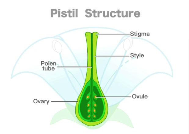
Regardless of architecture, every individual pistil has the same three parts:
- Stigma — the topmost, sticky receptive surface that catches and recognises pollen.
- Style — the slender neck (the "tunnel" through which the pollen tube grows).
- Ovary — the swollen base containing one or more ovules (the structures that will become seeds).
These three parts are evolutionarily and functionally analogous to a high-precision biological docking station — the stigma is the landing pad, the style is the corridor, and the ovary houses the cargo bay (the ovules).
Inside the Ovary — Ovule Number and Placentation
The ovary contains one or more ovules. The number varies wildly across species:
- One ovule per ovary — wheat, paddy, mango.
- Many ovules — papaya, watermelon; orchids have the most.
- Hundreds or thousands of ovules — orchids (record-holders, with up to 3 million ovules per ovary in some species).
After fertilisation, each ovule becomes a seed. So a single orchid capsule has thousands of seeds; a single mango has one. The ovary's internal cavities, called locules or chambers, house these ovules, attached to a swollen tissue called the placenta (yes, same word as in mammalian biology — convergent terminology).
NCERT-canonical phrase: "Inside the ovary is the ovarian cavity (locule). The placenta is located inside the ovarian cavity. Arising from the placenta are the megasporangia, commonly called ovules."
This is one of the most-quoted NCERT lines in Board exams. Memorise it verbatim — examiners love it.
The Ovule — Anatomy of the Female Megasporangium
\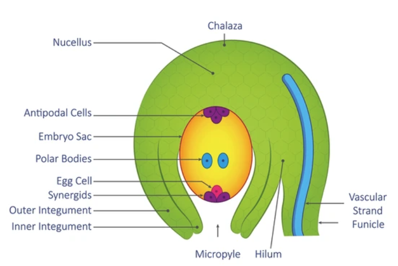
We zoom in further. Each ovule is itself a megasporangium (the female counterpart of the microsporangium). It has seven named parts — and you'll be tested on every one. Let's go through them systematically.
The seven parts of an ovule (memorise the order)
| # | Part | Description |
|---|---|---|
| 1 | Funicle | The "stalk" connecting the ovule to the placenta — like an umbilical cord. |
| 2 | Hilum | The point of attachment where funicle meets the ovule body. |
| 3 | Integuments | The outer protective coverings — usually two layers (outer + inner); a few species have one. |
| 4 | Micropyle | A small opening at the apex where the integuments don't quite meet. Pollen tube enters through here. |
| 5 | Chalaza | The basal end of the ovule, opposite the micropyle. |
| 6 | Nucellus | The large central mass of parenchymatous tissue — this is where the embryo sac develops. |
| 7 | Embryo sac | The female gametophyte, embedded in the nucellus. |
Anatropous ovule — the dominant orientation
In most angiosperms, the ovule is anatropous — meaning inverted. The micropyle and hilum end up on the same side because the ovule has bent 180° during development. Imagine taking an upright sausage and folding it back on its stalk — that's anatropous.
NCERT-canonical phrase: "Standard embryology (shown in the NCERT figure, not stated in its text): the most common ovule type in flowering plants is the anatropous ovule, in which the body of the ovule is completely inverted on the funicle so that the micropyle lies very close to the hilum."
Memorise this for Board 3-mark questions.
What this means functionally
The anatropous orientation places the micropyle right next to the funicle. So when the pollen tube reaches the ovule via the placenta and funicle, it has the shortest possible path to the micropyle. The geometry minimises the pollen tube's journey to the egg cell.
Megasporogenesis — Building a Functional Megaspore
Now the actual gametogenic event: how the ovule builds its female gametophyte. The process is called megasporogenesis, and it's the female counterpart to microsporogenesis (Section 2).
\
Step-by-step
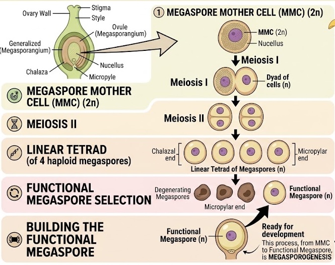
The process happens inside the nucellus of the ovule, near the micropylar end.
Step 1: The megaspore mother cell (MMC)
One cell in the nucellus — typically located near the micropylar end — differentiates into the megaspore mother cell (MMC). This cell is:
- Diploid ()
- Large, with conspicuous cytoplasm and a prominent nucleus.
Step 2: Meiosis
The MMC undergoes meiosis, producing 4 haploid daughter cells arranged in a linear tetrad (4 cells in a single straight line, end-to-end, from the micropylar pole to the chalazal pole).
Step 3: Survival of one — the functional megaspore
This is the key step that distinguishes megasporogenesis from microsporogenesis. In megasporogenesis, three of the four megaspores degenerate — only one survives. The surviving one is called the functional megaspore.
In most angiosperms, it's the chalazal-end megaspore that survives. The three at the micropylar end degenerate.
The functional megaspore then develops into the embryo sac (Section 5).
Microsporogenesis vs megasporogenesis — comparison
| Feature | Microsporogenesis | Megasporogenesis |
|---|---|---|
| Mother cell | PMC () | MMC () |
| Site | Inside microsporangia of anther | Inside nucellus of ovule |
| Products | 4 microspores (all ) | 4 megaspores (all ) |
| Tetrad arrangement | Tetrahedral | Linear |
| Survival | All 4 survive → 4 pollen grains | Only 1 survives (chalazal); 3 degenerate |
NCERT-canonical phrase: "This is termed as megasporogenesis. Generally, only one of the megaspores is functional while the other three degenerate."
Why does only one megaspore survive?
Plant biologists believe the reason is resource concentration. The female side invests heavily in just one well-provisioned egg cell, sacrificing the other 3 megaspores so that the surviving one has all the cytoplasmic resources it needs to build a large, multi-celled embryo sac. One big egg trumps four mediocre ones.
Small Memory Capsule — Section 4
Compact lock-in before the embryo sac itself.
Gynoecium architecture
- Monocarpellary = 1 pistil (pea, mango).
- Multicarpellary syncarpous = multiple carpels fused (Papaver, tomato).
- Multicarpellary apocarpous = multiple carpels free (rose, Michelia).
Three parts of a pistil
- Stigma (sticky landing pad) → Style (slender corridor) → Ovary (swollen base with ovules).
Seven parts of an ovule (memorise the order)
- Funicle — stalk.
- Hilum — funicle-ovule junction.
- Integuments — 2 protective layers.
- Micropyle — opening at the apex.
- Chalaza — basal end.
- Nucellus — central tissue.
- Embryo sac — female gametophyte inside nucellus.
Anatropous ovule
Inverted by 180° → micropyle ends up near hilum. Most common type in angiosperms.
Megasporogenesis
3 of 4 megaspores degenerate. The CHALAZAL one survives in most angiosperms.
Micro vs mega — the big difference
| Micro | Mega | |
|---|---|---|
| Tetrad | Tetrahedral | Linear |
| Survival | 4/4 | 1/4 (chalazal) |
One-line takeaway
The ovule (megasporangium) builds a single functional haploid megaspore via meiosis of an MMC and selective survival of one — the chalazal one — out of four daughter cells.
Solved Examples
Example 1: Naming the parts of a pistil
Sketch and label the parts of a typical pistil. State the function of each part.
Solution:
A pistil has three parts (from top to bottom):
| Part | Description | Function |
|---|---|---|
| Stigma | Topmost, sticky surface | Receives pollen; performs initial pollen recognition (compatibility check). |
| Style | Slender, elongated neck | Provides the corridor through which the pollen tube grows from stigma to ovary. |
| Ovary | Swollen base | Houses one or more ovules; after fertilisation, the ovary develops into the fruit. |
Answer: Stigma (receives pollen + recognition) → Style (pollen tube corridor) → Ovary (houses ovules → becomes fruit).
The big picture: The pistil is a precision biological docking station. Each part is engineered for a specific stage of the fertilisation process. No part is redundant.
[Board Important] Standard CBSE 2-mark or 3-mark question. Always pair part name with function for full marks.
Example 2: Identifying ovule parts from descriptions
Match each function to the correct part of the ovule:
(a) The opening through which the pollen tube enters. (b) The protective covering of the ovule. (c) The central tissue mass where the embryo sac develops. (d) The stalk attaching the ovule to the placenta. (e) The basal end of the ovule, opposite to the opening.
Solution:
We map each function to its part:
| Function | Part |
|---|---|
| (a) Opening for pollen tube entry | Micropyle |
| (b) Protective covering | Integuments (usually 2) |
| (c) Central tissue with embryo sac | Nucellus |
| (d) Stalk to placenta | Funicle |
| (e) Basal end opposite to opening | Chalaza |
Answer: (a) Micropyle, (b) Integuments, (c) Nucellus, (d) Funicle, (e) Chalaza.
Logic test: In an anatropous ovule, the micropyle and the hilum (where funicle attaches) end up on the same side. The chalaza is at the opposite end.
[NEET Important] "Match the column" questions on ovule anatomy appear frequently in NEET. Drill all seven part-function pairs.
Example 3: Megasporogenesis ploidy table
Determine the ploidy of: (a) Megaspore mother cell, (b) Megaspore tetrad cells, (c) Functional megaspore, (d) Nucellus, (e) Integuments.
Solution:
Apply the single-meiosis rule to the female line: meiosis happens only once, at the MMC → megaspore step.
| Cell | Ploidy | Reasoning |
|---|---|---|
| (a) Megaspore mother cell | Sporophytic; pre-meiosis. | |
| (b) Megaspore tetrad cells | Just produced by meiosis. | |
| (c) Functional megaspore | Same as tetrad — survived without ploidy change. | |
| (d) Nucellus | Sporophytic tissue of the ovule. | |
| (e) Integuments | Sporophytic protective layers. |
Answer: .
The big pattern (same as male side): Anything outside the gametophyte (i.e., outside the embryo sac) is . Anything inside the embryo sac (and the megaspores leading to it) is .
[NEET Important] Recurring NEET MCQ stem: "If the nucellus is 2n, then the functional megaspore is _" → Answer: n. Always.
Example 4: Why does only 1 megaspore survive?
In megasporogenesis, 1 megaspore mother cell produces 4 megaspores by meiosis, but only 1 survives. (a) Which one survives? (b) Why does the plant "throw away" the other 3? (c) How does this differ from microsporogenesis?
Solution:
(a) Which one survives?
In most angiosperms, the chalazal-end megaspore of the linear tetrad survives. The 3 megaspores at the micropylar end degenerate.
(b) Why throw away 3 of 4?
This is a resource-concentration strategy. Building a fully functional embryo sac (with 7 cells, 8 nuclei, abundant cytoplasm) is metabolically expensive. By sacrificing 3 of 4 megaspores, the plant pools all the cytoplasmic resources into the surviving one — producing a single robust, well-provisioned female gametophyte rather than four undersized ones.
This is the same logic as a mammal producing one large egg per ovulation rather than four small ones — quality over quantity, on the female side.
(c) How does this differ from microsporogenesis?
In microsporogenesis (male side), all 4 microspores survive and develop into 4 pollen grains. The strategy is opposite: produce abundance, scatter widely, accept that most pollen won't reach a stigma.
So the male and female strategies are evolutionarily complementary:
- Male side = mass production + dispersal (4 of 4 microspores survive).
- Female side = single high-investment unit (1 of 4 megaspores survives).
Answer: (a) The chalazal megaspore survives. (b) Resource concentration — one well-provisioned female gametophyte beats four mediocre ones. (c) Microsporogenesis preserves all 4 microspores (mass production strategy).
[NEET Important] A favourite "compare and contrast" question. The 4/4 vs 1/4 ratio is the key fact.
Example 5: The anatropous ovule and pollen-tube geometry
Why is the anatropous orientation of the ovule biologically advantageous?
Solution:
Recall that in an anatropous ovule, the body of the ovule is inverted by 180° on the funicle, so the micropyle ends up very close to the hilum (where the funicle attaches).
Why does this matter? Consider the pollen tube's journey:
- Pollen tube emerges from the pollen grain on the stigma.
- Grows down through the style.
- Enters the ovary.
- Travels along the placenta to reach the funicle.
- Travels along the funicle to reach the ovule.
- Must enter the ovule via the micropyle.
In an orthotropous (upright) ovule, the micropyle is at the FAR END from the funicle — so the pollen tube would have to grow halfway around the entire ovule to reach the micropyle.
In an anatropous ovule, the micropyle is right next to the funicle — so the pollen tube has a direct, minimum-distance path from funicle to micropyle.
So the anatropous orientation is an evolutionary optimisation: minimise the pollen tube's journey, maximise the chance of successful fertilisation.
Answer: The anatropous orientation places the micropyle next to the funicle, providing the pollen tube with the shortest possible path from the funicle into the ovule via the micropyle. This minimises tube growth distance and maximises fertilisation success.
[NEET Important] "Why is anatropous orientation important?" appears as a 2-mark Board reasoning question. Always frame the answer around pollen-tube geometry.