
Zooming into the Male Side
In Section 1, we mapped the four whorls of a flower. Now we focus exclusively on the androecium — the male reproductive whorl — and follow the journey from a sporophytic stem cell to a haploid pollen grain.
The androecium is made up of one or more stamens (also called microsporophylls, because each one carries microsporangia — the structures that produce pollen). A flower might have just one stamen (e.g., Canna) or several dozen (e.g., Hibiscus, where the stamens fuse around the style).
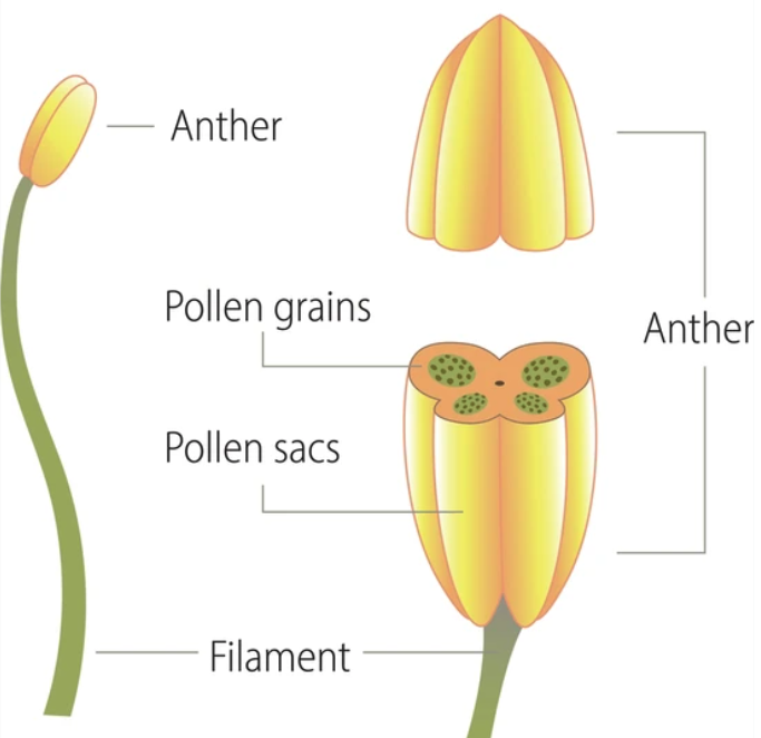
A stamen has just two parts
The architecture is simple:
- Filament — a long, slender stalk. Its proximal (base) end is attached either to the thalamus or directly to a petal (an attachment pattern called epipetalous, seen in Brinjal).
- Anther — the terminal swollen structure, where pollen is actually produced. Almost always yellow in colour due to its carotenoid pigments.
Most of what's interesting biologically happens inside the anther. We'll spend most of this section there.
The Anther — Bilobed, Dithecous, Tetrasporangiate
This three-word phrase is the single most important description of a typical angiosperm anther. NCERT states 'bilobed' and 'dithecous'; 'tetrasporangiate' is the standard derived term. NEET tests it nearly every year. Let's understand each word in plain English.
\
Bilobed
The anther has 2 lobes — clearly visible from the outside. You can think of these as two parallel cylinders fused at a connective strip in the middle. A longitudinal groove runs down between them.
Dithecous
Each lobe has 2 thecae (singular: theca; plural: thecae), giving 4 theca in total. Theca are the chambers visible if you cut a transverse section of the anther.
Tetrasporangiate
Inside each theca sits 1 microsporangium (a pollen sac). Since there are 4 theca, there are 4 microsporangia in total per anther.
The math: .
The longitudinal groove and dehiscence
The groove between the two lobes is more than a visual feature — it's the line of dehiscence. When the anther matures, mechanical stress (from drying + tissue stiffening) causes the wall to split open along this groove, releasing the pollen grains into the air or onto a visiting insect. This release is called anther dehiscence.
[NEET Important] "A typical angiosperm anther is bilobed, dithecous, tetrasporangiate." — memorise this exact NCERT phrase. NEET has tested all three terms in different years.
The Four Wall Layers of a Microsporangium
We've placed the microsporangia (4 of them) at the corners of the anther. Now let's look inside one and see how it's built.
\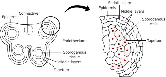
A transverse section through a single microsporangium reveals four concentric wall layers around a central mass of sporogenous tissue. The architecture is like a Russian doll — each layer wraps around the next.
The four wall layers, from outside to inside
| Order | Layer | Function |
|---|---|---|
| 1 | Epidermis (outermost) | Protective outer skin |
| 2 | Endothecium | Develops fibrous thickenings at maturity → aids dehiscence |
| 3 | Middle layers (1-3 rows) | Get crushed as pollen develops; almost invisible at maturity |
| 4 | Tapetum (innermost) | Nourishes developing pollen; secretes sporopollenin |
Mnemonic — "Eat Every Morning Toast"
To remember the order (outside → inside): Eat Every Morning Toast → Epidermis, Endothecium, Middle layers, Tapetum.
The outer three layers work as a team
- Protection during the long maturation of pollen (weeks to months).
- Dehiscence — the endothecium develops radially-oriented fibrous thickenings at maturity. As the anther dries out, these fibres pull on the wall and crack it open along the longitudinal groove.
- Middle layers are sacrificed — they don't have a final-state structure. They're crushed and absorbed as the pollen grains expand.
The tapetum is so important it gets its own section
(Next note.)
[NEET Important] "List the four wall layers of a microsporangium in correct order." Outside → inside: Epidermis → Endothecium → Middle layers → Tapetum. NEET asks both the order and the function of each.
The Tapetum — Anther's VIP Layer
The tapetum is the innermost of the four wall layers and arguably the single most biologically active tissue in the male reproductive structure. NEET spotlights it because it has unique features unlike any other plant cell.
What makes tapetum unusual
Take a microscope slide of a young anther, focus on the inner wall, and you'll see cells with three peculiar features:
- Dense cytoplasm. Tapetal cells are packed with ribosomes, ER, and metabolic machinery — far more than ordinary plant cells.
- Bi-nucleate (or even multinucleate). Most tapetal cells have 2 nuclei, sometimes more. This is a unique cellular state.
- Unique among the wall layers — neither protective like epidermis, nor fibrous like endothecium, nor sacrificial like middle layers. The tapetum has a job no other layer does: feeding the developing pollen grains.
How does a cell become bi-nucleate?
NCERT poses this exact question in its in-text section. Two mechanisms:
- Endomitosis — nuclear division without cytokinesis. The nucleus replicates, but the cell does not split. Result: one cell with 2 nuclei.
- Free nuclear division — repeated nuclear division without cell-wall formation, producing many nuclei in shared cytoplasm.
Either way, the biological reason for going multi-nucleate is the same: more nuclei = more gene expression = more metabolic output. The tapetum needs to manufacture nutrients and the polymer sporopollenin at enormous rates, and a single (diploid) nucleus would be the bottleneck.
What the tapetum actually produces
- Nutrients — sugars, amino acids, lipids — for developing pollen.
- Sporopollenin — the indestructible polymer that forms the outer wall (exine) of pollen. Without tapetum, no exine → no pollen survival.
- Hydrolytic enzymes — to break down the callose wall that initially surrounds the microspore tetrad.
- Mechanical support — keeps the developing pollen in place.
The tapetum self-destructs
By the time pollen is mature, the tapetum has completely consumed itself by programmed cell death (PCD). It is the perfect "altruistic tissue" — works hard, then dies, leaving behind well-developed pollen.
Why is the tapetum so heavily examined?
Because tapetum dysfunction = male sterility. In agriculture, hybrid seed production relies on creating "male sterile" lines, and almost all such mutations affect the tapetum. So this layer is biologically central, agriculturally important, and pedagogically rich — a triple threat for examiners.
[NEET Important] Memorise: "The tapetum is the innermost wall layer of the microsporangium. It nourishes developing pollen grains, secretes sporopollenin, and has cells that are densely cytoplasmic and bi-nucleate or multinucleate. It is the only wall layer that is gametophyte-supporting (not protective)."
Sporogenous Tissue, Pollen Mother Cells, and the Start of Meiosis
Now we go from the wall layers to the centre of the microsporangium. The cells we find there are the ones that will become pollen.
The sporogenous tissue
When the anther is young (still developing inside the bud), the centre of each microsporangium is filled with a compact, homogeneous mass of cells called the sporogenous tissue. These cells are:
- Diploid () — they are sporophytic.
- Identical-looking in young anthers.
- Compactly packed — no large spaces between them.
- Each capable of becoming a Pollen Mother Cell (PMC).
Pollen Mother Cells (PMCs)
As the anther matures, the sporogenous cells enlarge and mature into Pollen Mother Cells. The PMC is the cell that will actually undergo meiosis. It is still diploid (), but now it has the cytoplasmic resources and gene expression patterns required for meiosis.
NCERT also uses the term microspore mother cell (MMC) for the same thing. (Don't confuse with the female MMC — megaspore mother cell, in the ovule. Same abbreviation, different cell, different location, different gender. Context tells you which.)
How many PMCs per microsporangium?
Typically hundreds to thousands. If a microsporangium has 1000 PMCs, and each undergoes meiosis to give 4 microspores, you get 4000 pollen grains from that one microsporangium. Multiply by 4 microsporangia per anther → 16,000 pollen grains per anther. Multiply by all the anthers in all the stamens in all the flowers on a plant → a single plant can release millions of pollen grains per season.
This abundance is no accident. The huge numbers compensate for the inefficiency of pollination (especially wind pollination — covered in Section 6).
[NEET Important] "Which cell is the precursor to meiosis in the male reproductive line?" — Answer: the Pollen Mother Cell (PMC), also called the microspore mother cell.
Microsporogenesis — The Main Event
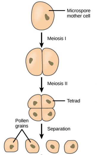
Now we reach the heart of this section. Microsporogenesis is the process by which a Pollen Mother Cell, through meiosis, gives rise to four microspores. Let's trace it carefully.
\
The definition (NCERT-canonical)
"The process of formation of microspores from a pollen mother cell through meiosis is called microsporogenesis."
The key word here is meiosis — specifically, one cycle of meiosis (which includes Meiosis I + Meiosis II). One PMC → one round of meiosis → four haploid microspores. This 1:4 ratio is absolute, dictated by the meiotic machinery itself.
Step by step
Sporogenous cell → PMC. A diploid sporogenous cell stops dividing mitotically and matures into a Pollen Mother Cell (, with cytoplasm and gene expression primed for meiosis).
Meiosis I — the reduction division.
- Homologous chromosomes pair up.
- Crossing over occurs at chiasmata — generating new combinations of alleles.
- Homologues separate.
- Result: 2 daughter cells, each with chromosomes (but each chromosome still has 2 chromatids, since DNA was replicated before Meiosis I).
- Meiosis II — the equational division.
- Sister chromatids separate.
- Result: 4 haploid cells with single chromatids each.
Tetrad formation. The 4 daughter cells stay attached as a microspore tetrad, surrounded by a temporary callose wall that the tapetum will later dissolve. The tetrad is typically arranged in a tetrahedral pattern (4 cells touching each other at a central point).
Tetrad dissociation. As the anther matures and dries, the callose wall is enzymatically dissolved (the tapetum's enzymes do this), and the four microspores separate. Each microspore is now free to develop independently into a pollen grain.
Why this matters for variation
Two things happen during meiosis that are crucial for genetic diversity:
- Crossing over in Meiosis I — homologous chromosomes exchange segments, creating new allele combinations.
- Independent assortment of chromosomes during Meiosis I — each daughter cell gets a random mix of maternal and paternal chromosomes.
These mechanisms ensure that every pollen grain a plant produces is genetically unique. When you multiply this by millions of pollen grains per plant per season, you get the raw material for evolution.
The numerical takeaway
Multiply by PMCs per microsporangium → microspores per microsporangium. Multiply by 4 microsporangia per anther → microspores per anther. NEET frequently tests this multiplication.
The Master Ploidy Map (Male Line)
This is the single most-tested concept in this entire section. Every NEET paper has at least one ploidy question. Master this map.
The ploidy of every cell in the male reproductive line
| Cell or tissue | Ploidy | Why |
|---|---|---|
| Cells of anther wall layers (epidermis, endothecium, middle layers, tapetum) | All sporophytic | |
| Sporogenous tissue | Sporophytic precursor | |
| Pollen Mother Cell (PMC) | The cell that undergoes meiosis | |
| Cells of microspore tetrad (immediately after meiosis) | Meiosis halved them | |
| Free microspores | Same as tetrad cells | |
| Pollen grain (male gametophyte) | Haploid throughout | |
| Vegetative cell + generative cell of pollen | Both haploid | |
| Male gametes (formed by mitosis of generative cell) | Two haploid male gametes per pollen grain |
The one rule to remember
Only ONE step changes ploidy — meiosis at the PMC stage. Everything before is . Everything after is .
This is symmetric on the female side (covered in Section 4) — only one ploidy change there too.
NEET classic trap — the tapetum
A common NEET trap: students assume that because tapetum is adjacent to haploid microspores, it must be haploid too. Wrong. The tapetum is part of the sporophyte (anther wall) — it is diploid. Haploidy starts only at the microspore stage.
[NEET Important] Memorise the rule: "In the male reproductive line, ploidy changes only at meiosis (PMC → microspore). The tapetum is diploid; the pollen grain is haploid."
Small Memory Capsule — Section 2
Lock-in summary before solved examples.
Stamen architecture
- Stamen = filament + anther.
- Filament attaches to thalamus (or, in epipetalous flowers, to a petal).
- Anther: bilobed, dithecous, tetrasporangiate — 2 lobes × 2 theca/lobe × 1 microsporangium/theca = 4 microsporangia per anther.
- Longitudinal groove between the lobes = line of dehiscence.
4 wall layers (outside → inside)
"Eat Every Morning Toast" → Epidermis · Endothecium · Middle layers · Tapetum
| Layer | Function |
|---|---|
| Epidermis | Outer protection |
| Endothecium | Fibrous thickenings → dehiscence |
| Middle layers | Crushed as pollen develops |
| Tapetum | Nourishes pollen; bi-nucleate; secretes sporopollenin |
Tapetum highlights (NEET high-yield)
- Bi-nucleate or multinucleate — via endomitosis or free nuclear division.
- Dense cytoplasm — for heavy metabolic output.
- Secretes: nutrients, sporopollenin, hydrolytic enzymes.
- Self-destructs by PCD when pollen is mature.
- Ploidy: diploid () — sporophytic, not haploid.
Microsporogenesis — the master arrow
- PMC → microspores (fixed by meiosis).
- PMCs → microspores per microsporangium.
- 4 microsporangia per anther → microspores per anther.
Master ploidy map (male line)
| Cell | Ploidy |
|---|---|
| Anther wall (all 4 layers, including tapetum) | |
| Sporogenous tissue | |
| PMC | |
| Microspores onwards (pollen, vegetative cell, generative cell, male gametes) |
Only ONE ploidy change → at meiosis (PMC → microspores).
One-line takeaway
"Inside the 4-microsporangia of a bilobed anther, sporogenous cells become PMCs that undergo meiosis to give 4 haploid microspores per PMC; the bi-nucleate diploid tapetum nourishes the developing pollen and synthesises the sporopollenin of its exine."
Solved Examples
Example 1: The defining triple-term description
A NEET MCQ asks: "A typical angiosperm anther is described as:" with options (a) Unilobed, monothecous, monosporangiate; (b) Bilobed, dithecous, tetrasporangiate; (c) Trilobed, trithecous, hexasporangiate; (d) Bilobed, monothecous, bisporangiate. Pick the correct option and justify each term.
Solution:
Apply each term to the standard anther:
- Bilobed: ✓ — every typical anther has 2 lobes visible externally (left lobe and right lobe, separated by a longitudinal groove).
- Dithecous: ✓ — each lobe has 2 theca (chambers visible in T.S.). 2 lobes × 2 theca = 4 theca total.
- Tetrasporangiate: ✓ — each theca contains 1 microsporangium. 4 theca × 1 microsporangium = 4 microsporangia total.
Option (b) matches.
The other options either undercount (a, d) or overcount (c) the number of structural units.
Answer: (b) Bilobed, dithecous, tetrasporangiate.
[NEET Important] This phrasing is a frequent NEET target. The three-word NCERT phrase is the most NEET-tested description in the entire chapter. Memorise it verbatim.
Example 2: List and function of wall layers
State the four wall layers of a microsporangium in correct order from outside to inside, and assign each its primary function.
Solution:
Apply the mnemonic "Eat Every Morning Toast" for the order:
| # | Layer | Function |
|---|---|---|
| 1 | Epidermis | Outermost protective covering of the microsporangium |
| 2 | Endothecium | Develops fibrous (radial) thickenings; aids in dehiscence (opening of the anther) when mature |
| 3 | Middle layers | 1-3 thin rows; get crushed and consumed as the pollen grains expand inside |
| 4 | Tapetum | Innermost; nourishes developing pollen; bi-nucleate; secretes sporopollenin for pollen exine |
Why each function?
- Epidermis is the boundary with the outside world → naturally protective.
- Endothecium's fibrous bands → like a rubber-band mechanism that releases tension at maturity, splitting the anther open.
- Middle layers are sacrificial → they don't have a long-term role; they're spaceholders.
- Tapetum is closest to the developing germ cells → it makes biological sense for the food source to be adjacent.
Answer: Epidermis → Endothecium → Middle layers → Tapetum, with functions as above.
[Board Important] A standard 3-mark CBSE question. Always list the layers outside-to-in (reverse order loses marks).
Example 3: Why is the tapetum bi-nucleate? (NCERT in-text)
NCERT explicitly asks: "How could tapetal cells become bi-nucleate?" Answer in 3-4 lines and state the biological significance.
Solution:
Tapetal cells become bi-nucleate primarily through one of two cellular mechanisms:
- Endomitosis — the nuclear DNA replicates and the nucleus divides mitotically, but cytokinesis does not follow. The cell ends up with two daughter nuclei sharing one cytoplasm.
- Free nuclear division — repeated rounds of nuclear division without cell wall formation, producing 2, 4, or even more nuclei in shared cytoplasm. (At the extreme this gives multinucleate cells.)
Biological significance:
Both mechanisms achieve the same outcome — they multiply the genetic / transcriptional capacity of the tapetal cell without splitting it into smaller cells. With twice (or more) the number of nuclei, the tapetum can:
- Synthesise nutrients at twice the rate.
- Produce sporopollenin (a chemically complex polymer) more efficiently.
- Sustain heavy gene expression during the brief window of pollen development.
A single haploid (or even diploid) nucleus would be a bottleneck given how much the tapetum has to manufacture. Bi-nucleation is evolution's solution.
Answer: Endomitosis (mitosis without cytokinesis) or free nuclear division (repeated nuclear division without cell wall formation) produces the bi/multinucleate state. The biological purpose is to amplify metabolic capacity for nourishing pollen and secreting sporopollenin.
[Board Important] A direct NCERT in-text question — appears as a 2-mark CBSE classic.
Example 4: Numerical — pollen output of one anther
A typical anther has 4 microsporangia. If each microsporangium contains 200 PMCs, calculate the total number of pollen grains produced by one anther.
Solution:
Step 1: How many microspores does one PMC produce?
From microsporogenesis (1 PMC → meiosis → 4 microspores):
Step 2: How many microspores per microsporangium?
If there are 200 PMCs per microsporangium:
Step 3: How many microspores per anther?
If there are 4 microsporangia per anther:
Step 4: Microspores → pollen grains.
Each microspore matures into one pollen grain (no further division at this step). So:
Answer: 3,200 pollen grains per anther.
Key formula to remember: If PMCs per microsporangium, total pollen per anther = .
In our problem: . Same answer.
[NEET Important] Numerical pollen-count questions are NEET classics. Master the formula: .
Example 5: The ploidy trap
State the ploidy of each of the following:
(a) Tapetum (b) PMC (c) Microspore tetrad cell (d) Male gamete (e) Endothecium
Solution:
Apply the master rule: only ONE ploidy change in the male line — at meiosis (PMC → microspore). Everything before is ; everything after is .
| Cell | Ploidy | Reason |
|---|---|---|
| (a) Tapetum | Anther wall layer = sporophytic | |
| (b) PMC | Pre-meiosis precursor | |
| (c) Microspore tetrad cell | Just produced by meiosis | |
| (d) Male gamete | Produced by mitosis of generative cell — mitosis doesn't change ploidy | |
| (e) Endothecium | Anther wall layer = sporophytic |
Answer: .
The trap: Students often assume tapetum is haploid because it's "next to" the haploid microspores. WRONG. Adjacency does not equal ploidy. The tapetum is sporophytic (built before meiosis), so it remains .
Answer key shorthand: All four wall layers + sporogenous tissue + PMC = . Microspores onwards = .
[NEET Important] This is the highest-frequency NEET MCQ pattern in the entire section. Memorise the rule and you'll never miss it.
Example 6: Comparing microsporogenesis with megasporogenesis (preview)
Without yet studying megasporogenesis in detail (Section 4), state two ways in which microsporogenesis differs from megasporogenesis in its outcomes.
Solution:
Both processes involve a diploid mother cell () undergoing meiosis to give 4 haploid daughter cells. But the outcomes differ in two key ways:
Difference 1: Survival of products
- Microsporogenesis: All 4 daughter cells (microspores) survive and develop into pollen grains. So 1 PMC → 4 functional gametophytes.
- Megasporogenesis: Only 1 of the 4 daughter cells survives (the "functional megaspore"). The other 3 degenerate. So 1 MMC → 1 functional gametophyte.
Difference 2: Arrangement of the tetrad
- Microsporogenesis: Tetrad is typically tetrahedral (4 cells touching at a central point, like a 3D pyramid).
- Megasporogenesis: Tetrad is linear (4 cells arranged in a single straight line, end-to-end).
Why these differences?
- Asymmetry in resource allocation — the female plant invests heavily in one egg cell with abundant cytoplasm and nutrients, sacrificing 3 daughter cells to consolidate resources. The male side opts for many small gametophytes, maximising the chance that at least one reaches a stigma.
- Geometric arrangement is a clue to the developmental fate — the chalazal-end cell of the linear tetrad becomes the functional megaspore, taking the dominant position by virtue of geometry.
Answer: (1) Microsporogenesis produces 4 surviving gametophytes; megasporogenesis produces only 1. (2) Microspore tetrad is tetrahedral; megaspore tetrad is linear.
[NEET Important] A frequent NEET MCQ pattern is to compare micro- and megasporogenesis. Memorise both differences.