
After Fertilisation — The Story Continues
Section 8 ended with two products sitting inside the embryo sac:
- A zygote (diploid, ) at the micropylar end.
- A PEN (Primary Endosperm Nucleus, triploid, ) at the centre.
Both will now develop in parallel. The PEN becomes the endosperm. The zygote becomes the embryo. Together, they form the future seed.
This section traces both developments.
One critical sequence rule
Endosperm forms BEFORE the embryo.
Why? Because the embryo needs a food supply ready when it starts dividing. If the embryo developed first, it would have nothing to feed on. So the PEN starts dividing immediately after fertilisation — building the endosperm — while the zygote pauses for a while before starting its development.
NCERT-canonical phrase: "Endosperm development precedes embryo development because the developing embryo derives its nutrition from the endosperm."
This sequence — endosperm first, embryo second — is exam-critical.
Endosperm Development — The Free-Nuclear Stage
The PEN starts dividing immediately. But here's the interesting part: its first many divisions are nuclear divisions without cell wall formation (free-nuclear mitosis).
Step-by-step
Step 1: Free-nuclear endosperm
- PEN () divides repeatedly by mitosis.
- Each division produces 2 nuclei → 4 nuclei → 8 nuclei → 16 → 32 → … → hundreds of nuclei.
- No cell walls form during this stage. All the nuclei share a common cytoplasm in one big multi-nucleate cell.
Step 2: Cellularisation
- Eventually, cell walls form around each nucleus.
- The multi-nucleate mass becomes a multicellular endosperm tissue.
- Each cell of the endosperm is (triploid).
The famous example: COCONUT WATER
Here's where biology gets surprisingly delicious. Coconut water (the clear liquid inside a tender coconut) is the free-nuclear endosperm of the coconut!
NCERT-canonical phrase: "Per NCERT, coconut water is free-nuclear endosperm (thousands of nuclei) and the surrounding white kernel is cellular endosperm. The cellular endosperm forms the white edible meat of the coconut."
So:
- Coconut WATER = free-nuclear endosperm (liquid, multinucleate, nutritive).
- Coconut WHITE MEAT = cellular endosperm (solid, formed by cellularisation).
Both are endosperm — same tissue at different stages of development. Memorise this — it's a guaranteed Board MCQ.
A short note on consumption
When you drink coconut water, you're drinking the multi-nucleate, triploid endosperm of a developing seed. When you eat coconut white meat, you're eating the cellular triploid endosperm. Both are tissues nourishing the (still-developing) coconut embryo.
Embryo Development — Embryogeny
The zygote waits until the endosperm has begun to form (so the food supply is ready), then starts its own development. This process is called embryogeny.
Dicot embryo development
\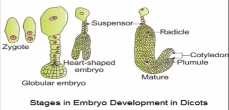
The dicot embryo develops through a characteristic sequence of stages:
Stage 1: Zygote — single diploid cell at the micropylar end.
Stage 2: Pro-embryo — early multicellular stage; a short filament with two ends. The end near the micropyle becomes the suspensor; the other end (toward chalaza) becomes the embryo proper.
Stage 3: Globular embryo — small spherical cluster of cells.
Stage 4: Heart-shaped embryo — two lobes appear on either side of the embryo axis (the future cotyledons).
Stage 5: Mature embryo — has 2 cotyledons + a clear embryonic axis:
- Plumule — future shoot, at the top of the axis (between the cotyledons).
- Radicle — future root, at the bottom (toward the micropyle).
- The portion of the axis between the cotyledons and the radicle is called the hypocotyl (below cotyledons); the part between the cotyledons and the plumule is the epicotyl (above cotyledons).
NCERT-canonical phrase: "The early stages of embryo development are similar in both monocots and dicots. The zygote divides to form the proembryo, which subsequently develops into globular, heart-shaped, and finally mature embryo."
Two cotyledons — defining feature
The presence of two cotyledons is the defining feature of dicots (hence "DI-cot"). Examples: pea, gram, sunflower, mustard.
Monocot Embryo Development
\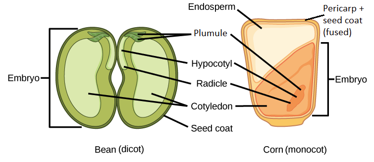
Monocot embryos have a simpler architecture but with some specialised parts.
Key differences from dicot
- Only 1 cotyledon (hence MONO-cot) — called the SCUTELLUM (large, shield-shaped, on one side of the embryo axis).
- Plumule is protected by a sheath called the COLEOPTILE (a hollow tube around the developing shoot).
- Radicle is protected by a sheath called the COLEORRHIZA (a tubular covering of the root tip).
The coleoptile and coleorhiza are unique to monocots — adaptations for pushing through the soil during germination.
A complete monocot embryo (e.g., maize)
| Part | Description |
|---|---|
| Scutellum | Single shield-shaped cotyledon |
| Coleoptile | Protective sheath of plumule |
| Plumule | Future shoot (inside coleoptile) |
| Coleorhiza | Protective sheath of radicle |
| Radicle | Future root |
| Aleurone layer | Outer protein-rich layer of endosperm (gives nutritive function) |
A famous monocot embryo function
In germinating maize/wheat, the coleoptile breaks through the soil surface first, protecting the delicate plumule inside. Only after reaching air does the plumule emerge and start photosynthesis. This is why monocot seedlings (like grass shoots) appear as sharp green spikes pushing up — that's the coleoptile in action.
Dicot vs Monocot embryo — comparison table
| Feature | Dicot | Monocot |
|---|---|---|
| Cotyledons | 2 | 1 (scutellum) |
| Plumule covering | Naked (between cotyledons) | Coleoptile |
| Radicle covering | Naked | Coleorhiza |
| Example | Pea, sunflower | Maize, wheat, rice |
Small Memory Capsule — Section 9
Lock-in before the seed and fruit.
The sequence rule
Endosperm forms FIRST → then embryo develops.
Endosperm provides food for the developing embryo.
Endosperm development stages
The COCONUT example (NEET-essential)
- Coconut water = free-nuclear (multi-nucleate) endosperm — liquid.
- Coconut white meat = cellular endosperm — solid.
- Both are TRIPLOID ().
Dicot embryo stages
Mature dicot: 2 cotyledons + plumule (top) + radicle (bottom) + suspensor.
Monocot embryo — key features
- 1 cotyledon = SCUTELLUM (shield-shaped, on one side).
- Plumule covered by COLEOPTILE.
- Radicle covered by COLEORRHIZA.
- Aleurone layer = outer protein-rich layer of endosperm.
- Examples: maize, wheat, rice.
Dicot vs Monocot — drilled
| Dicot | Monocot | |
|---|---|---|
| Cotyledons | 2 | 1 (scutellum) |
| Plumule | Naked | In coleoptile |
| Radicle | Naked | In coleorhiza |
One-line takeaway
After double fertilisation, the PEN develops first into a free-nuclear then cellular triploid endosperm (e.g., coconut water → coconut meat); the zygote then develops through proembryo-globular-heart-shaped stages into a mature embryo with 2 cotyledons (dicot) or 1 scutellum + coleoptile + coleorhiza (monocot).
Solved Examples
Example 1: The endosperm-before-embryo rule
Why does endosperm development precede embryo development in angiosperms? Explain the biological logic.
Solution:
The rule: In every angiosperm, the PEN starts dividing IMMEDIATELY after triple fusion, while the zygote pauses for a while before starting its development.
The biological logic:
The embryo, once it starts developing, needs a constant supply of nutrition — sugars, amino acids, lipids, minerals. Without a ready food supply, the embryo would starve before it could grow.
The endosperm is that food supply.
So evolution has built the timing as follows:
- PEN forms → endosperm starts developing immediately.
- Free-nuclear stage → many nuclei accumulate quickly (no cell-wall building delay).
- By the time the zygote starts dividing, a rich nutritive endosperm is already in place.
- The embryo absorbs nutrients from the endosperm as it grows.
This is just-in-time nutrient delivery. The endosperm is built first specifically because the embryo needs it ready before it begins its own development.
Answer: Endosperm forms first because the developing embryo derives its nutrition from the endosperm. By the time the zygote starts dividing, a ready food supply is already in place.
[NEET Important] A favourite NEET MCQ stem: "Why does endosperm form before the embryo?" — always answer with the nutrition argument.
Example 2: Coconut water — what is it really?
What is coconut water? Why is it sweet? Why is it described as a "natural saline solution rich in nutrients"?
Solution:
What coconut water is:
Coconut water is the free-nuclear endosperm of the developing coconut (the seed of Cocos nucifera). It is a liquid, multi-nucleate cytoplasm with many nuclei suspended but no cell walls yet formed.
In a tender coconut, this watery endosperm fills most of the seed cavity. As the coconut matures further, the endosperm undergoes cellularisation, becoming the solid white meat (kernel) that we eat.
NCERT-canonical phrase: "In the developing endosperm of coconut, only the free-nuclear stage may be seen. The cellular endosperm forms the white edible meat of the coconut."
Why it's sweet:
The endosperm is a nutritive tissue rich in:
- Sugars (glucose, fructose, sucrose) — provides energy.
- Amino acids and proteins — for protein synthesis.
- Vitamins (B-complex, vitamin C).
- Minerals (potassium, sodium, magnesium, calcium).
- Fats (in the form of fatty acids and small amounts of triglycerides).
The sugar content is why coconut water tastes sweet.
Why it's a natural saline solution:
Coconut water is a nutritive fluid rich in sugars and minerals.
Ploidy:
Every nucleus in coconut water is triploid () — because the endosperm formed from the triploid PEN. So when you drink coconut water, you're consuming thousands of triploid nuclei (NCERT)!
Answer: Coconut water = free-nuclear, triploid endosperm of the coconut seed. Sweet due to sugars, saline-like due to natural minerals. Eventually undergoes cellularisation to form the white solid edible meat of the mature coconut.
[Board Important] Recurring CBSE 2-mark question. Always mention: free-nuclear stage, triploid, nutritive tissue.
Example 3: Stages of dicot embryo development
List the stages of dicot embryo development from zygote to mature embryo. Briefly describe each.
Solution:
| Stage | Description |
|---|---|
| 1. Zygote () | Single diploid cell at the micropylar end of the embryo sac. Pauses briefly while endosperm starts forming. |
| 2. Pro-embryo | Zygote divides to form a small filament of cells, with the suspensor at one end (toward micropyle) and the embryo proper at the other end (toward chalaza). |
| 3. Globular embryo | A small spherical cluster of cells — the proper embryo (separate from the suspensor). |
| 4. Heart-shaped embryo | The globular embryo develops two lobes on either side of an emerging axis — these lobes are the developing cotyledons. |
| 5. Mature embryo | Has 2 cotyledons + an embryonic axis with plumule (future shoot, at top) and radicle (future root, at bottom). |
Mnemonic for the sequence: "Zygote → Pro → Globular → Heart → Mature" (Z-P-G-H-M).
Key parts of the mature dicot embryo:
- Cotyledons (2) — store food in non-albuminous seeds (e.g., pea) or transfer endosperm food (in albuminous seeds).
- Plumule — future shoot (gives leaves, stem).
- Radicle — future root.
- Epicotyl — part of axis ABOVE cotyledons (becomes stem above ground).
- Hypocotyl — part of axis BELOW cotyledons (becomes the lower stem and root connection).
- Suspensor — pushes the embryo deeper into the nutritive endosperm; also provides nutrient transfer.
Answer: Zygote → Proembryo → Globular embryo → Heart-shaped embryo → Mature embryo with 2 cotyledons, plumule, and radicle.
[Board Important] Classic 5-mark CBSE diagram question. Memorise the 5-stage sequence and the parts of the mature embryo.
Example 4: Monocot embryo — identifying parts
Identify the function of each of the following monocot embryo parts: (a) Scutellum, (b) Coleoptile, (c) Coleorhiza, (d) Aleurone layer.
Solution:
| Part | Function |
|---|---|
| (a) Scutellum | The single cotyledon of a monocot — shield-shaped, lies on one side of the embryo axis. Function: Absorbs nutrients from the surrounding endosperm and transfers them to the developing embryo and seedling. |
| (b) Coleoptile | A protective sheath surrounding the plumule (future shoot). Function: Protects the delicate young shoot as it pushes through the soil during germination. Bursts open once it reaches air, releasing the plumule. |
| (c) Coleorhiza | A protective sheath surrounding the radicle (future root). Function: Protects the young root as it grows downward through the soil during germination. |
| (d) Aleurone layer | The outer protein-rich layer of the endosperm in monocot grains (e.g., maize, wheat). Function: Stores protein reserves; also releases enzymes (especially α-amylase) during germination to digest stored starch in the endosperm. |
Why these parts exist in monocots:
Monocot embryos are tightly packed inside a small grain (e.g., a wheat or rice grain). The embryo doesn't have room for large fleshy cotyledons. So instead:
- The endosperm carries the food (not the cotyledons).
- The single scutellum is a thin absorber that transfers food from the endosperm to the embryo.
- The coleoptile/coleorhiza protect the plumule/radicle as they push through the soil.
Dicot embryos, by contrast, have 2 large fleshy cotyledons that often replace the endosperm as the food store, with no sheath needed for plumule or radicle.
Answer: (a) Scutellum = absorbing cotyledon. (b) Coleoptile = protective sheath of plumule. (c) Coleorhiza = protective sheath of radicle. (d) Aleurone layer = outer protein-rich endosperm layer releasing enzymes during germination.
[Board Important] Standard 3-mark CBSE question on monocot embryo. Always pair name with function.
Example 5: Dicot vs monocot embryo — compare
Compare the embryo structure of a dicot (e.g., pea) with that of a monocot (e.g., maize). Mention at least 4 differences.
Solution:
| Feature | Dicot embryo (pea) | Monocot embryo (maize) |
|---|---|---|
| Number of cotyledons | 2 (large, fleshy) | 1 = scutellum (thin, shield-shaped) |
| Position of cotyledons | On either side of the embryo axis | One side of the embryo axis |
| Plumule covering | No protective sheath | Coleoptile (protective sheath) |
| Radicle covering | No protective sheath | Coleorhiza (protective sheath) |
| Endosperm role at maturity | Usually consumed by cotyledons (non-albuminous seeds — pea) | Persists; main food source (albuminous seeds — maize) |
| Cotyledon function | Food storage (replacing endosperm) | Absorption (transferring endosperm food to embryo) |
| Aleurone layer | Absent or thin | Prominent — outer layer of endosperm with α-amylase |
The big functional difference:
- Dicot strategy: Cotyledons absorb the endosperm during seed maturation and become the main food reserves. Cotyledons are fleshy and large.
- Monocot strategy: Endosperm persists into the mature seed. The single scutellum is a thin absorber that, during germination, releases enzymes to digest the endosperm and transfer food to the growing embryo.
So dicots concentrate food in cotyledons; monocots leave it in the endosperm.
Answer: Dicot: 2 fleshy cotyledons, no plumule/radicle sheaths, endosperm consumed during maturation. Monocot: 1 scutellum, coleoptile + coleorhiza sheaths, persistent endosperm.
[NEET Important] Recurring NEET MCQ pattern — drill the 4 main differences.