
Blood Is the Medium, Haemoglobin Is the Carrier
Blood is the medium of transport for oxygen and carbon dioxide. Once a gas has crossed the diffusion membrane it still has to travel, and the blood is what carries it.
For oxygen the split is lopsided and the two figures are asked as a pair.
- About 97 per cent of oxygen is transported by RBCs in the blood.
- The remaining 3 per cent of oxygen is carried in a dissolved state through the plasma.
Almost all the oxygen rides inside a red blood cell, and it rides on one particular molecule.
Haemoglobin is a red coloured, iron containing pigment present in the RBCs. Three descriptive words, all examinable: red coloured, iron containing, and present in the RBCs.
Oxygen can bind with haemoglobin in a reversible manner to form oxyhaemoglobin.
The word reversible is the whole point. A carrier that bound oxygen permanently would be useless - haemoglobin has to pick oxygen up in the lungs and put it down in the tissues, and it can do that only because the binding can be undone.
Each haemoglobin molecule can carry a maximum of four molecules of oxygen. Not one, not two - four, because the molecule has four haem groups, each with its own iron.
[NEET Important] Three numbers from this block are asked directly: 97 per cent by RBCs, 3 per cent dissolved in plasma, and a maximum of four oxygen molecules per haemoglobin. The distractors come from the carbon dioxide list in the next section - 7 per cent and 20-25 per cent and 70 per cent - so keep the oxygen pair and the carbon dioxide trio separate in your head. And the binding is reversible, never permanent.
What Decides Whether Oxygen Binds, and the Curve That Shows It
Binding of oxygen with haemoglobin is primarily related to the partial pressure of oxygen, .
Read the word primarily. It ranks the factors for you. is the main factor; the others only modify what it does.
Partial pressure of carbon dioxide, hydrogen ion concentration and temperature are the other factors which can interfere with this binding.
So the list has one head and three modifiers:
| Rank | Factor | Effect on binding |
|---|---|---|
| Primary | partial pressure of oxygen, | the main determinant of how much oxygen binds |
| Other | partial pressure of carbon dioxide, | interferes with the binding |
| Other | hydrogen ion concentration | interferes with the binding |
| Other | temperature | interferes with the binding |
The oxygen dissociation curve
A sigmoid curve is obtained when percentage saturation of haemoglobin with oxygen is plotted against .
This curve is called the oxygen dissociation curve. On the graph, percentage saturation of haemoglobin with oxygen goes up the vertical axis, marked off at 0, 20, 40, 60, 80 and 100, and the partial pressure of oxygen in mm Hg runs along the horizontal axis, marked off at the same values.
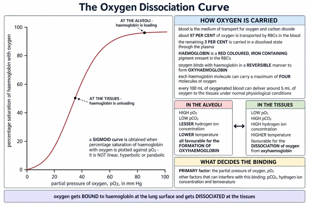
The curve is highly useful in studying the effect of factors like and hydrogen ion concentration on the binding of oxygen with haemoglobin. That is the chapter's stated purpose for it - it is not decoration, it is the tool with which the modifying factors are read off.
The shape is S-shaped, not a straight line. A straight line would mean saturation rises in step with all the way; the S says something more interesting is happening, and the reason is taken up in the solved examples.
[NEET Important] Get the axes the right way round. Percentage saturation of haemoglobin with oxygen is plotted against , not the other way about, and the curve that results is sigmoid. Options offering linear, hyperbolic or parabolic are all standing distractors. The primary factor is ; carbon dioxide, hydrogen ion concentration and temperature are the other three - an item that calls the primary factor is testing exactly that word.
The Two Opposed Sets - Alveoli Against Tissues
This is the most examined comparison in the chapter. The chapter gives four conditions at each of two places, and the two lists are exact opposites of each other.
| Condition | In the alveoli | In the tissues |
|---|---|---|
| Partial pressure of oxygen, | high | low |
| Partial pressure of carbon dioxide, | low | high |
| Hydrogen ion concentration | lesser | high |
| Temperature | lower | higher |
| What the conditions favour | the formation of oxyhaemoglobin | the dissociation of oxygen from oxyhaemoglobin |
In the chapter's own words: in the alveoli, where there is high , low , lesser hydrogen ion concentration and lower temperature, the factors are all favourable for the formation of oxyhaemoglobin; whereas in the tissues, where low , high , high hydrogen ion concentration and higher temperature exist, the conditions are favourable for dissociation of oxygen from the oxyhaemoglobin.
Learn them as one set and its mirror image. If you can write the alveolar column, the tissue column is every entry flipped. Trying to memorise eight separate facts is how students lose this mark.
This clearly indicates that oxygen gets bound to haemoglobin at the lung surface and gets dissociated at the tissues.
That single sentence is the conclusion of the whole topic, and it is exactly what the body needs - loading where the air is, unloading where the cells are.
And the amount delivered
Every 100 mL of oxygenated blood can deliver around 5 mL of oxygen to the tissues under normal physiological conditions.
Note the qualifier "under normal physiological conditions". Blood leaves the lungs nearly saturated and comes back still carrying a good deal of oxygen; only about 5 mL out of every 100 mL is actually handed over at rest, which is why there is a reserve to draw on during exercise.
[NEET Important] The four-condition table is asked as a set, and always with one entry flipped. The commonest planted error is "high hydrogen ion concentration in the alveoli" or "lower temperature in the tissues". Remember the delivery figure too - 5 mL of oxygen per 100 mL of oxygenated blood - and do not confuse it with 4 mL, which belongs to carbon dioxide.
Quick Recap
- Blood is the medium of transport for oxygen and carbon dioxide.
- About 97 per cent of oxygen is transported by RBCs; the remaining 3 per cent is carried in a dissolved state through the plasma.
- Haemoglobin is a red coloured, iron containing pigment present in the RBCs.
- Oxygen binds with haemoglobin in a reversible manner to form oxyhaemoglobin.
- Each haemoglobin molecule can carry a maximum of four molecules of oxygen.
- Binding of oxygen with haemoglobin is primarily related to .
- The other factors that can interfere with this binding are , hydrogen ion concentration and temperature.
- A sigmoid curve is obtained when percentage saturation of haemoglobin with oxygen is plotted against ; it is called the oxygen dissociation curve.
- The curve is highly useful in studying the effect of factors like and hydrogen ion concentration on the binding of oxygen with haemoglobin.
- In the alveoli: high , low , lesser hydrogen ion concentration, lower temperature - all favourable for the formation of oxyhaemoglobin.
- In the tissues: low , high , high hydrogen ion concentration, higher temperature - favourable for the dissociation of oxygen from oxyhaemoglobin.
- Oxygen gets bound to haemoglobin at the lung surface and gets dissociated at the tissues.
- Every 100 mL of oxygenated blood can deliver around 5 mL of oxygen to the tissues under normal physiological conditions.
Solved Examples
Question 1
Q. How is oxygen carried in the blood, and in what proportions?
Answer. About 97 per cent of oxygen is transported by RBCs in the blood, bound to haemoglobin. The remaining 3 per cent is carried in a dissolved state through the plasma.
Question 2
Q. Describe haemoglobin in the chapter's own words.
Answer. A red coloured, iron containing pigment present in the RBCs. All three parts of that description carry marks.
Question 3
Q. What is oxyhaemoglobin, and why does the binding have to be reversible?
Answer. Oxyhaemoglobin is the compound formed when oxygen binds with haemoglobin. The binding is reversible. It has to be, because haemoglobin must pick oxygen up at the lung surface and release it again at the tissues - a permanent bond would carry oxygen around the body and never deliver it.
Question 4
Q. How many molecules of oxygen can one molecule of haemoglobin carry?
Answer. A maximum of four molecules of oxygen, one for each of its four haem groups.
Question 5
Q. Which factor primarily governs the binding of oxygen with haemoglobin, and which are the others?
Answer. The binding is primarily related to the partial pressure of oxygen, . The other factors which can interfere with this binding are the partial pressure of carbon dioxide, the hydrogen ion concentration and the temperature. One primary factor and three modifiers - the word primarily is doing the ranking.
Question 6
Q. Define oxygen dissociation curve. Can you suggest any reason for its sigmoidal pattern? This is one of the chapter-end exercises.
Answer. The oxygen dissociation curve is the curve obtained when the percentage saturation of haemoglobin with oxygen is plotted against the partial pressure of oxygen, . The curve obtained is sigmoid, that is S-shaped, and it is highly useful in studying the effect of factors like and hydrogen ion concentration on the binding of oxygen with haemoglobin.
Why the shape is sigmoid.
- Haemoglobin has four haem groups and binds a maximum of four molecules of oxygen.
- The binding is co-operative. The binding of the first oxygen molecule changes the shape of the haemoglobin molecule so that the remaining sites bind oxygen more readily.
- At very low the binding is slow to start, because the first oxygen molecule is the hardest one to attach - so the curve begins almost flat.
- Once binding has begun, each further oxygen goes on more easily, and saturation climbs steeply - that is the steep middle of the S.
- At high nearly all the sites are already filled, so extra pressure adds very little - the curve flattens off at the top.
A flat start, a steep middle and a flat top give the S shape.
And the shape is useful, not accidental.
- The steep middle means a small fall in at the tissues unloads a large amount of oxygen - exactly where it is wanted.
- The flat top means saturation stays nearly complete even if the alveolar drops somewhat, so loading in the lungs is protected against small changes.
Question 7
Q. List the four conditions in the alveoli and say what they favour.
Answer. High , low , lesser hydrogen ion concentration and lower temperature. All these factors are favourable for the formation of oxyhaemoglobin.
Question 8
Q. List the four conditions in the tissues and say what they favour.
Answer. Low , high , high hydrogen ion concentration and higher temperature. These conditions are favourable for the dissociation of oxygen from oxyhaemoglobin.
Question 9
Q. What is the effect of pCO2 on oxygen transport? This is one of the chapter-end exercises.
Answer. Carbon dioxide is one of the factors that interfere with the binding of oxygen with haemoglobin, and its effect is to push haemoglobin towards giving up its oxygen.
- A high favours the dissociation of oxygen from oxyhaemoglobin. This is the situation in the tissues, where low , high , high hydrogen ion concentration and higher temperature all act together, so oxygen is unloaded exactly where the cells are using it.
- A low , as in the alveoli, favours the formation of oxyhaemoglobin. With high , low , lesser hydrogen ion concentration and lower temperature, haemoglobin loads oxygen at the lung surface.
- Read on the graph, a rise in shifts the oxygen dissociation curve so that haemoglobin gives up more oxygen at the same - saturation at any given is lower than it would otherwise be.
So the sense of the effect is helpful, not harmful. A tissue that is working hard makes more carbon dioxide, and the extra carbon dioxide makes the blood hand over more oxygen to that very tissue. Carbon dioxide acts alongside hydrogen ion concentration and temperature, all three pulling the same way.
Question 10
Q. Where does oxygen get bound to haemoglobin and where does it get dissociated?
Answer. Oxygen gets bound to haemoglobin at the lung surface and gets dissociated at the tissues. The four conditions at each place explain why.
Question 11
Q. How much oxygen does the blood actually deliver to the tissues?
Answer. Every 100 mL of oxygenated blood can deliver around 5 mL of oxygen to the tissues under normal physiological conditions. The blood returning to the lungs is therefore still carrying oxygen - a reserve that can be drawn on when the tissues need more.
Question 12
Q. What is the oxygen dissociation curve used for?
Answer. It is highly useful in studying the effect of factors like and hydrogen ion concentration on the binding of oxygen with haemoglobin. Instead of listing effects in words, the curve lets you read the saturation at any and see how a change in one of those factors moves it.
Question 13
Q. A muscle during heavy exercise becomes warmer, more acidic and richer in carbon dioxide. What does this do to oxygen delivery there?
Answer. It increases it. Higher temperature, higher hydrogen ion concentration and higher are exactly the tissue conditions that favour the dissociation of oxygen from oxyhaemoglobin. So the harder the muscle works, the more readily the blood passing through it gives up oxygen - the three modifying factors all act in the same direction.
Question 14
Q. Why would a linear graph of saturation against be worse for the body than the sigmoid one?
Answer. Because a straight line would give the same small change in saturation for every equal change in . The sigmoid curve instead has a steep middle and a flat top, so a small fall in at the tissues releases a large amount of oxygen, while saturation in the lungs stays nearly complete even if alveolar falls somewhat. The S shape both protects loading and sharpens unloading.