
A Packaging Problem
We now know what DNA looks like — a long, antiparallel double helix. But here is a problem the cell has to solve. If you took all the DNA from a single mammalian cell and stretched it end to end, it would measure about 2.2 metres.
Where does that number come from? Adjacent base pairs sit 0.34 nm apart, and a diploid mammalian cell carries about 6.6 × 10⁹ bp. Multiply the two:
Now compare that with the nucleus, which is only about 10⁻⁶ m (one micrometre) across. Fitting 2.2 metres of thread inside a sphere that small is like stuffing about 150 kilometres of rope into a tennis ball — without tangling it so badly that it can never be read. The rest of this section is the cell's elegant answer.
Packaging in Prokaryotes — The Nucleoid
A prokaryote such as E. coli has no defined nucleus, yet its DNA is not left floating loosely all over the cell. It is gathered into a region called the nucleoid.
How is it held together? DNA carries many phosphate groups, so it is negatively charged. The cell uses positively charged proteins to grip this negatively charged DNA, folding it into large loops. Opposite charges attract — that simple electrostatic pull keeps the loops organised and compact.
So even without a nucleus, a bacterium packages its DNA neatly. The eukaryotic version of this idea is far more elaborate, and that is where we turn next.
Histones — The Positively Charged Spools
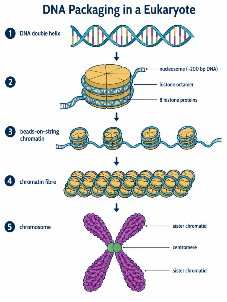
Eukaryotes use a set of small, basic proteins called histones to package their DNA. A protein gains its charge from the amino acids in it: histones are unusually rich in lysine and arginine, two basic amino acid residues whose side chains carry positive charges.
That positive charge is the whole point. The negatively charged DNA is drawn to the positively charged histones, exactly the same opposites-attract logic we saw in the prokaryotic nucleoid — just used far more systematically.
Histones do not act alone. Eight histone molecules organise themselves into a single unit called a histone octamer. This octamer is the spool around which the DNA is about to be wound.
[NEET Tip] Two facts examiners love: histones are basic / positively charged, rich in lysine and arginine; and the octamer = 8 histone molecules.
The Nucleosome and Chromatin
Now we assemble the first level of packaging. The negatively charged DNA wraps around the positively charged histone octamer, and the resulting structure is called a nucleosome. A typical nucleosome holds about 200 bp of DNA helix wound around its octamer.
A single nucleosome is just one bead. String many of them together along the DNA and you build the next level: chromatin, the thread-like, stainable (coloured) material seen inside the nucleus. The nucleosome is therefore the repeating unit of chromatin.
Under the electron microscope (EM), chromatin looks exactly like its description suggests — a row of beads on a string, the 'beads-on-string' appearance, where each bead is a nucleosome and the connecting string is the linker DNA running between them.
Build-up so far: DNA → wraps around histone octamer → nucleosome (≈200 bp) → many nucleosomes → chromatin (beads-on-string under EM).
Higher-Order Coiling — and Two Kinds of Chromatin
The beads-on-string structure is only the beginning. Chromatin is further packaged into chromatin fibres, which then coil and condense during the metaphase stage of cell division to form the compact chromosomes we can see under a light microscope.
This higher level of packaging needs extra help. A set of additional proteins, collectively called Non-histone Chromosomal (NHC) proteins, carry out this higher-order folding (histones do the first level; NHC proteins handle the levels above it).
Within a typical nucleus, not all chromatin is packed equally tightly, and this gives us two types:
| Feature | Euchromatin | Heterochromatin |
|---|---|---|
| Packing | Loosely packed | Densely packed |
| Staining | Stains light | Stains dark |
| Activity | Transcriptionally active | Transcriptionally inactive |
The link is intuitive: loosely packed DNA is open for the machinery to read (active), while tightly packed DNA is shut away and cannot easily be transcribed (inactive).
Memory Capsule — Section 3
- DNA in one mammalian cell ≈ 2.2 m long, from 6.6 × 10⁹ bp × 0.34 nm/bp; nucleus is only ≈ 10⁻⁶ m.
- Prokaryotes: DNA (negatively charged) held by positively charged proteins in large loops within the nucleoid (no defined nucleus).
- Histones: basic, positively charged, rich in lysine & arginine; 8 molecules = histone octamer.
- Nucleosome = DNA wrapped around a histone octamer, ≈ 200 bp; it is the repeating unit of chromatin.
- Chromatin → 'beads-on-string' under EM → chromatin fibres → coil/condense at metaphase → chromosomes.
- NHC proteins drive higher-order packaging.
- Euchromatin = loose, light-staining, active; Heterochromatin = dense, dark-staining, inactive.
Solved Examples — Section 3
Q1. Roughly how long is the DNA in a typical mammalian cell, and how is this length calculated?
Answer: About 2.2 metres. It is found by multiplying the total base pairs (≈ 6.6 × 10⁹ bp) by the distance between adjacent base pairs (0.34 nm), since 6.6 × 10⁹ × 0.34 × 10⁻⁹ m ≈ 2.2 m.
Q2. How is DNA packaged in a prokaryote like E. coli if it has no nucleus?
Answer: The negatively charged DNA is held by positively charged proteins and folded into large loops within a region called the nucleoid. Opposite charges attract, keeping the DNA organised without a membrane-bound nucleus.
Q3. Why are histones positively charged, and why does this matter?
Answer: Histones are rich in the basic amino acid residues lysine and arginine, whose side chains carry positive charges. This positive charge lets them bind the negatively charged DNA, which is essential for wrapping DNA into nucleosomes.
Q4. What is a nucleosome, and how much DNA does a typical one contain?
Answer: A nucleosome is the structure formed when DNA wraps around a histone octamer (8 histone molecules). A typical nucleosome contains about 200 bp of DNA helix and is the repeating unit of chromatin.
Q5. What does chromatin look like under an electron microscope, and what does each part represent?
Answer: It appears as a 'beads-on-string' structure. Each bead is a nucleosome (DNA wound on a histone octamer) and the string is the linker DNA connecting consecutive nucleosomes.
Q6. Distinguish euchromatin from heterochromatin.
Answer: Euchromatin is loosely packed, stains light and is transcriptionally active. Heterochromatin is densely packed, stains dark and is transcriptionally inactive. Loose packing leaves DNA accessible for transcription; tight packing shuts it down.