
Genetic Disorders — Two Major Categories
A genetic disorder is a disease that traces back to an abnormality in a person's DNA — either in a single gene (a Mendelian disorder) or in a whole chromosome or set of chromosomes (a chromosomal disorder).
Why these earn a chapter of their own:
- They're a major cause of illness and death in modern medicine.
- They tie Mendel's laws directly to human health.
- They put inheritance patterns, mutation types, and genetic counselling into a real-world setting.
- And they come up heavily in NEET and Board exams.
The two main categories:
A. Mendelian disorders — caused by a change in a single gene and passed down in the classic Mendelian patterns (autosomal dominant, autosomal recessive, X-linked).
- Sickle-cell anaemia (autosomal recessive)
- Thalassemia (autosomal recessive)
- Phenylketonuria / PKU (autosomal recessive)
- Haemophilia (X-linked recessive — see Section 11)
- Colour blindness (X-linked recessive — see Section 11)
B. Chromosomal disorders — caused by an abnormality in chromosome number or structure, so they affect many genes at once.
- Down syndrome (Trisomy 21)
- Klinefelter syndrome (XXY)
- Turner syndrome (XO)
These arise from non-disjunction during meiosis — chromosomes that fail to separate the way they should.
[Big idea] A Mendelian disorder is a single faulty gene, so in principle gene therapy could one day fix it. A chromosomal disorder is a whole extra or missing chromosome — and you can't simply replace one of those, which is why gene therapy can't cure it.
Mendelian Disorders — Single-Gene Diseases
1. Sickle-cell anaemia (autosomal recessive)
- Cause: a point mutation in the beta-globin gene (codon 6: GAG → GTG; Glu → Val).
- Affected genotype: HbS HbS — homozygous for the sickle allele.
- Carrier (sickle trait): HbA HbS — heterozygous, usually with no symptoms.
- Symptoms: anaemia, fatigue, painful vaso-occlusive crises, joint pain, organ damage (kidney, spleen, lungs) and a higher infection risk.
- Treatment: painkillers, blood transfusion, hydroxyurea (which raises fetal haemoglobin), and a bone marrow or stem cell transplant — the only cure available so far.
- The heterozygote advantage: people with sickle-cell trait resist malaria, which is why the HbS allele stays common wherever malaria is endemic — sub-Saharan Africa, the Mediterranean, and parts of India, tribal populations especially.
- In India: common in the tribal populations of central India — Madhya Pradesh, Maharashtra and Odisha.
2. Thalassemia (autosomal recessive)
- Cause: a fault in making the α-globin or β-globin chains of haemoglobin. Note the difference from sickle-cell, where the chain is built but mis-shaped.
- α-thalassemia: a mutation in the α-globin gene on chromosome 16; severity depends on how many of the four α-globin alleles are hit.
- β-thalassemia: a mutation in the β-globin gene on chromosome 11; ranges from minor (heterozygous, mild) to major (homozygous, severe).
- Symptoms: severe anaemia needing lifelong transfusions, bone deformities from expanded marrow, iron overload from those transfusions, and an enlarged spleen and liver.
- Treatment: regular transfusions plus iron chelation; a bone marrow transplant is the only cure.
- In India: about 3-4% of people are carriers, with the highest frequency in Punjab, Gujarat, Sindhi and Bengali populations.
3. Phenylketonuria (PKU, autosomal recessive)
- Cause: a deficiency of the enzyme phenylalanine hydroxylase (PAH), which normally converts phenylalanine to tyrosine.
- Affected: homozygous recessive (pp).
- Symptoms: phenylalanine piles up and turns toxic to the brain, causing intellectual disability; less melanin is made, so skin, hair and eyes are lighter; phenylketones in urine and sweat give a musty odour; eczema can appear too.
- It's pleiotropic — one enzyme defect, many effects.
- Diagnosis: newborn screening (the Guthrie test) at birth, which is why catching it early matters so much.
- Treatment: a lifelong phenylalanine-restricted diet — no meat, dairy, eggs or aspartame. Start it in infancy and the child can develop normal intelligence.
Why PKU stands out: it was the first genetic disease where managing the diet could prevent the disability, and it launched the whole study of inborn errors of metabolism.
Chromosomal Disorders — Whole-Chromosome Abnormalities
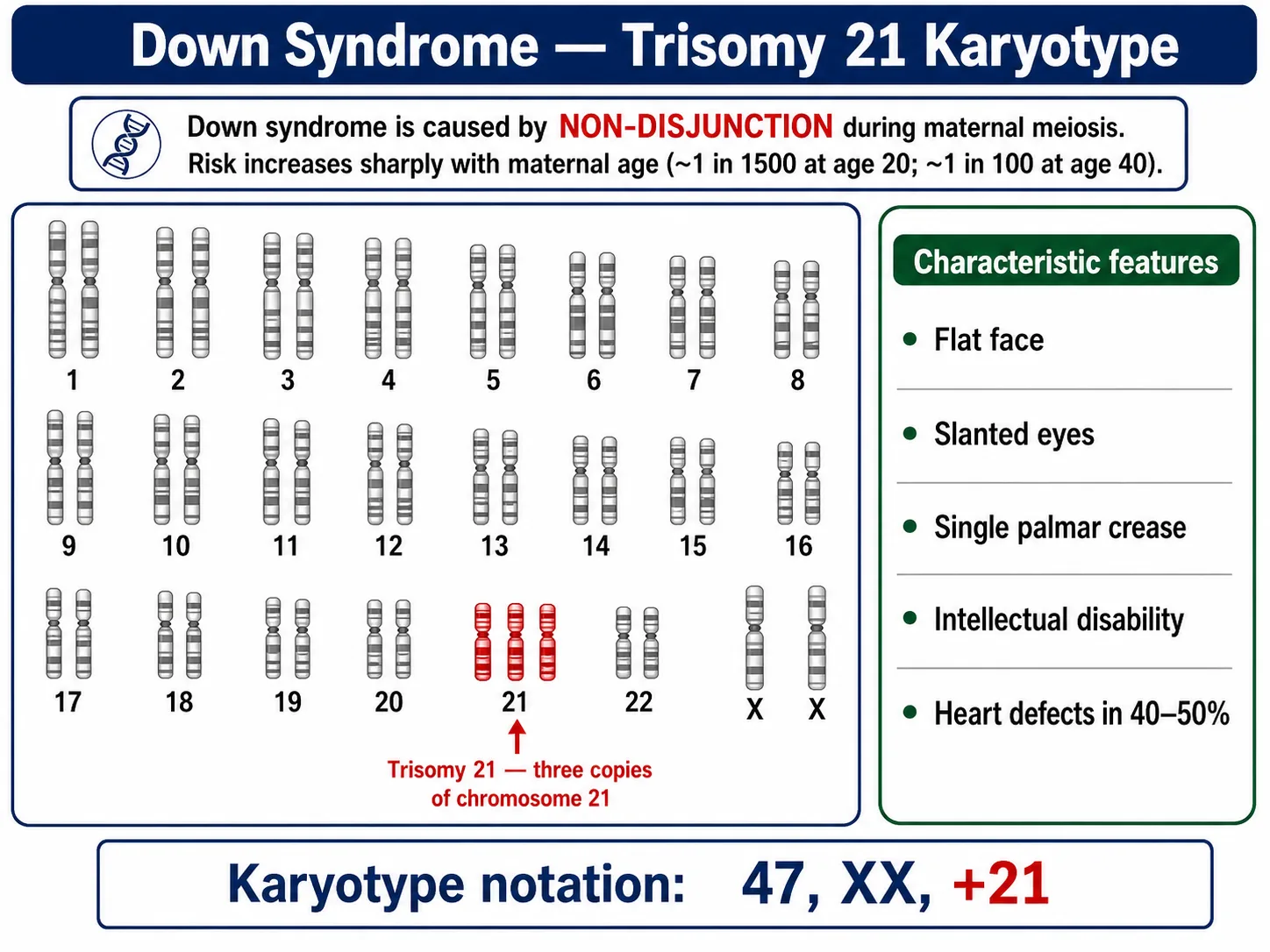
1. Down syndrome (Trisomy 21)
- Karyotype: 47, XX, +21 (female) or 47, XY, +21 (male) — three copies of chromosome 21.
- Cause: non-disjunction during maternal meiosis in about 95% of cases. The risk climbs with maternal age — minimal at 20, roughly 1 in 100 at 40, and about 1 in 30 at 45.
- Features:
- Short stature, a broad flat face, a small round head.
- Upward-slanting eyelid (palpebral) fissures and small ears.
- A single transverse palmar crease (the simian crease).
- A big, often protruding and furrowed tongue, and a partly open mouth.
- Physical, psychomotor and mental retardation — the intellectual disability ranges from moderate to severe.
- Congenital heart defects in about 40-50% of cases.
- A raised risk of leukaemia in childhood.
- Treatment: no cure. Care is supportive — physical and speech therapy, educational support, and treatment of the associated medical problems.
Why chromosome 21? It's the smallest human chromosome and carries relatively few genes, so an extra copy can be tolerated — most other trisomies are lethal before birth. That makes Down syndrome the most common viable autosomal trisomy.
History: John Langdon Down described the clinical picture in 1866; Jerome Lejeune pinned down the chromosomal cause in 1959.
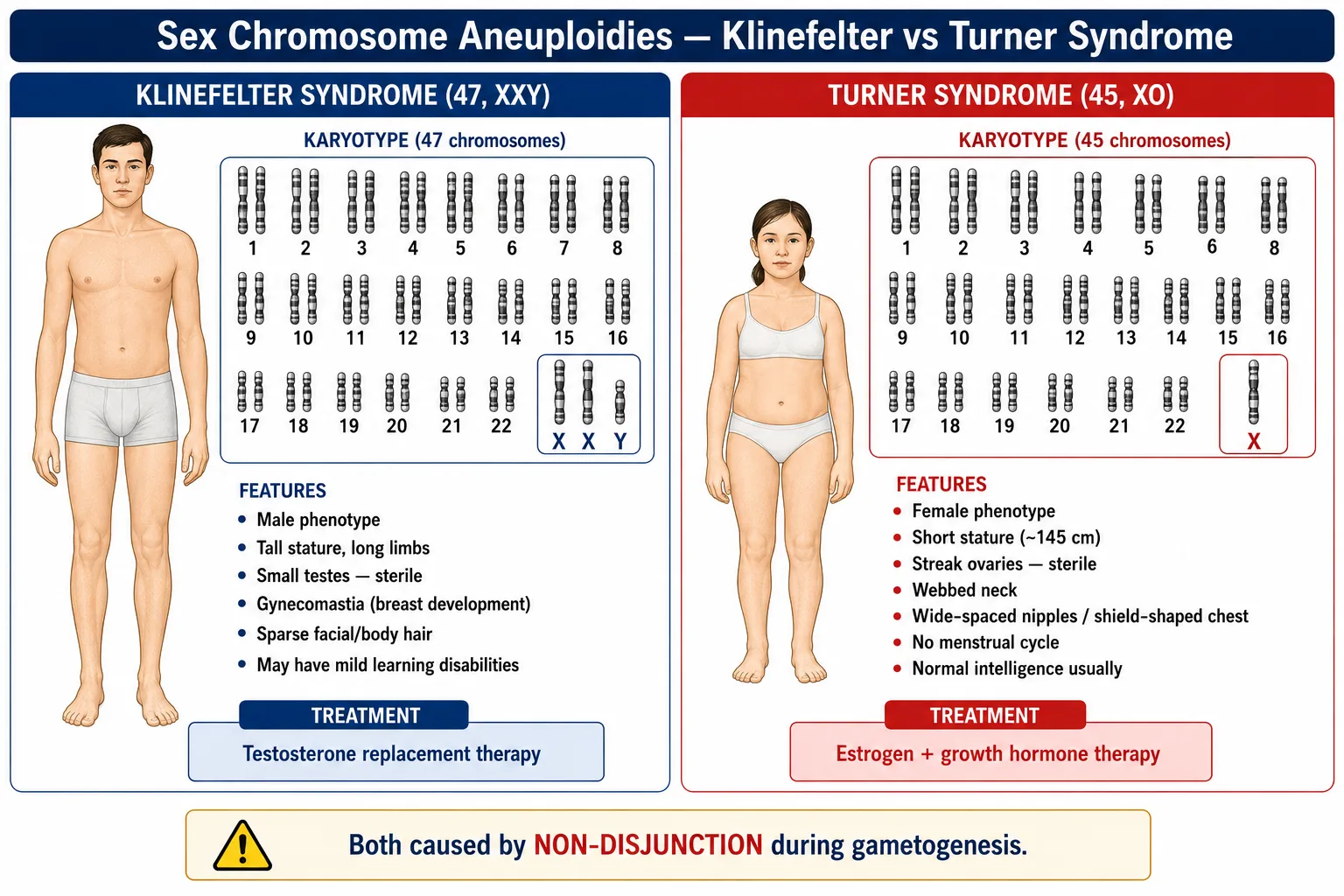
2. Klinefelter syndrome (47, XXY)
- Karyotype: 47, XXY — an extra X in a male. Rarer variants run to 48, XXXY or 49, XXXXY.
- Cause: non-disjunction in either parent's meiosis, producing an XX egg or an XY sperm.
- Phenotype: male, because the Y is present → SRY → testis formation.
- Features:
- Tall stature with overall masculine development.
- Small, underdeveloped testes and reduced testosterone.
- Sterility — no sperm production.
- Gynaecomastia (breast development), from the relative excess of oestrogen.
- Sparse body and facial hair.
- Usually normal intelligence, though mild learning difficulties are possible.
- Treatment: testosterone replacement improves muscle development and libido and reduces the gynaecomastia.
3. Turner syndrome (45, XO)
- Karyotype: 45, XO — a single X and no second sex chromosome.
- Cause: non-disjunction during gametogenesis, leaving an egg or sperm with no sex chromosome.
- Phenotype: female — with no Y there's no SRY, so development defaults to female.
- Features:
- Short stature and underdeveloped feminine characters.
- Underdeveloped ovaries (streak gonads) → sterile, no menstruation, no secondary sexual characters.
- A webbed neck (an extra fold of skin from shoulder to neck).
- Wide-spaced nipples and a shield-shaped chest.
- Cubitus valgus — forearms that angle outward.
- Usually normal intelligence, with some specific spatial-perception deficits.
- Congenital heart defects (coarctation of the aorta especially) and kidney abnormalities are common.
- Treatment: growth hormone for stature, oestrogen for secondary sex characters and bone health, and counselling about fertility — pregnancy with donor eggs is possible.
Why just these few:
- Most other trisomies and monosomies are lethal in utero; the embryo miscarries.
- Down (Trisomy 21), Klinefelter (XXY) and Turner (XO) survive because the chromosomes involved carry relatively little genetic cargo.
Memory Capsule — Section 13
Six things to lock in:
Mendelian disorders (single-gene):
Sickle-cell anaemia (autosomal recessive): a point mutation, GAG → GTG (Glu → Val) at codon 6 of β-globin. Heterozygotes resist malaria.
Thalassemia (autosomal recessive): faulty synthesis of α- or β-globin chains, transfusion-dependent. Common in Punjab, Gujarat, Sindhi and Bengali populations of India.
Phenylketonuria (PKU) (autosomal recessive): faulty phenylalanine hydroxylase. Treatable with a phenylalanine-restricted diet from infancy; causes brain damage if missed.
Cystic fibrosis (autosomal recessive): a defective chloride-transport protein makes body secretions thick and sticky — recurrent lung infections and digestive problems. On NCERT's list of common Mendelian disorders.
Chromosomal disorders (whole-chromosome):
Down syndrome (Trisomy 21): an extra chromosome 21, 47 in all. Flat face, slanting eyes, a single palmar crease, intellectual disability, often heart defects. Risk rises with maternal age. The most common autosomal trisomy.
Klinefelter syndrome (47, XXY): an extra X in a male. Masculine overall but with gynaecomastia; tall; sterile. Testosterone therapy helps.
Turner syndrome (45, XO): a single X, no second. Short stature; sterile (ovaries rudimentary); no secondary sexual characters. Oestrogen plus growth hormone therapy.
The big distinction: a Mendelian disorder is one gene defect, in principle treatable by gene therapy; a chromosomal disorder is a whole chromosome out of place, which gene therapy can't fix.
One Indian-context fact: sickle-cell is common in tribal central India; thalassemia in Punjab, Gujarat, Sindhi and Bengali populations.
Solved Examples — Section 13
Q1. Describe sickle-cell anaemia in terms of its (a) cause, (b) mode of inheritance, (c) symptoms and (d) treatment.
Answer: (a) A single point mutation in the beta-globin gene on chromosome 11 — codon 6 changes GAG → GTG, so Glu → Val at position 6 of the β-chain, giving HbS that polymerises and sickles the red cells at low oxygen. (b) Autosomal recessive: HbS HbS is affected, HbA HbS is a healthy carrier, HbA HbA is normal. (c) Anaemia, fatigue, painful vaso-occlusive crises, organ damage and frequent infections. (d) Painkillers and transfusions, hydroxyurea, and a bone marrow transplant as the only cure. Carriers resist malaria, which keeps the allele common in malaria-endemic regions.
Q2. What is phenylketonuria (PKU), and why does early dietary treatment matter so much?
Answer: PKU is an autosomal recessive disorder caused by a deficiency of phenylalanine hydroxylase, the enzyme that turns phenylalanine into tyrosine. Phenylalanine then builds up and is toxic to the developing brain, while reduced melanin lightens skin, hair and eyes and phenylketones give a musty odour. Because most brain development happens in the first few years, a phenylalanine-restricted diet started in infancy (after the Guthrie newborn screen) allows normal intelligence — but if it's missed, the brain damage is irreversible.
Q3. Describe Down syndrome in terms of its (a) chromosomal abnormality, (b) features, (c) cause and (d) treatment.
Answer: (a) Trisomy of chromosome 21 — karyotype 47, XX, +21 or 47, XY, +21; the most common viable autosomal trisomy. (b) Short stature, a flat face, upward-slanting eyes, a single palmar crease, a large protruding tongue, intellectual disability, and heart defects in 40-50% of cases. (c) Non-disjunction of chromosome 21 in meiosis (usually maternal), with risk rising sharply with maternal age; a few cases come from a Robertsonian (chr 14-21) translocation. (d) No cure — only supportive therapy, special education and treatment of the associated conditions. John Langdon Down described it in 1866; Jerome Lejeune identified the cause in 1959.
Q4. Differentiate between Klinefelter syndrome and Turner syndrome.
Answer: Klinefelter is an extra X in a male (47, XXY); Turner is a missing X in a female (45, XO). Both are sex-chromosome aneuploidies and both cause sterility.
| Feature | Klinefelter (47, XXY) | Turner (45, XO) |
|---|---|---|
| Sex | Male | Female |
| Stature | Tall | Short |
| Gonads | Small testes | Streak ovaries |
| Other | Gynaecomastia, sparse hair | Webbed neck, shield chest, cubitus valgus |
| Treatment | Testosterone | Oestrogen + growth hormone |
Q5. What causes thalassemia, and how does it differ from sickle-cell anaemia?
Answer: Thalassemia (autosomal recessive) comes from mutations that reduce the synthesis of the α-globin (chromosome 16) or β-globin (chromosome 11) chains, so the α:β ratio is imbalanced, the haemoglobin is unstable, and severe anaemia follows. The key difference: thalassemia is a quantitative defect — too little of a normal chain is made — whereas sickle-cell is a qualitative defect, where the β-chain is made but structurally wrong (Glu → Val at position 6).