
Sex-Linked Inheritance — Genes on the Sex Chromosomes
Up to now we've quietly assumed that every gene sits on an autosome — one of the 22 non-sex chromosome pairs. But plenty of genes ride on the X and Y chromosomes, and those genes follow their own rules. We call this sex-linked inheritance.
What makes it different:
- The X chromosome is large and carries roughly 800-900 genes, most of which have nothing to do with sex itself.
- The Y chromosome is tiny and carries only about 50-70 genes, mostly tied to male development.
- A female (XX) has two copies of every X-linked gene. A male (XY) has just one — there is no second X to fall back on.
- The upshot: the inheritance pattern looks different in males than in females. That asymmetry is the whole signature of sex linkage.
The categories you'll meet:
- X-linked recessive — by far the most common and the one exams love. Haemophilia and colour blindness both belong here.
- X-linked dominant — rare; hypophosphatemic rickets is an example.
- Y-linked — rarer still; passed strictly father to son, never to daughters.
The rest of this section stays with X-linked recessive inheritance, since that's what your syllabus leans on.
Why X-linked recessive disorders show up more in males:
- In a female (XX), even if one X carries the faulty allele, the other X usually carries a working copy. The normal allele covers for the defect, so she's typically a carrier but stays healthy.
- In a male (XY), if his single X carries the faulty allele, there's nothing to cover for it — the Y has no working version of the gene. He's affected straight away.
So males end up expressing recessive X-linked traits far more often than females do.
Haemophilia & Colour Blindness — Two Classic Examples
Haemophilia is a blood-clotting disorder, inherited as X-linked recessive.
- It comes from mutations in clotting-factor genes on the X chromosome — Factor VIII gives Haemophilia A, Factor IX gives Haemophilia B.
- With little or no clotting factor, an affected person bleeds for a long time even after a minor cut.
- A male carrying the allele (X^h Y) is affected.
- A female with just one copy (X^H X^h) is a carrier — usually healthy, because the X^H allele makes enough functional clotting factor.
- A female with two copies (X^h X^h) is affected, but this is rare: both parents have to pass on the allele.
The textbook pedigree: Queen Victoria (1819-1901) of England.
- She herself was a carrier (X^H X^h).
- Some of her sons — Prince Leopold among them — had haemophilia.
- Several of her daughters and granddaughters were carriers and carried the allele into the royal houses of Europe. The best-known case is Tsarevich Alexei of Russia, whose haemophilia fed into the political crisis surrounding the Russian Revolution.
Colour blindness (red-green type) is the inability to tell red from green, also X-linked recessive.
- It results from mutations in the opsin pigment genes on the X chromosome.
- It shows up in about 8% of men but only about 0.4% of women in most populations — women need two defective X alleles before they're affected.
- The inheritance pattern is the same as haemophilia.
- A colour-blind father crossed with a carrier mother gives 50% colour-blind sons and 50% colour-blind daughters (the other half of the daughters being carriers).
The rules of X-linked recessive inheritance:
- An affected father passes the allele to all his daughters (they get his X) but to none of his sons (they get his Y).
- An affected mother (X^h X^h) passes it to all her sons (they get her X). Her daughters get one X^h from her plus an X^H from a normal father, so they end up carriers.
- A carrier mother (X^H X^h) hands X^H to half her children and X^h to the other half. Sons who get X^h are affected; daughters who get it are carriers.
- The trait can skip a generation: a carrier mother passes it to an affected son, but his own son won't be affected — that grandson gets his X from his mother, not from his father.
[NEET high-yield] Father-to-son transmission of an X-linked trait is impossible — a son gets the Y, not the X, from his father. If a pedigree shows it, the trait can't be X-linked.
Pedigree Analysis — Reading a Family Tree
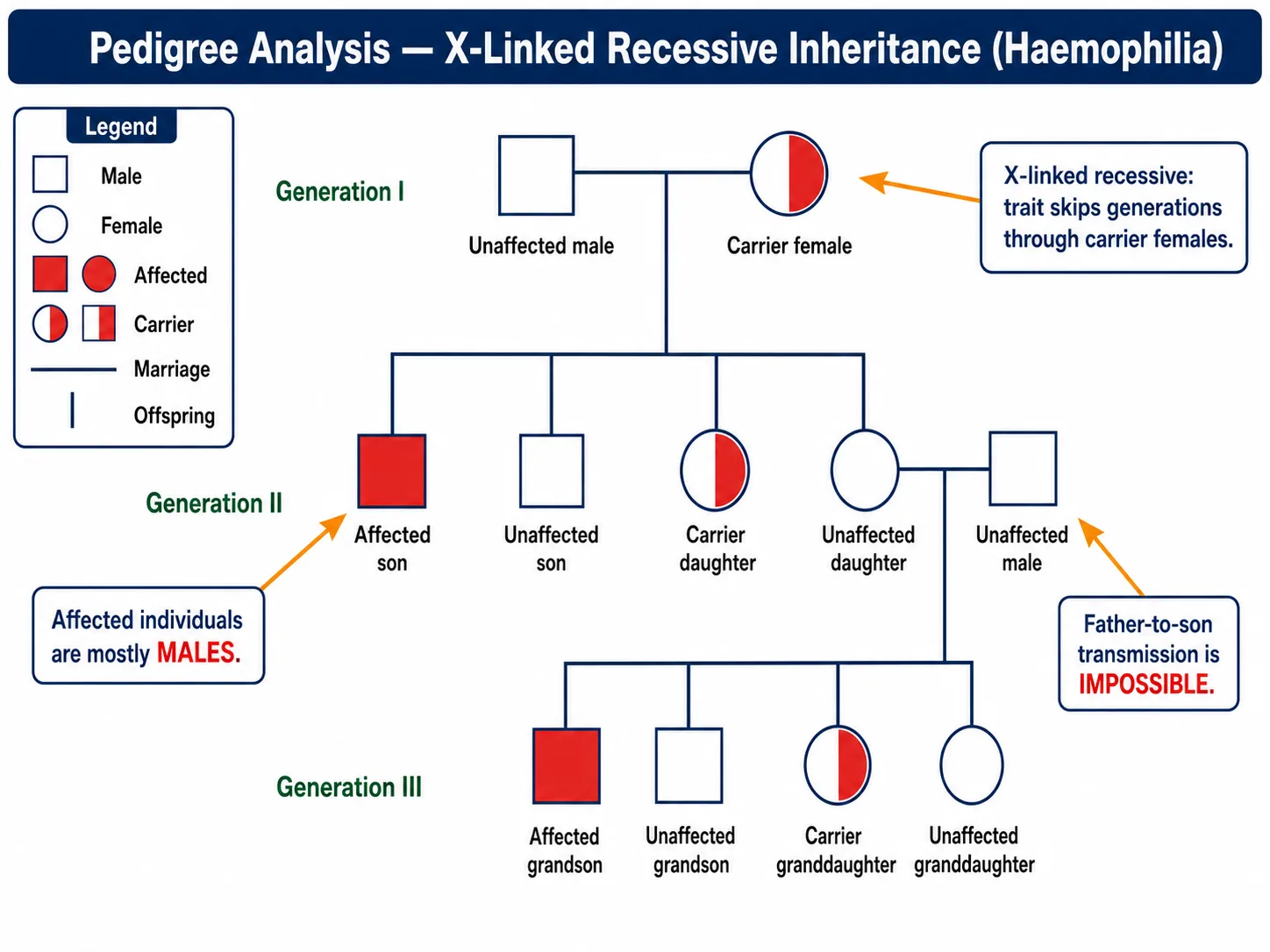
A pedigree is just a family tree drawn to track how a trait moves through several generations. Genetic counsellors lean on it constantly, and once you know the symbols, you can read one at a glance.
The standard symbols:
| Symbol | Meaning |
|---|---|
| □ Square | Male |
| ○ Circle | Female |
| ■ ● Filled symbol | Affected individual (shows the trait) |
| ▢ Half-filled | Heterozygous carrier (usually X-linked recessive females) |
| Horizontal line between two symbols | Marriage / mating |
| Vertical line below | Offspring |
| Roman numerals (I, II, III) on the left | Generation number |
| Arabic numerals (1, 2, 3) below symbols | Sibling order within a generation |
Five questions to ask of any pedigree:
Dominant or recessive? Two unaffected parents with an affected child means recessive. One affected parent passing it to about half the children means dominant.
Autosomal or sex-linked? Roughly equal numbers of affected males and females point to autosomal. A heavy male bias points to X-linked recessive — especially if the trait also skips generations.
Any carriers? For recessive traits, look for healthy people who must be carrying the allele — the unaffected parents of an affected child, for instance.
Any father-to-son transmission? If you see it, the trait is not X-linked, because sons inherit the Y, not the X, from their fathers. This one test rules X-linkage out on the spot.
Does it skip generations? A trait that vanishes for a generation and resurfaces later is usually recessive — the carriers in between simply don't show it.
Worked example (haemophilia, X-linked recessive):
Say a pedigree shows:
- Generation I: an unaffected father and a carrier mother.
- Generation II: two sons (one affected, one not) and two unaffected daughters.
- Generation III: a grandson, born to one of those daughters, is affected.
Reading it:
- The Generation I mother has to be X^H X^h — she's a carrier; that's the only way she could have an affected son.
- In Generation II, the affected son is X^h Y and the unaffected son is X^H Y. Each daughter has a 50% chance of being a carrier (X^H X^h), even though both look healthy.
- In Generation III, the affected grandson must have received X^h from his mother — one of the carrier daughters.
More affected males than females, plus a trait skipping through carrier females — that combination is the fingerprint of X-linked recessive inheritance.
[Board / NEET tip] To name the inheritance pattern from a pedigree, check three things first: are males and females affected in roughly equal numbers, is there any father-to-son transmission, and do two unaffected parents produce an affected child? Those three checks rule out most of the alternatives.
Memory Capsule — Section 11
Six things to lock in:
X-linked recessive disorders are far more common in males. A male (XY) has only one X — if it's defective, there's no backup. A female (XX) has two, and usually one is normal.
Haemophilia (X-linked recessive) — a clotting-factor deficiency. The classic case is Queen Victoria's descendants.
Colour blindness (X-linked recessive) — red-green difficulty. About 8% of males, only 0.4% of females.
The X-linked rules:
- An affected father makes all his daughters carriers (or affected, if a daughter also gets an X^h from her mother).
- A carrier mother gives 50% affected sons and 50% carrier daughters.
- Father-to-son transmission of an X-linked trait is impossible — sons get the Y, not the X.
Pedigree symbols: square = male, circle = female, filled = affected, half-filled = carrier, horizontal line = marriage, vertical line = offspring.
The five questions: dominant or recessive, autosomal or sex-linked, carriers present, any father-to-son transmission, does it skip generations?
The one idea to keep: X-linked recessive means males are affected more often, and the trait travels from a carrier mother to her affected son.
Solved Examples — Section 11
Q1. Why is haemophilia more common in males than in females?
Answer: Because haemophilia is X-linked recessive. A male (X^h Y) has only one X, so a single faulty allele makes him affected — the Y has no working copy to compensate. A female needs two faulty alleles (X^h X^h) to be affected, which is rare; with one she's just a healthy carrier.
Q2. A colour-blind man marries a woman with normal vision who is a carrier. What is the probability that their (a) first daughter is colour-blind, (b) first son is colour-blind?
Answer: 50% each. Cross X^c Y × X^C X^c:
| X^C | X^c | |
|---|---|---|
| X^c | X^C X^c (carrier daughter) | X^c X^c (colour-blind daughter) |
| Y | X^C Y (normal son) | X^c Y (colour-blind son) |
Half the daughters and half the sons are colour-blind.
Q3. Explain the principles of pedigree analysis. What symbols are used?
Answer: A pedigree charts a trait across generations of a family. Square = male, circle = female, filled = affected, half-filled = carrier; a horizontal line marks a marriage, a vertical line the offspring; Roman numerals number the generations and Arabic numerals the siblings. From the chart you work out whether the trait is dominant or recessive, autosomal or sex-linked, who the carriers are, and the likely genotypes — the backbone of genetic counselling.
Q4. A man with haemophilia marries a woman whose family has no history of it. Give the genotypes of the parents and of their sons and daughters.
Answer: Father X^h Y, mother X^H X^H. Cross X^h Y × X^H X^H:
| X^H | X^H | |
|---|---|---|
| X^h | X^H X^h | X^H X^h |
| Y | X^H Y | X^H Y |
All daughters are carriers (X^H X^h) and all sons are normal (X^H Y). The trait resurfaces later through the carrier daughters' sons.
Q5. In a pedigree, two unaffected parents have an affected son, there is no father-to-son transmission, and the trait skips generations through unaffected females. Identify the inheritance pattern and justify it.
Answer: X-linked recessive. Unaffected parents with an affected child means recessive; no father-to-son transmission rules out autosomal and points to the X; and the skipping through healthy carrier females (X^H X^h) clinches it. Haemophilia, colour blindness and Duchenne muscular dystrophy all fit this pattern.