
The Four Parts of the Human Excretory System
In humans the excretory system consists of a pair of KIDNEYS, one pair of URETERS, a URINARY BLADDER and a URETHRA. Learn that as a list of four, because it is asked as a list.
- A pair of KIDNEYS - the organs that actually make the urine.
- One pair of URETERS - one tube leaving each kidney.
- A URINARY BLADDER - a single sac in which urine is stored.
- A URETHRA - the single tube through which urine finally leaves the body.
Count as you read: two, two, one, one. The kidneys and the ureters are paired; the bladder and the urethra are single. An option list that offers you "a pair of urethras" or "two urinary bladders" is testing exactly that.
Where the kidneys sit. Kidneys are reddish brown, bean shaped structures, situated between the levels of the LAST THORACIC and the THIRD LUMBAR vertebra, close to the DORSAL INNER WALL of the abdominal cavity.
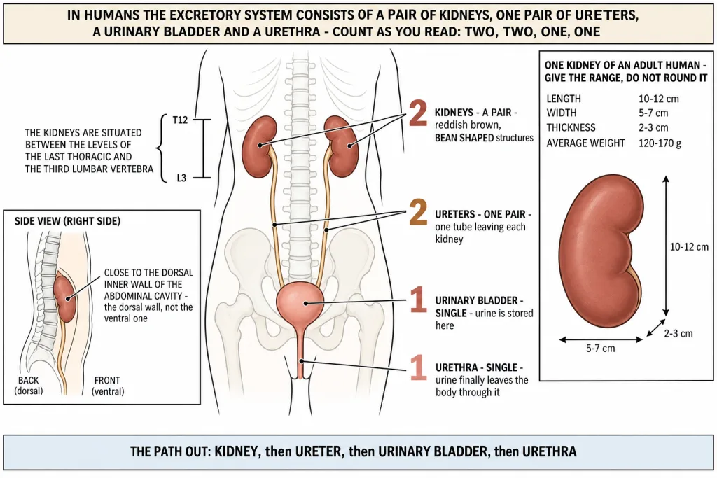
How big a kidney is. The chapter gives four figures for an adult human kidney, and every one of them is a range. Reproduce the range - do not round it and do not average it.
| Measurement | Value in an adult human |
|---|---|
| Length | 10-12 cm |
| Width | 5-7 cm |
| Thickness | 2-3 cm |
| Average weight | 120-170 g |
The three lengths fall in a tidy order - 10-12, then 5-7, then 2-3 - so if you remember that the kidney is longest top to bottom and thinnest front to back, you can rebuild the table from one figure.
[NEET Important] The vertebral levels are asked word for word: LAST THORACIC to THIRD LUMBAR. The usual distractor swaps in "first thoracic" or "last lumbar". The other examined word is DORSAL - the kidneys lie against the dorsal inner wall of the abdominal cavity, not the ventral wall.
Working In From the Outside - Capsule, Hilum, Pelvis and Calyces
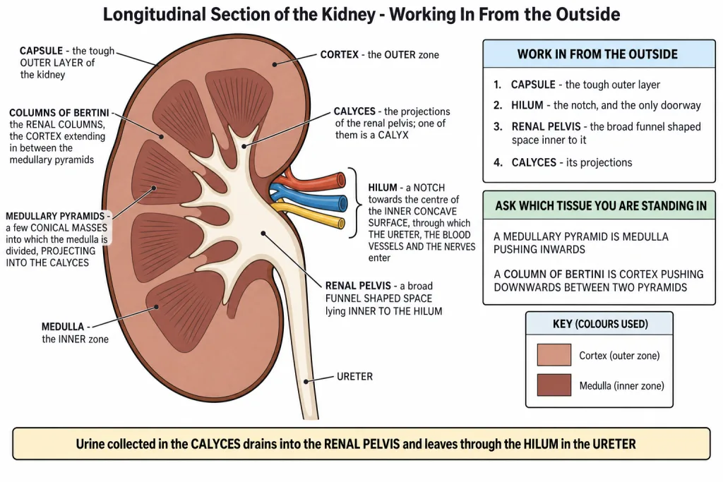
The outer layer of the kidney is a tough CAPSULE. That is the wrapping, and it is the first thing you meet coming in from outside.
Towards the centre of the inner concave surface of the kidney is a notch called the HILUM, through which the ureter, blood vessels and nerves enter. The kidney is bean shaped, so one side is convex and the other is concave - the hilum is the dent in the concave side, and it is the only doorway.
Inner to the hilum is a broad funnel shaped space called the RENAL PELVIS, with projections called CALYCES (singular: calyx).
| Structure | The one fact it is asked on |
|---|---|
| Capsule | the tough outer layer of the kidney |
| Hilum | the notch on the inner concave surface; the ureter, blood vessels and nerves enter through it |
| Renal pelvis | the broad funnel shaped space inner to the hilum |
| Calyces | the projections of the renal pelvis (singular: calyx) |
Read that inward order once more: capsule, then hilum, then renal pelvis, then calyces. Urine collected in the calyces drains into the renal pelvis and leaves through the hilum in the ureter.
[NEET Important] Hilum and renal pelvis are routinely swapped. The hilum is a NOTCH - a doorway in the wall. The renal pelvis is a SPACE - the broad funnel lying inner to that doorway. And the thing that enters at the hilum is not just the ureter - the ureter, the blood vessels AND the nerves all pass through it.
Inside the Kidney - Cortex, Medulla, Pyramids and the Columns of Bertini
Inside the kidney there are two zones - an outer CORTEX and an inner MEDULLA. That single sentence carries a whole set of later questions, because every part of the nephron has to be placed in one zone or the other.
The medulla is divided into a few conical masses, the MEDULLARY PYRAMIDS, projecting into the calyces. So the pyramids are medullary tissue pointing inwards, and their tips sit in the cup shaped calyces that drain them.
The cortex extends in between the medullary pyramids as renal columns called COLUMNS OF BERTINI.
| Structure | The one fact it is asked on |
|---|---|
| Cortex | the outer zone of the kidney |
| Medulla | the inner zone of the kidney |
| Medullary pyramids | a few conical masses into which the medulla is divided, projecting into the calyces |
| Columns of Bertini | the renal columns - extensions of the CORTEX running down between the medullary pyramids |
Hold the two apart by asking which tissue you are standing in. A pyramid is medulla pushing inwards. A column of Bertini is cortex pushing downwards between two pyramids. They interlock like fingers, which is why a diagram question can point at either one.
[NEET Important] "Name the cortical portions projecting between the medullary pyramids" is a set question, and the answer is the renal columns, the COLUMNS OF BERTINI. The distractor offered is almost always "medullary pyramids" itself, or "calyces". Remember that the columns of Bertini belong to the cortex, not to the medulla, even though they lie deep inside the kidney.
Quick Recap
- In humans the excretory system consists of a pair of KIDNEYS, one pair of URETERS, a URINARY BLADDER and a URETHRA.
- Kidneys are reddish brown, bean shaped structures.
- They are situated between the levels of the LAST THORACIC and the THIRD LUMBAR vertebra, close to the DORSAL INNER WALL of the abdominal cavity.
- Each kidney of an adult human measures 10-12 cm in length, 5-7 cm in width and 2-3 cm in thickness, with an average weight of 120-170 g.
- Towards the centre of the inner concave surface is a notch called the HILUM, through which the ureter, blood vessels and nerves enter.
- Inner to the hilum is a broad funnel shaped space called the RENAL PELVIS, with projections called CALYCES (singular: calyx).
- The outer layer of the kidney is a tough CAPSULE.
- Inside the kidney there are two zones - an outer CORTEX and an inner MEDULLA.
- The medulla is divided into a few conical masses, the MEDULLARY PYRAMIDS, projecting into the calyces.
- The cortex extends in between the medullary pyramids as renal columns called COLUMNS OF BERTINI.
Solved Examples
Question 1
Q. Name the parts that make up the human excretory system.
Answer. A pair of kidneys, one pair of ureters, a urinary bladder and a urethra. The kidneys and ureters are paired; the bladder and the urethra are single.
Question 2
Q. Describe the colour and shape of the kidneys.
Answer. Kidneys are reddish brown, bean shaped structures. The bean shape matters for the next question - one side is convex and the other is concave, and the notch called the hilum sits in the concave side.
Question 3
Q. Where exactly are the kidneys placed in the body?
Answer. They are situated between the levels of the last thoracic and the third lumbar vertebra, close to the dorsal inner wall of the abdominal cavity. Both halves of that answer are marked - the vertebral levels and the dorsal inner wall.
Question 4
Q. Give the dimensions and the weight of a kidney of an adult human.
Answer. 10-12 cm in length, 5-7 cm in width and 2-3 cm in thickness, with an average weight of 120-170 g. Give the ranges as they stand - a single rounded value loses the mark.
Question 5
Q. What is the hilum, and what passes through it?
Answer. The hilum is a notch towards the centre of the inner concave surface of the kidney. The ureter, blood vessels and nerves enter through it. It is the kidney's only doorway.
Question 6
Q. What lies inner to the hilum, and what are its projections called?
Answer. Inner to the hilum is a broad funnel shaped space called the renal pelvis. Its projections are called calyces, and one of them is a calyx.
Question 7
Q. What is the outer layer of the kidney called?
Answer. A tough capsule.
Question 8
Q. Name the two zones seen inside a kidney.
Answer. An outer cortex and an inner medulla. Every part of the nephron is later placed in one of these two zones, so fix which is outer and which is inner now.
Question 9
Q. What are the medullary pyramids, and where do they project?
Answer. The medulla is divided into a few conical masses called the medullary pyramids, and they project into the calyces.
Question 10
Q. Name the cortical portions projecting between the medullary pyramids in the human kidney. This is one of the chapter-end exercises.
Answer. The renal columns, called the COLUMNS OF BERTINI.
- They are extensions of the cortex. The cortex is the outer zone of the kidney, and it extends in between the medullary pyramids instead of stopping at the boundary between the two zones.
- Those downward extensions running between the pyramids are the renal columns, named the columns of Bertini.
- So although a column of Bertini lies deep inside the kidney, surrounded by medulla on both sides, the tissue itself is cortical tissue, not medullary tissue. A medullary pyramid is medulla pushing inwards; a column of Bertini is cortex pushing downwards between two pyramids.
Question 11
Q. Trace the path urine takes from the kidney to the outside of the body.
Answer. Kidney - ureter - urinary bladder - urethra. Inside the kidney the urine is first collected in the calyces, drains into the renal pelvis, and leaves through the hilum in the ureter.
Question 12
Q. Distinguish between a medullary pyramid and a column of Bertini.
Answer. A medullary pyramid is one of the few conical masses into which the medulla is divided, and it projects into a calyx. A column of Bertini is a renal column - an extension of the cortex that runs down between two medullary pyramids. One is medulla, the other is cortex, and they interlock.
Question 13
Q. Which structures of the kidney can be seen only when it is cut open in a longitudinal section?
Answer. The cortex and the medulla, the medullary pyramids, the columns of Bertini, the calyces and the renal pelvis. From outside you can see only the tough capsule, the bean shape and the notch at the hilum with the ureter, blood vessels and nerves entering there.