
The Nephron - Nearly One Million Per Kidney
Each kidney has nearly ONE MILLION complex tubular structures called NEPHRONS, which are the functional units. Everything the kidney does, one nephron does in miniature, a million times over.
Each nephron has two parts - the GLOMERULUS and the RENAL TUBULE. Hold on to that two-part split, because the glomerulus is made of blood vessels and the renal tubule is not.
The glomerulus is a tuft of capillaries formed by the AFFERENT ARTERIOLE, a fine branch of the renal artery. Blood from the glomerulus is carried away by an EFFERENT ARTERIOLE.
- Afferent - the arteriole that brings blood IN and breaks up into the tuft.
- Efferent - the arteriole that carries blood AWAY from the tuft.
- Both are arterioles. A capillary bed sitting between two arterioles is unusual, and it is the reason the glomerulus can be kept under pressure.
The renal tubule begins with a double walled cup-like structure called BOWMAN'S CAPSULE, which encloses the glomerulus. Picture a soft cup with the tuft of capillaries pushed into it, so the cup wraps around the tuft in two layers.
The glomerulus along with Bowman's capsule is called the MALPIGHIAN BODY or RENAL CORPUSCLE.
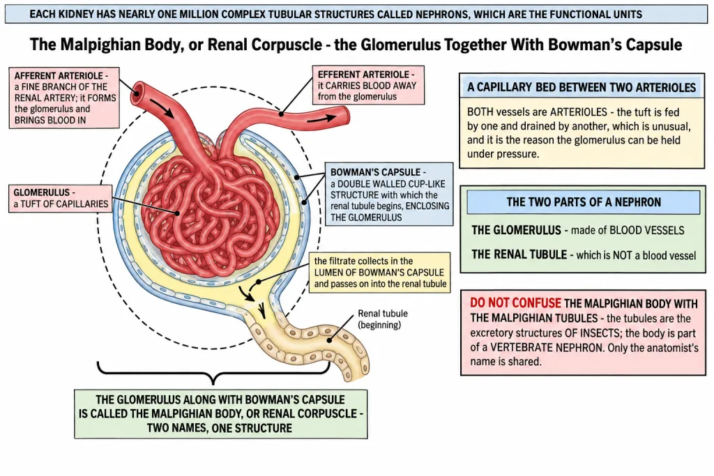
[NEET Important] Two names, one structure: MALPIGHIAN BODY and RENAL CORPUSCLE are the same thing - the glomerulus plus Bowman's capsule. Do not confuse it with the Malpighian tubules, which are the excretory structures of insects. The other examined pair is afferent against efferent: the afferent arteriole forms the glomerulus, the efferent arteriole drains it.
Walking Down the Renal Tubule
The tubule is one continuous pipe, and the chapter names its segments in order. Learn the order as a walk, not as a list of words.
Bowman's capsule -> PCT -> descending limb of Henle's loop -> ascending limb of Henle's loop -> DCT -> collecting duct -> renal pelvis.
The tubule continues as a highly coiled network, the PROXIMAL CONVOLUTED TUBULE (PCT). A hairpin shaped HENLE'S LOOP is the next part, with a DESCENDING and an ASCENDING limb. The ascending limb continues as another highly coiled region, the DISTAL CONVOLUTED TUBULE (DCT). The DCTs of many nephrons open into a straight tube called the COLLECTING DUCT, many of which converge and open into the renal pelvis through the medullary pyramids in the calyces.
| Step | Segment | Its shape or defining feature |
|---|---|---|
| 1 | Bowman's capsule | double walled cup-like structure enclosing the glomerulus |
| 2 | Proximal convoluted tubule (PCT) | a highly coiled network |
| 3 | Descending limb of Henle's loop | the arm going down into the medulla |
| 4 | Ascending limb of Henle's loop | the arm coming back up; the loop as a whole is hairpin shaped |
| 5 | Distal convoluted tubule (DCT) | another highly coiled region |
| 6 | Collecting duct | a straight tube that many DCTs open into |
| 7 | Renal pelvis | reached by many collecting ducts converging through the medullary pyramids in the calyces |
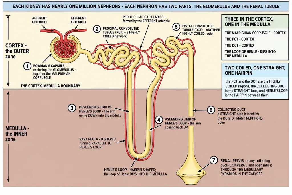
Two of the seven are coiled and one is straight. The PCT and the DCT are the highly coiled regions; the collecting duct is the straight tube. Henle's loop is the hairpin between them.
[NEET Important] The order itself is a question. "Which part follows the ascending limb?" is answered DCT; "which part do the DCTs open into?" is answered the collecting duct; and the collecting ducts open into the renal pelvis through the medullary pyramids in the calyces - not straight into the ureter.
Which Part Sits Where - Cortex or Medulla
This is a direct one-mark question, and it has only two possible answers for any part you are given.
The Malpighian corpuscle, the PCT and the DCT are situated in the CORTICAL region, whereas the loop of Henle dips into the MEDULLA.
| Part of the nephron | Where it is situated |
|---|---|
| Malpighian corpuscle (glomerulus with Bowman's capsule) | cortex |
| Proximal convoluted tubule (PCT) | cortex |
| Distal convoluted tubule (DCT) | cortex |
| Loop of Henle | dips into the MEDULLA |
Three in the cortex, one in the medulla. The loop is the only part of the nephron proper that leaves the outer zone, which is why the medulla can be given a job the cortex cannot do.
How far the loop dips is what separates the two kinds of nephron.
| CORTICAL nephrons | JUXTA MEDULLARY nephrons | |
|---|---|---|
| How common | the majority of nephrons | some of the nephrons |
| Loop of Henle | too short, and extends only very little into the medulla | very long, and runs deep into the medulla |
| Vasa recta | absent or highly reduced | present, running parallel to the long loop |
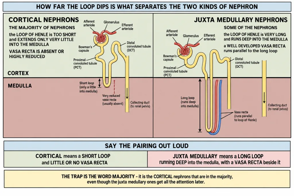
[NEET Important] Say the pairing out loud: CORTICAL means SHORT loop and little or no vasa recta; JUXTA MEDULLARY means LONG loop running deep into the medulla. The trap is the word "majority" - it is the cortical nephrons that are in the majority, even though the juxta medullary ones get all the attention later in the chapter.
The Blood Route - Peritubular Capillaries and Vasa Recta
The efferent arteriole emerging from the glomerulus forms a fine capillary network around the renal tubule called the PERITUBULAR CAPILLARIES.
A minute vessel of this network runs parallel to Henle's loop forming a U shaped VASA RECTA.
Vasa recta is absent or highly reduced in cortical nephrons.
The blood route, in order: renal artery -> afferent arteriole -> glomerulus -> efferent arteriole -> peritubular capillaries and vasa recta.
Now put the two routes side by side, because students merge them. There are two tubes running alongside each other - one carrying filtrate and one carrying blood.
| The filtrate route (the tubule) | The blood route (the vessels) | |
|---|---|---|
| Starts at | Bowman's capsule | the renal artery |
| Runs through | PCT, descending limb, ascending limb, DCT | afferent arteriole, glomerulus, efferent arteriole, peritubular capillaries |
| Alongside the loop | the descending and ascending limbs of Henle's loop | the U shaped vasa recta |
| Ends at | the collecting duct and then the renal pelvis | it drains away in the renal venous system |
The vasa recta is a blood vessel, not a part of the tubule. It only looks like part of the loop because it is shaped like one and lies right beside it.
[NEET Important] "Vasa recta is absent or highly reduced in cortical nephrons" is the most examined negative in this section. Learn it together with its partner - the cortical nephron has a short loop, so there is little for a vasa recta to run alongside, while the juxta medullary nephron has a long loop and a vasa recta beside it. The other trap is the origin: the vasa recta and the peritubular capillaries come from the EFFERENT arteriole, not the afferent one.
Quick Recap
- Each kidney has nearly ONE MILLION complex tubular structures called NEPHRONS, which are the functional units.
- Each nephron has two parts - the GLOMERULUS and the RENAL TUBULE.
- The glomerulus is a tuft of capillaries formed by the AFFERENT ARTERIOLE, a fine branch of the renal artery. Blood from the glomerulus is carried away by an EFFERENT ARTERIOLE.
- The renal tubule begins with a double walled cup-like structure called BOWMAN'S CAPSULE, which encloses the glomerulus.
- The glomerulus along with Bowman's capsule is called the MALPIGHIAN BODY or RENAL CORPUSCLE.
- The tubule continues as a highly coiled network, the PROXIMAL CONVOLUTED TUBULE (PCT).
- A hairpin shaped HENLE'S LOOP is the next part, with a DESCENDING and an ASCENDING limb.
- The ascending limb continues as another highly coiled region, the DISTAL CONVOLUTED TUBULE (DCT).
- The DCTs of many nephrons open into a straight tube called the COLLECTING DUCT, many of which converge and open into the renal pelvis through the medullary pyramids in the calyces.
- Order of the tubule: Bowman's capsule - PCT - descending limb - ascending limb - DCT - collecting duct - renal pelvis.
- The Malpighian corpuscle, the PCT and the DCT are situated in the CORTICAL region, whereas the loop of Henle dips into the MEDULLA.
- CORTICAL nephrons are the majority; in them the loop of Henle is too short and extends only very little into the medulla.
- JUXTA MEDULLARY nephrons are some of the nephrons; in them the loop of Henle is very long and runs deep into the medulla.
- The efferent arteriole emerging from the glomerulus forms a fine capillary network around the renal tubule called the PERITUBULAR CAPILLARIES.
- A minute vessel of this network runs parallel to Henle's loop forming a U shaped VASA RECTA.
- Vasa recta is absent or highly reduced in cortical nephrons.
- Blood route: renal artery - afferent arteriole - glomerulus - efferent arteriole - peritubular capillaries and vasa recta.
Solved Examples
Question 1
Q. How many nephrons does each kidney have, and what are they called?
Answer. Nearly one million. They are complex tubular structures called nephrons, and they are the functional units of the kidney.
Question 2
Q. Name the two parts of a nephron.
Answer. The glomerulus and the renal tubule.
Question 3
Q. What is the glomerulus, and which vessel forms it?
Answer. The glomerulus is a tuft of capillaries. It is formed by the afferent arteriole, a fine branch of the renal artery.
Question 4
Q. Which vessel carries blood away from the glomerulus?
Answer. The efferent arteriole. So the tuft sits between two arterioles - the afferent arteriole brings blood in and the efferent arteriole takes it away.
Question 5
Q. Describe Bowman's capsule.
Answer. It is a double walled cup-like structure with which the renal tubule begins, and it encloses the glomerulus.
Question 6
Q. What is the Malpighian body, and what else is it called?
Answer. The glomerulus along with Bowman's capsule. It is also called the renal corpuscle. Do not mix it up with the Malpighian tubules of insects - only the anatomist's name is shared.
Question 7
Q. Name the parts of the renal tubule in order, from Bowman's capsule to the renal pelvis.
Answer. Bowman's capsule - proximal convoluted tubule - descending limb of Henle's loop - ascending limb of Henle's loop - distal convoluted tubule - collecting duct - renal pelvis. The PCT and the DCT are the highly coiled regions, the collecting duct is a straight tube, and Henle's loop is the hairpin in between.
Question 8
Q. Describe Henle's loop.
Answer. It is a hairpin shaped part of the tubule that comes after the PCT, and it has a descending limb and an ascending limb. The ascending limb continues as the distal convoluted tubule.
Question 9
Q. Where do the DCTs of many nephrons open, and where does that tube lead?
Answer. They open into a straight tube called the collecting duct. Many collecting ducts converge and open into the renal pelvis through the medullary pyramids in the calyces.
Question 10
Q. Which parts of the nephron lie in the cortex, and which part dips into the medulla?
Answer. The Malpighian corpuscle, the PCT and the DCT are situated in the cortical region of the kidney, whereas the loop of Henle dips into the medulla. Three parts in the cortex, one in the medulla.
Question 11
Q. Distinguish between cortical nephrons and juxta medullary nephrons.
Answer. In the majority of nephrons the loop of Henle is too short and extends only very little into the medulla - these are the cortical nephrons. In some of the nephrons the loop of Henle is very long and runs deep into the medulla - these are the juxta medullary nephrons. The vasa recta is absent or highly reduced in cortical nephrons, while the juxta medullary nephron has one running alongside its long loop.
Question 12
Q. Name a loop of capillary running parallel to the Henle's loop. This is one of the chapter-end exercises.
Answer. The VASA RECTA.
- The efferent arteriole emerging from the glomerulus forms a fine capillary network around the renal tubule, called the peritubular capillaries.
- A minute vessel of this network runs parallel to Henle's loop, forming a U shaped vasa recta. The name simply describes it - a straight vessel folded into a U beside the loop.
- Vasa recta is absent or highly reduced in cortical nephrons, because in those nephrons the loop of Henle is too short for a long capillary to run beside. It is well developed in the juxta medullary nephrons, whose loops run deep into the medulla.
- Note that the vasa recta is a blood vessel, not a part of the renal tubule. It only looks like one because it copies the shape of the loop it lies beside.
Question 13
Q. What are the peritubular capillaries, and which vessel forms them?
Answer. A fine capillary network around the renal tubule. They are formed by the efferent arteriole emerging from the glomerulus - the same vessel that drained the tuft.
Question 14
Q. Students often merge the tubule with the blood vessels beside it. How do you keep them apart?
Answer. Treat them as two tubes running side by side - one carrying filtrate and one carrying blood.
- The filtrate route is the tubule: Bowman's capsule - PCT - descending limb - ascending limb - DCT - collecting duct - renal pelvis.
- The blood route is the vessels: renal artery - afferent arteriole - glomerulus - efferent arteriole - peritubular capillaries and vasa recta.
- The glomerulus and the vasa recta belong to the blood route; Henle's loop and the DCT belong to the filtrate route. They lie against each other along their whole length, which is what lets material pass from one to the other.