
One Cardiac Cycle, Step by Step
The heart does the same thing over and over, and the chapter describes exactly one turn of it. Walk through the seven steps in order, because the order IS the answer - almost every question on this section is really asking "what happens next?"
Joint diastole - all four chambers relaxed. To begin with, all the four chambers of the heart are in a relaxed state, that is, they are in JOINT DIASTOLE. The tricuspid and bicuspid valves are OPEN, so blood from the pulmonary veins and the vena cava flows into the left and the right ventricle respectively through the left and right atria. The semilunar valves are CLOSED at this stage.
Atrial systole. The SAN now generates an action potential which stimulates both the atria to undergo a simultaneous contraction - the ATRIAL SYSTOLE. This increases the flow of blood into the ventricles by about 30 per cent.
The impulse reaches the ventricles - ventricular systole. The action potential is conducted to the ventricular side by the AVN and the AV bundle, from where the bundle of His transmits it through the entire ventricular musculature. This causes the ventricular muscles to contract - the VENTRICULAR SYSTOLE - while the atria undergo relaxation (diastole), coinciding with the ventricular systole.
The tricuspid and bicuspid valves shut. Ventricular systole increases the ventricular pressure, causing the closure of the tricuspid and bicuspid valves due to the attempted backflow of blood into the atria.
The semilunar valves are forced open. As the ventricular pressure increases further, the semilunar valves guarding the pulmonary artery (right side) and the aorta (left side) are FORCED OPEN, allowing the blood in the ventricles to flow through these vessels into the circulatory pathways.
Ventricular diastole. The ventricles now relax - the VENTRICULAR DIASTOLE - and the ventricular pressure falls, causing the closure of the semilunar valves, which prevents the backflow of blood into the ventricles.
Back to joint diastole. As the ventricular pressure declines further, the tricuspid and bicuspid valves are pushed open by the pressure in the atria exerted by the blood which was being emptied into them by the veins. Blood now moves freely into the ventricles and the heart is once again in joint diastole, and soon the SAN generates a new action potential and the events above are repeated in the same sequence.
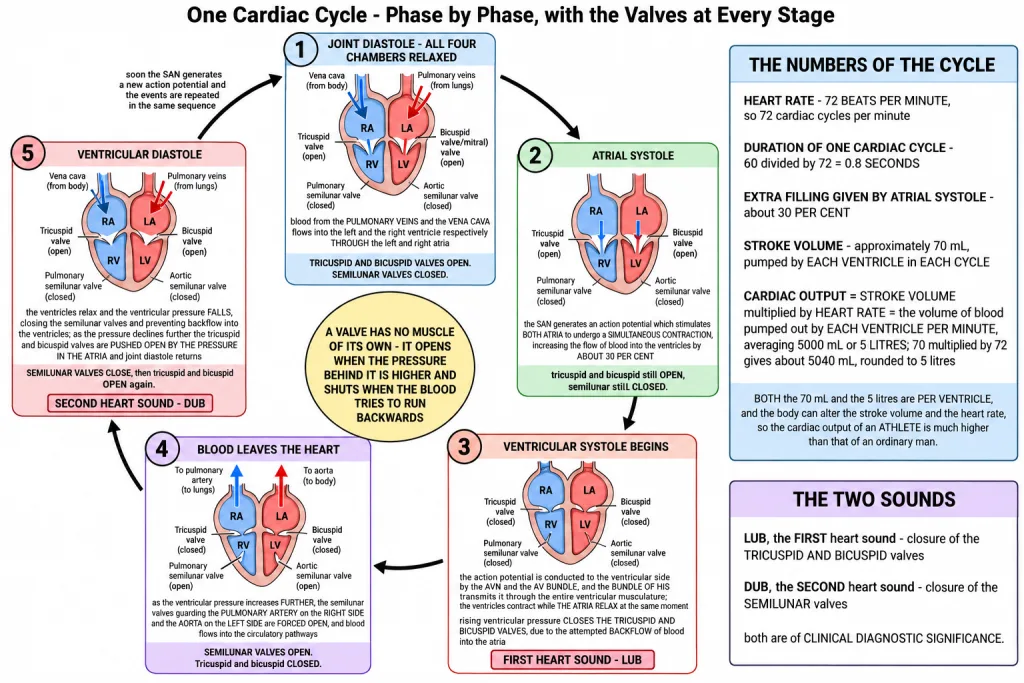
Notice what actually moves the valves: pressure, never muscle of their own. A valve opens when the pressure behind it is higher and shuts when the blood tries to run backwards. That single idea explains steps 4, 5, 6 and 7 together.
[NEET Important] The commonest question form is a freeze frame - "at this moment, which valves are open?" Hold the two extremes and you can answer any of them: in JOINT DIASTOLE the tricuspid and bicuspid valves are OPEN and the semilunar valves are CLOSED, and during VENTRICULAR SYSTOLE it is the exact reverse. The standard wrong option puts the semilunar valves open during joint diastole.
The Cardiac Cycle and Its Numbers
This sequential event in the heart, cyclically repeated, is called the CARDIAC CYCLE, and it consists of the systole and diastole of both the atria and the ventricles. That is the definition to write down, and the marks after it are for the arithmetic.
How the duration of 0.8 seconds is arrived at. Do not memorise the answer, derive it - the chapter-end exercise asks you to define the cycle, and this derivation is the mark.
- The heart beats 72 times per minute, so 72 cardiac cycles are performed per minute.
- One minute is 60 seconds, so the duration of one cycle is 60 divided by 72.
- 60 divided by 72 = 0.8, so the duration of a cardiac cycle is 0.8 SECONDS.
Stroke volume. During each cardiac cycle each ventricle pumps out approximately 70 mL of blood, which is called the STROKE VOLUME. Read the words "each ventricle" carefully - it is not the total the heart pushes out, it is what one ventricle delivers in one beat.
Cardiac output. The stroke volume multiplied by the heart rate, that is the number of beats per minute, gives the CARDIAC OUTPUT. Cardiac output can therefore be defined as the volume of blood pumped out by EACH VENTRICLE PER MINUTE, and it averages 5000 mL or 5 litres in a healthy individual.
- 70 mL multiplied by 72 beats per minute = 5040 mL per minute, and the chapter gives this rounded figure as 5000 mL or 5 litres.
And the output is not fixed. The body has the ability to alter the stroke volume as well as the heart rate, and thereby the cardiac output. The cardiac output of an athlete will be much higher than that of an ordinary man - a trained heart pushes more blood per beat and can beat faster when it has to.
| Quantity | Value |
|---|---|
| Heart rate | 72 beats per minute |
| Duration of one cardiac cycle | 0.8 seconds |
| Extra filling given by atrial systole | about 30 per cent |
| Stroke volume | approximately 70 mL per ventricle per cycle |
| Cardiac output | 5000 mL or 5 litres per minute |
[NEET Important] Every one of those five figures is asked on its own, and the wrong options are simply the other four. The two that get mixed up most are 70 mL, which is per beat, and 5000 mL, which is per minute - and both are per VENTRICLE, not for the whole heart. Cardiac output = stroke volume multiplied by heart rate is the one formula in this chapter.
The Two Heart Sounds
During each cardiac cycle two prominent sounds are produced, which can be easily heard through a stethoscope. They are not the muscle contracting - each sound is a set of valves slamming shut.
| Sound | Which valves close | Where it sits in the cycle |
|---|---|---|
| First heart sound - LUB | the TRICUSPID AND BICUSPID valves | as ventricular systole begins and the ventricular pressure shuts them (step 4) |
| Second heart sound - DUB | the SEMILUNAR valves | as ventricular diastole begins and the falling pressure shuts them (step 6) |
These sounds are of clinical diagnostic significance. A doctor listening at the chest is listening to valves, so a sound that is muffled, split or replaced by a murmur points to a valve that is not closing or opening properly.
[NEET Important] This is the most swapped pair in the whole section: LUB goes with the TRICUSPID AND BICUSPID valves, DUB goes with the SEMILUNAR valves. Fix it by the order of the cycle - the atrio-ventricular valves shut first, so the first sound is theirs. The standard wrong option hands lub to the semilunar valves, and the second commonest one says the sounds come from the contraction of the heart muscle rather than from the closure of valves.
Quick Recap
- A cardiac cycle is the sequential event in the heart which is cyclically repeated, consisting of the systole and diastole of both the atria and the ventricles.
- Joint diastole: all four chambers relaxed, tricuspid and bicuspid valves OPEN, semilunar valves CLOSED; blood from the pulmonary veins and the vena cava flows into the left and right ventricle respectively through the left and right atria.
- The SAN generates an action potential - both atria contract simultaneously (atrial systole) - and the flow of blood into the ventricles rises by about 30 per cent.
- The AVN and AV bundle carry the impulse to the ventricular side, and the bundle of His transmits it through the entire ventricular musculature - ventricular systole, while the atria relax (diastole) at the same time.
- Rising ventricular pressure closes the tricuspid and bicuspid valves because blood attempts to flow back into the atria.
- A further rise forces open the semilunar valves guarding the pulmonary artery (right side) and the aorta (left side), and blood leaves for the circulatory pathways.
- Ventricular diastole lowers the ventricular pressure, closing the semilunar valves and preventing backflow into the ventricles.
- A further fall lets atrial pressure push the tricuspid and bicuspid valves open, blood moves freely into the ventricles, joint diastole returns, and the SAN fires again.
- The heart beats 72 times per minute, so 72 cycles occur per minute; 60 divided by 72 gives a cycle duration of 0.8 seconds.
- Stroke volume: approximately 70 mL pumped by each ventricle per cardiac cycle.
- Cardiac output = stroke volume multiplied by heart rate = the volume of blood pumped out by each ventricle per minute, averaging 5000 mL or 5 litres in a healthy individual; 70 multiplied by 72 gives about 5040 mL, rounded to 5 litres.
- The body can alter stroke volume and heart rate, and so the cardiac output - an athlete's cardiac output is much higher than that of an ordinary man.
- First heart sound (lub) - closure of the tricuspid and bicuspid valves. Second heart sound (dub) - closure of the semilunar valves. Both are of clinical diagnostic significance.
Solved Examples
Question 1
Q. In one sentence, what is a cardiac cycle?
Answer. The sequential event in the heart which is cyclically repeated, and it consists of the systole and diastole of both the atria and the ventricles.
Question 2
Q. What is joint diastole, and which valves are open and which are closed during it?
Answer. Joint diastole is the state in which all the four chambers of the heart are relaxed. The tricuspid and bicuspid valves are OPEN and the semilunar valves are CLOSED. Because the atrio-ventricular valves are open, blood from the pulmonary veins and the vena cava flows into the left and the right ventricle respectively through the left and right atria.
Question 3
Q. Which structure starts the cardiac cycle, and what does atrial systole achieve?
Answer. The SAN generates an action potential, and this stimulates both the atria to undergo a simultaneous contraction - the atrial systole. It increases the flow of blood into the ventricles by about 30 per cent, topping up ventricles that were already filling passively.
Question 4
Q. Trace the path of the action potential from the atria to the whole of the ventricular muscle.
Answer. The action potential is conducted to the ventricular side by the AVN and the AV bundle, and from there the bundle of His transmits it through the entire ventricular musculature. The result is ventricular systole - the contraction of the ventricular muscles.
Question 5
Q. What are the atria doing while the ventricles are contracting?
Answer. They are relaxing. The atria undergo relaxation (diastole) coinciding with the ventricular systole - the two happen at the same time, which is why a single cycle contains a systole and a diastole for both pairs of chambers.
Question 6
Q. Why do the tricuspid and bicuspid valves close, and what would happen if they did not?
Answer. Ventricular systole increases the ventricular pressure, and the blood attempts to flow back into the atria, which closes the tricuspid and bicuspid valves. If they failed to close, blood would be pushed backwards into the atria instead of into the arteries, and the ventricle would waste its work.
Question 7
Q. When are the semilunar valves forced open, and where does the blood go?
Answer. As the ventricular pressure increases further during ventricular systole. The semilunar valves guard the pulmonary artery on the right side and the aorta on the left side, so once they are forced open the blood in the ventricles flows through these vessels into the circulatory pathways.
Question 8
Q. What closes the semilunar valves, and what does that closure prevent?
Answer. Ventricular diastole. The ventricles relax and the ventricular pressure falls, which closes the semilunar valves, and this prevents the backflow of blood into the ventricles from the aorta and the pulmonary artery.
Question 9
Q. How does the heart return to joint diastole at the end of a cycle?
Answer. As the ventricular pressure declines further, the tricuspid and bicuspid valves are pushed open by the pressure in the atria, which has been built up by the blood emptied into them by the veins. Blood then moves freely into the ventricles, and the ventricles and atria are once again in the relaxed joint diastole state. Soon the SAN generates a new action potential and the whole sequence repeats.
Question 10
Q. Write the differences between Systole and Diastole. This is one of the chapter-end exercises.
Answer. They are the two halves of every cardiac cycle - the working phase and the filling phase of a chamber.
| Feature | Systole | Diastole |
|---|---|---|
| What the muscle does | the muscle of the chamber CONTRACTS | the muscle of the chamber RELAXES |
| Pressure in the chamber | rises | falls |
| Valves | the valve leading INTO the chamber closes and the valve leading OUT is forced open - in ventricular systole the tricuspid and bicuspid close and the semilunar are forced open | the valve leading OUT closes and the valve leading IN opens - in ventricular diastole the semilunar close and the tricuspid and bicuspid are pushed open |
| Blood movement | blood is PUSHED OUT of the chamber | blood is RECEIVED INTO the chamber |
| Example in the cycle | atrial systole raises ventricular filling by about 30 per cent; ventricular systole sends blood into the aorta and pulmonary artery | joint diastole fills the ventricles from the pulmonary veins and vena cava |
Remember that the two overlap in time in the two pairs of chambers: while the ventricles are in systole the atria are in diastole.
Question 11
Q. Define a cardiac cycle and the cardiac output. This is one of the chapter-end exercises.
Answer.
Cardiac cycle. The sequential event in the heart which is cyclically repeated is called the cardiac cycle, and it consists of the systole and diastole of both the atria and the ventricles. Its duration follows from the heart rate:
- The heart beats 72 times per minute, so 72 cardiac cycles are performed per minute.
- One minute is 60 seconds, so one cycle takes 60 divided by 72.
- 60 divided by 72 = 0.8, so the duration of one cardiac cycle is 0.8 seconds.
Cardiac output. The volume of blood pumped out by each ventricle per minute. It is obtained as stroke volume multiplied by heart rate, where the stroke volume is the approximately 70 mL of blood each ventricle pumps out during one cardiac cycle.
- 70 mL multiplied by 72 beats per minute = 5040 mL per minute.
- The chapter gives this as an average of 5000 mL or 5 litres in a healthy individual.
The body can alter both the stroke volume and the heart rate, and so the cardiac output - the cardiac output of an athlete will be much higher than that of an ordinary man.
Question 12
Q. Explain heart sounds. This is one of the chapter-end exercises.
Answer. During each cardiac cycle two prominent sounds are produced, and they can be easily heard through a stethoscope. Each is made by valves closing, not by the muscle contracting.
- The FIRST heart sound, lub, is associated with the closure of the TRICUSPID AND BICUSPID valves. These shut when ventricular systole raises the ventricular pressure and blood attempts to flow back into the atria, so lub marks the start of ventricular systole.
- The SECOND heart sound, dub, is associated with the closure of the SEMILUNAR valves. These shut when ventricular diastole lets the ventricular pressure fall, which prevents the backflow of blood into the ventricles, so dub marks the start of ventricular diastole.
These sounds are of clinical diagnostic significance. Because each sound reports on a named set of valves, a changed, muffled or extra sound tells a doctor which valve is not working properly.
Question 13
Q. What is stroke volume?
Answer. The volume of blood pumped out by each ventricle during one cardiac cycle - approximately 70 mL.
Question 14
Q. A person has a heart rate of 72 beats per minute and a stroke volume of 70 mL. Work out the cardiac output.
Answer. Cardiac output = stroke volume multiplied by heart rate = 70 mL multiplied by 72 = 5040 mL per minute. This is the volume pumped out by each ventricle per minute, and it matches the average of 5000 mL or 5 litres given for a healthy individual.
Question 15
Q. Why is the cardiac output of an athlete much higher than that of an ordinary man?
Answer. Because the body has the ability to alter the stroke volume as well as the heart rate, and thereby the cardiac output. A trained heart pushes out more blood per beat and can raise its rate when the body demands more, and since cardiac output is stroke volume multiplied by heart rate, raising either one raises the output.