
The Human Circulatory System and Where the Heart Sits
The human circulatory system, also called the BLOOD VASCULAR SYSTEM, consists of a muscular chambered heart, a network of closed branching blood vessels, and blood, the fluid which is circulated. Three parts, and the question that asks you to "name the components of the human circulatory system" wants all three.
Now the heart itself, address first.
- The heart is the MESODERMALLY DERIVED organ - like every other connective and muscular structure, it comes from the middle germ layer.
- It is situated in the thoracic cavity, in between the two lungs, slightly tilted to the LEFT.
- It has the size of a clenched fist.
- It is protected by a double walled membranous bag, the PERICARDIUM, enclosing the PERICARDIAL FLUID.
Two words in that list carry marks by themselves. DOUBLE WALLED - the pericardium is not a single sheet, and the fluid lies between its two walls. And PERICARDIAL FLUID - the fluid has its own name, and it is what lets the beating heart move without friction.
[NEET Important] Mesodermal origin, thoracic cavity between the two lungs, tilted to the LEFT, the size of a clenched fist, and a double walled pericardium holding pericardial fluid - five facts, all one-liners, all asked. The wrong option usually tilts the heart to the right or calls the covering the pleura, which belongs to the lungs.
Four Chambers and Three Septa
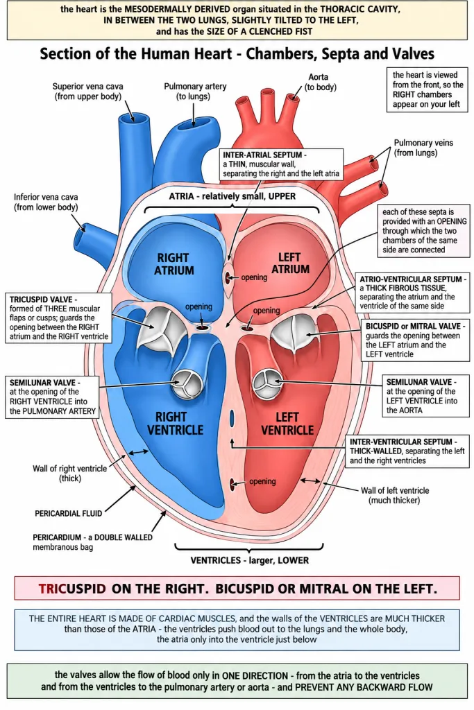
Our heart has four chambers - two relatively SMALL UPPER chambers called ATRIA and two LARGER LOWER chambers called VENTRICLES. Keep the two adjectives with the two names: atria small and upper, ventricles larger and lower.
Three septa divide those four chambers, and the adjective on each septum is examined as hard as its name.
| Septum | What it separates | Its description in the chapter |
|---|---|---|
| Inter-atrial septum | the right and the left atria | a THIN, muscular wall |
| Inter-ventricular septum | the left and the right ventricles | THICK-WALLED |
| Atrio-ventricular septum | the atrium and the ventricle of the same side | a THICK FIBROUS TISSUE |
Read the three adjectives as a set: thin between the atria, thick between the ventricles, and fibrous between an atrium and its ventricle. They are the commonest swap in this part of the chapter.
And then the sentence that keeps the heart working: each of these septa is provided with an opening through which the two chambers of the same side are connected. The septa divide the heart, but they do not seal it off - blood on the right side must still get from the right atrium into the right ventricle, and blood on the left side from the left atrium into the left ventricle.
[NEET Important] THIN inter-atrial, THICK-WALLED inter-ventricular, THICK FIBROUS atrio-ventricular. Learn the adjectives; a question that gives you "thick fibrous tissue" is asking for the atrio-ventricular septum, not the inter-ventricular one.
The Valves - and Which Side Each One Is On
Every opening in the heart is guarded by a valve, and the single most swapped pair in this chapter is the tricuspid and the bicuspid. Fix them in a table and never guess again.
| Opening | Valve guarding it | Side |
|---|---|---|
| Right atrium into the right ventricle | TRICUSPID VALVE - formed of three muscular flaps or cusps | RIGHT |
| Left atrium into the left ventricle | BICUSPID or MITRAL VALVE | LEFT |
| Right ventricle into the PULMONARY ARTERY | SEMILUNAR VALVE | RIGHT |
| Left ventricle into the AORTA | SEMILUNAR VALVE | LEFT |
Say the pairing out loud once: TRICUSPID on the RIGHT, BICUSPID on the LEFT. Tri and right for the side with three cusps; bi and left for the side with two, which is also called the mitral valve. And the openings of the right and the left ventricles into the pulmonary artery and the aorta respectively are provided with the SEMILUNAR VALVES - note the order of that sentence: right ventricle to pulmonary artery, left ventricle to aorta.
What all four valves are for is one sentence. The valves in the heart allow the flow of blood only in ONE DIRECTION - from the atria to the ventricles and from the ventricles to the pulmonary artery or aorta. These valves prevent any backward flow.
[NEET Important] Tricuspid RIGHT, bicuspid or mitral LEFT is the pairing NEET swaps most often in this chapter. The second guaranteed item is the function - one way flow, atria to ventricles and ventricles to the pulmonary artery or aorta, with no backward flow.
Cardiac Muscle and the Thickness of the Walls
The entire heart is made of CARDIAC MUSCLES. That is why it can contract on and on without tiring, and why the chapter treats the heart as a muscular organ rather than a passive bag.
And within that muscle the thickness is not even: the walls of the ventricles are much thicker than those of the atria.
The reason is what each chamber has to do. The ventricles must push blood right out of the heart - to the lungs on the right and to the whole body on the left - so they need far more muscle behind each contraction. The atria only pass blood down into the ventricle just below them, a very short journey against very little resistance, so a thinner wall is enough.
[NEET Important] The entire heart is made of cardiac muscle, and the ventricular walls are much thicker than the atrial walls. If a question asks why, the marks are for the distance and resistance against which each chamber pumps - ventricles out to the lungs and the body, atria only into the ventricle below.
Quick Recap
- The human circulatory system, also called the blood vascular system, consists of a muscular chambered heart, a network of closed branching blood vessels, and blood, the fluid which is circulated.
- The heart is the mesodermally derived organ, situated in the thoracic cavity, in between the two lungs, slightly tilted to the left, and has the size of a clenched fist.
- It is protected by a double walled membranous bag, the pericardium, enclosing the pericardial fluid.
- Four chambers: two relatively small upper chambers, the atria, and two larger lower chambers, the ventricles.
- A thin, muscular wall called the inter-atrial septum separates the right and the left atria.
- A thick-walled inter-ventricular septum separates the left and the right ventricles.
- The atrium and the ventricle of the same side are separated by a thick fibrous tissue, the atrio-ventricular septum.
- Each of these septa is provided with an opening through which the two chambers of the same side are connected.
- The opening between the RIGHT atrium and the RIGHT ventricle is guarded by the TRICUSPID valve, formed of three muscular flaps or cusps.
- The opening between the LEFT atrium and the LEFT ventricle is guarded by the BICUSPID or MITRAL valve.
- The openings of the right and the left ventricles into the pulmonary artery and the aorta respectively are provided with the SEMILUNAR valves.
- The valves allow the flow of blood only in one direction - atria to ventricles, ventricles to the pulmonary artery or aorta - and prevent any backward flow.
- The entire heart is made of cardiac muscles, and the walls of the ventricles are much thicker than those of the atria.
Solved Examples
Question 1
Q. Name the three components of the human circulatory system.
Answer. A muscular chambered heart, a network of closed branching blood vessels, and blood, the fluid which is circulated. The system is also called the blood vascular system.
Question 2
Q. From which germ layer is the heart derived?
Answer. The mesoderm - the heart is the mesodermally derived organ.
Question 3
Q. Where exactly is the human heart situated?
Answer. In the thoracic cavity, in between the two lungs, slightly tilted to the LEFT. It has the size of a clenched fist.
Question 4
Q. What covers and protects the heart, and what does it contain?
Answer. A double walled membranous bag called the PERICARDIUM, which encloses the PERICARDIAL FLUID. The fluid lets the beating heart move without rubbing against the structures around it.
Question 5
Q. Name the four chambers of the heart and give their positions and relative sizes.
Answer. Two relatively small upper chambers called ATRIA - the right atrium and the left atrium - and two larger lower chambers called VENTRICLES - the right ventricle and the left ventricle.
Question 6
Q. Name the three septa of the heart and say what each separates.
Answer.
| Septum | Separates |
|---|---|
| Inter-atrial septum - a thin, muscular wall | the right and the left atria |
| Inter-ventricular septum - thick-walled | the left and the right ventricles |
| Atrio-ventricular septum - a thick fibrous tissue | the atrium and the ventricle of the same side |
Question 7
Q. Which septum is described as a thick fibrous tissue?
Answer. The ATRIO-VENTRICULAR septum, which separates the atrium and the ventricle of the same side. The inter-atrial septum is thin and muscular and the inter-ventricular septum is thick-walled.
Question 8
Q. If the septa separate the chambers, how does blood get from an atrium to a ventricle?
Answer. Because each of these septa is provided with an opening through which the two chambers of the same side are connected. The septa divide the heart into chambers but leave a guarded opening on each side, so right atrium connects to right ventricle and left atrium to left ventricle.
Question 9
Q. Which valve guards the opening between the right atrium and the right ventricle, and what is it made of?
Answer. The TRICUSPID valve, formed of three muscular flaps or cusps.
Question 10
Q. Which valve guards the opening between the left atrium and the left ventricle?
Answer. The BICUSPID valve, also called the MITRAL valve. Remember the pairing as tricuspid on the RIGHT and bicuspid on the LEFT - it is the commonest swap in the chapter.
Question 11
Q. Which valves guard the openings of the ventricles into the great arteries?
Answer. The SEMILUNAR valves. The openings of the right and the left ventricles into the pulmonary artery and the aorta respectively are provided with the semilunar valves - right ventricle into the pulmonary artery, left ventricle into the aorta.
Question 12
Q. What is the function of the valves of the heart?
Answer. They allow the flow of blood only in ONE DIRECTION - from the atria to the ventricles, and from the ventricles to the pulmonary artery or aorta. These valves prevent any backward flow, so blood cannot slip back into the chamber it has just left.
Question 13
Q. The entire heart is made of which tissue, and why are the ventricular walls thicker than the atrial walls?
Answer. The entire heart is made of CARDIAC MUSCLES. The walls of the ventricles are much thicker than those of the atria because the ventricles have to push blood out of the heart altogether - to the lungs on the right side and to the whole body on the left - while the atria only pass blood into the ventricle just below them, a short journey against very little resistance.
Question 14
Q. A patient's mitral valve fails to close properly. Which opening leaks, and in which direction?
Answer. The opening between the LEFT atrium and the LEFT ventricle, because the bicuspid or mitral valve guards it. Since the valves prevent any backward flow, a mitral valve that will not close lets blood flow backwards from the left ventricle into the left atrium instead of moving forward into the aorta.