
The Nodal Tissue and Its Two Addresses
A specialised cardiac musculature called the NODAL TISSUE is distributed in the heart. It is still cardiac muscle, but it does a different job - instead of pumping, it generates and carries the electrical signal that tells the rest of the heart when to contract.
Two patches of it have names, and both questions about them are address questions.
| Nodal mass | Where it lies, exactly |
|---|---|
| Sino-atrial node (SAN) | a patch of nodal tissue in the RIGHT UPPER CORNER of the RIGHT ATRIUM |
| Atrio-ventricular node (AVN) | a mass of nodal tissue in the LOWER LEFT CORNER of the RIGHT ATRIUM, close to the atrio-ventricular septum |
Read the second row twice. BOTH nodes are in the RIGHT ATRIUM. The AVN is in the lower LEFT corner - but that is the lower left corner of the right atrium, not of the left atrium. That single phrase catches more students than anything else in this part of the chapter, because the word "left" is sitting inside the address of a right-sided structure.
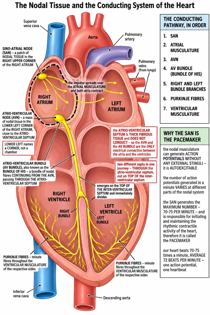
[NEET Important] SAN - right upper corner of the right atrium. AVN - lower left corner of the right atrium, close to the atrio-ventricular septum. The standard wrong option puts the AVN in the left atrium. Both nodes are in the right atrium; only their corners differ.
From the AVN to the Purkinje Fibres
The signal has to reach the ventricles, and there is exactly one road for it.
- The ATRIO-VENTRICULAR BUNDLE (AV bundle) is a bundle of nodal fibres continuing from the AVN. It is also known as the bundle of His.
- It passes through the atrio-ventricular septa to emerge on the TOP OF THE INTER-VENTRICULAR SEPTUM and immediately divides into a RIGHT and a LEFT BUNDLE.
- These branches give rise to minute fibres throughout the ventricular musculature of the respective sides, called PURKINJE FIBRES.
Now put the whole conducting system in order. Learn it as a numbered chain, because questions often give you two links and ask for the one in between.
- SAN - the signal starts here.
- Atrial musculature - the impulse spreads over both atria and makes them contract.
- AVN - picks the signal up at the lower left corner of the right atrium.
- AV bundle (bundle of His) - carries it through the atrio-ventricular septum.
- Right and left bundle branches - on top of the inter-ventricular septum, one branch to each side.
- Purkinje fibres - minute fibres spreading through the ventricular musculature.
- Ventricular musculature - the ventricles contract.
Why the road matters: the atrio-ventricular septum is a thick FIBROUS tissue and does not conduct the impulse, so the signal cannot simply cross from atrial muscle into ventricular muscle. The AVN and the AV bundle are the only electrical connection between the atria and the ventricles, and the small delay along that route is what makes the ventricles contract after the atria rather than along with them.
[NEET Important] The sequence is asked as a sequence. SAN, atrial musculature, AVN, AV bundle, right and left bundle branches, Purkinje fibres, ventricular musculature. Note that the AV bundle emerges on the top of the INTER-VENTRICULAR septum after passing through the ATRIO-VENTRICULAR septum - two different septa in one sentence, and NEET swaps them.
Autoexcitability and the Pacemaker
The nodal musculature has the ability to generate action potentials WITHOUT ANY EXTERNAL STIMULI - that is, it is AUTOEXCITABLE. No nerve has to tell it to fire. This is why a heart keeps beating with its nerve supply cut, and why the heartbeat is called myogenic rather than driven from outside.
But not every part of the nodal system fires at the same rate. The number of action potentials that could be generated in a minute varies at different parts of the nodal system.
And that is what settles the pacemaker question. The SAN can generate the maximum number of action potentials - 70-75 per minute - and is responsible for initiating and maintaining the rhythmic contractile activity of the heart. Therefore it is called the PACEMAKER. The fastest part of the system sets the rhythm that every slower part simply follows.
Two numbers, and they are deliberately the same.
- The SAN generates 70-75 action potentials per minute.
- Our heart normally beats 70-75 times in a minute, average 72 beats per minute.
They match because the second is caused by the first - each action potential from the SAN produces one heartbeat.
[NEET Important] Autoexcitable, maximum number of action potentials, 70-75 per minute, pacemaker - those four go together in every version of this question. The wrong options offer the AVN as the pacemaker, or claim the SAN needs a nerve impulse to fire, which contradicts without any external stimuli.
Quick Recap
- A specialised cardiac musculature called the nodal tissue is distributed in the heart.
- The sino-atrial node (SAN) is a patch of this tissue present in the RIGHT UPPER CORNER of the RIGHT ATRIUM.
- The atrio-ventricular node (AVN) is another mass of this tissue in the LOWER LEFT CORNER of the RIGHT ATRIUM, close to the atrio-ventricular septum.
- Both the SAN and the AVN lie in the RIGHT atrium.
- The atrio-ventricular bundle (AV bundle), also called the bundle of His, is a bundle of nodal fibres continuing from the AVN.
- It passes through the atrio-ventricular septa to emerge on the top of the inter-ventricular septum and immediately divides into a right and a left bundle.
- These branches give rise to minute fibres throughout the ventricular musculature of the respective sides, called PURKINJE FIBRES.
- The conducting pathway: SAN, atrial musculature, AVN, AV bundle, right and left bundle branches, Purkinje fibres, ventricular musculature.
- The nodal musculature can generate action potentials without any external stimuli - it is AUTOEXCITABLE.
- The number of action potentials generated in a minute varies at different parts of the nodal system.
- The SAN generates the maximum number, 70-75 per minute, and is responsible for initiating and maintaining the rhythmic contractile activity of the heart - hence it is the PACEMAKER.
- The heart normally beats 70-75 times in a minute, average 72 beats per minute.
- The atrio-ventricular septum is fibrous and does not conduct, so the AVN and the AV bundle are the only electrical connection between the atria and the ventricles.
Solved Examples
Question 1
Q. What is nodal tissue?
Answer. A specialised cardiac musculature distributed in the heart. It does not pump; it generates and conducts the impulses that set off each heartbeat.
Question 2
Q. Where exactly is the sino-atrial node located?
Answer. In the RIGHT UPPER CORNER of the RIGHT ATRIUM, as a patch of nodal tissue.
Question 3
Q. Where exactly is the atrio-ventricular node located?
Answer. In the LOWER LEFT CORNER of the RIGHT ATRIUM, close to the atrio-ventricular septum. The corner is "left", but the chamber is still the right atrium.
Question 4
Q. In which chamber do both the SAN and the AVN lie?
Answer. Both lie in the RIGHT ATRIUM. Only their corners differ - the SAN in the right upper corner, the AVN in the lower left corner near the atrio-ventricular septum.
Question 5
Q. What is the atrio-ventricular bundle, and what path does it take?
Answer. A bundle of nodal fibres continuing from the AVN, also called the bundle of His. It passes through the atrio-ventricular septa to emerge on the top of the inter-ventricular septum and immediately divides into a right and a left bundle.
Question 6
Q. What are Purkinje fibres?
Answer. The minute fibres that the right and left bundle branches give rise to throughout the ventricular musculature of the respective sides. They carry the impulse into the muscle of the ventricles themselves.
Question 7
Q. Write the conducting pathway of the heart in order.
Answer. SAN, then the atrial musculature, then the AVN, then the AV bundle (bundle of His), then the right and left bundle branches, then the Purkinje fibres, and finally the ventricular musculature.
Question 8
Q. What does autoexcitable mean, and which tissue of the heart is described that way?
Answer. The NODAL musculature is autoexcitable. It means the tissue has the ability to generate action potentials without any external stimuli - no nerve impulse is needed to start a beat.
Question 9
Q. How many action potentials can the SAN generate in a minute, and how does that compare with the rest of the nodal system?
Answer. 70-75 per minute, which is the maximum number of any part of the nodal system. The number of action potentials that could be generated in a minute varies at different parts of the nodal system, and the SAN is the fastest of them.
Question 10
Q. How many times does the human heart normally beat in a minute?
Answer. 70-75 times in a minute, with an average of 72 beats per minute.
Question 11
Q. Sino-atrial node is called the pacemaker of our heart. Why? This is one of the chapter-end exercises.
Answer. Because it sets the rate that the whole heart follows, and it does so for three linked reasons.
- It is AUTOEXCITABLE. The nodal musculature has the ability to generate action potentials without any external stimuli, so the SAN starts a beat on its own without waiting for a nerve impulse.
- It generates the maximum number of action potentials of any part of the nodal system - 70-75 per minute. The number of action potentials that could be generated in a minute varies at different parts of the nodal system, and since the SAN is the fastest, its signal always arrives before any slower part can fire on its own.
- It is therefore responsible for initiating and maintaining the rhythmic contractile activity of the heart.
Because it initiates each beat and sets the rhythm every other part of the nodal system must follow, the SAN is called the PACEMAKER. This is also why the heart beats 70-75 times in a minute, average 72 beats per minute - the rate of the heart is simply the rate of the SAN.
Question 12
Q. What is the significance of the atrio-ventricular node and the atrio-ventricular bundle in the functioning of the heart? This is one of the chapter-end exercises.
Answer. They are the route by which the signal that starts in the atria reaches the ventricles.
- The atrio-ventricular node (AVN) lies in the lower left corner of the right atrium, close to the atrio-ventricular septum. It picks up the action potential that has spread over the atrial musculature from the SAN and passes it on.
- The atrio-ventricular bundle (AV bundle) is a bundle of nodal fibres continuing from the AVN. It carries the impulse through the atrio-ventricular septum to emerge on the top of the inter-ventricular septum, where it divides into a right and a left bundle. These branches give rise to the Purkinje fibres, which spread the signal throughout the ventricular musculature of both sides, so the whole ventricular muscle is excited and contracts together.
Why this route matters, plainly: the atrio-ventricular septum is a thick fibrous tissue and does not conduct the impulse. The atrial muscle and the ventricular muscle are therefore electrically separated, and the AVN and the AV bundle are the ONLY electrical connection between the atria and the ventricles. Without them the ventricles would never receive the signal at all. And because the impulse has to travel this one path, the ventricles contract after the atria rather than at the same time - which is exactly what the heart needs, since the atria must finish emptying into the ventricles before the ventricles squeeze.
Question 13
Q. If the AV bundle were cut, what would happen to the beating of the heart?
Answer. The atria would go on beating at the rate set by the SAN, but the signal would not reach the ventricles by the normal route. The atrio-ventricular septum is fibrous and does not conduct, so the AVN and the AV bundle are the only electrical connection between the atria and the ventricles - cut the bundle and the coordinated sequence of atria first, ventricles after, is lost.