
The Theory in One Sentence
The mechanism of muscle contraction is best explained by the SLIDING FILAMENT THEORY, which states that the contraction of a muscle fibre takes place by the SLIDING OF THE THIN FILAMENTS OVER THE THICK FILAMENTS.
That one sentence is a chapter-end exercise on its own, so learn it exactly as it stands. Everything else in this section is the detail of how that sliding is brought about.
The name of the theory carries the whole idea. The filaments SLIDE - they do not shorten. The thin filament is ACTIN and the thick filament is MYOSIN, and during contraction the thin filaments slide over the thick ones, towards the centre of the sarcomere. Both sets keep their own length throughout. The muscle gets shorter only because the two sets of filaments come to overlap more than they did before.
[NEET Important] Reproduce the definition with all three of its parts - the sliding filament theory, the thin filaments, sliding over the thick filaments. The commonest wrong version says the filaments themselves contract or shorten. Nothing shortens by contracting. The second trap is swapping the pair: thin is actin, thick is myosin, and it is the thin filament that does the sliding.
The Steps of Muscle Contraction, In Order
This is a sequence, and it is asked as a sequence - "describe the important steps in muscle contraction" is one of the chapter-end exercises. Walk the ten steps in order, and do not skip the ones that look like housekeeping, because the two ATP events are where most of the marks hide.
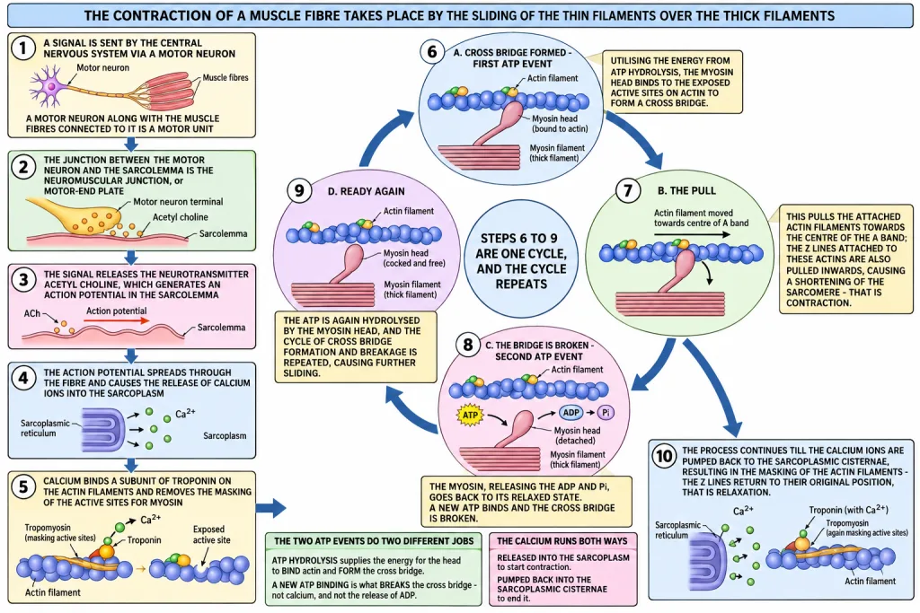
- Muscle contraction is initiated by a signal sent by the CENTRAL NERVOUS SYSTEM (CNS) via a MOTOR NEURON. A motor neuron along with the muscle fibres connected to it constitutes a MOTOR UNIT.
- The junction between a motor neuron and the sarcolemma of the muscle fibre is called the NEUROMUSCULAR JUNCTION or MOTOR-END PLATE.
- A neural signal reaching this junction releases a neurotransmitter, ACETYL CHOLINE, which generates an ACTION POTENTIAL in the sarcolemma.
- This action potential spreads through the muscle fibre and causes the release of CALCIUM IONS into the sarcoplasm.
- The increase in level leads to the binding of calcium with a SUBUNIT OF TROPONIN on the actin filaments, and thereby REMOVES THE MASKING of the active sites for myosin.
- Utilising the energy from ATP HYDROLYSIS, the myosin head binds to the exposed active sites on actin to form a CROSS BRIDGE.
- This PULLS the attached actin filaments towards the centre of the 'A' band. The 'Z' lines attached to these actins are also pulled inwards, causing a SHORTENING OF THE SARCOMERE - that is, contraction.
- The myosin, releasing the ADP and Pi, goes back to its relaxed state. A NEW ATP BINDS and the cross bridge is BROKEN.
- The ATP is again hydrolysed by the myosin head, and the cycle of cross bridge formation and breakage is repeated, causing further sliding.
- The process continues till the ions are PUMPED BACK to the sarcoplasmic cisternae, resulting in the MASKING of the actin filaments. This causes the return of the 'Z' lines to their original position - that is, RELAXATION.
Steps 6 to 9 are one cycle, and that cycle repeats. A single cross bridge pulls the actin only a short distance; the muscle shortens as much as it does because the cycle of cross bridge formation and breakage is repeated again and again, causing further sliding each time.
[NEET Important] ATP is used twice in the cycle, and for two different jobs. ATP hydrolysis supplies the energy for the myosin head to bind actin and form the cross bridge, and then the binding of a NEW ATP is what BREAKS the cross bridge. A question asking what breaks the cross bridge is answered by a new ATP binding, not by calcium and not by ADP release. Note also the direction of the calcium: released into the sarcoplasm to start contraction, pumped back into the sarcoplasmic cisternae to end it.
What Changes in the Bands - and What Does Not
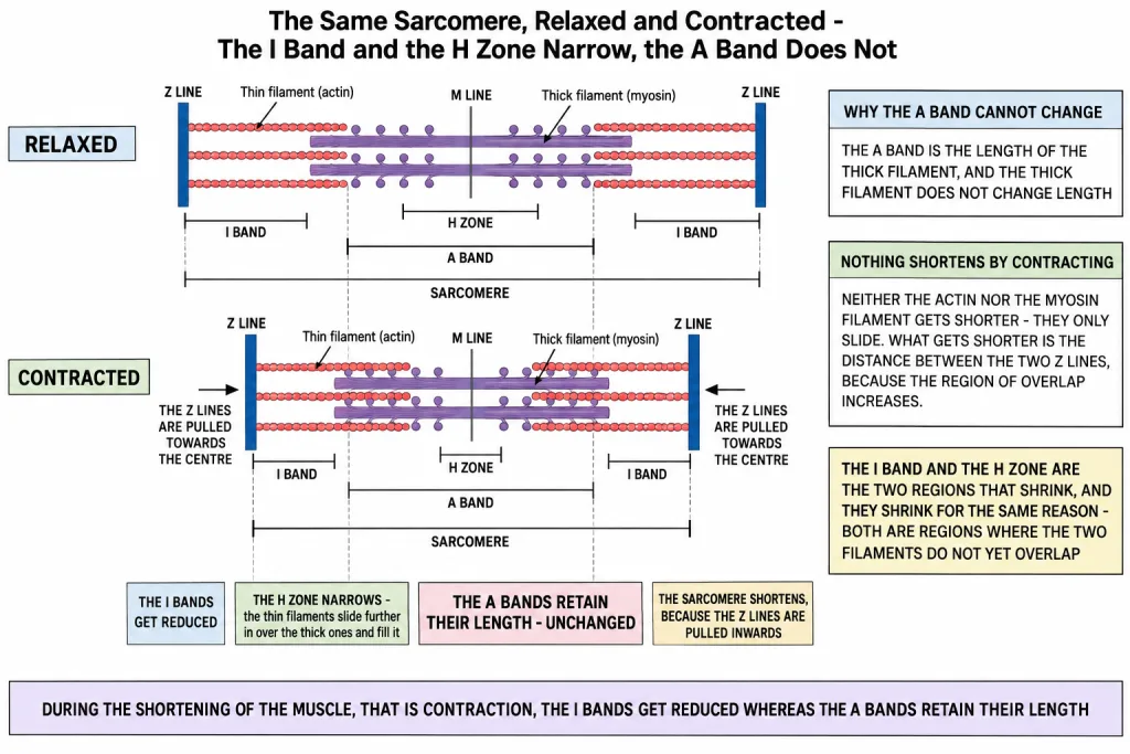
During the shortening of the muscle, that is contraction, the 'I' bands get REDUCED whereas the 'A' bands RETAIN THEIR LENGTH.
| Region of the sarcomere | During contraction | Why |
|---|---|---|
| 'I' band | gets REDUCED | it is the stretch of thin filament not yet overlapped by myosin, and the overlap grows |
| 'A' band | RETAINS ITS LENGTH | it measures the thick filament, and the thick filament does not change length |
| 'H' zone | NARROWS | the thin filaments slide further in over the thick ones and fill it |
| Sarcomere, 'Z' line to 'Z' line | SHORTENS | the 'Z' lines are pulled inwards towards the centre |
| The filaments themselves | KEEP THEIR LENGTH | nothing shortens by contracting - the filaments only slide |
That last row is the whole point of the theory's name, and it is the commonest misconception in the chapter. Neither the actin nor the myosin filament gets shorter. What gets shorter is the distance between the two 'Z' lines, and it does so because the region of overlap between the two sets of filaments increases. The 'I' band and the 'H' zone are the two regions of the sarcomere that shrink, and they shrink for the same reason - both are regions where the two filaments do not yet overlap.
[NEET Important] 'I' band reduced, 'A' band unchanged. That pairing is asked directly, and the wrong option always says the 'A' band shortens. Remember the reason and you will never swap them: the 'A' band is the length of the thick filament, and the thick filament never changes length. The 'H' zone narrowing is the third fact in the same figure and is asked as the harder version of the same question.
Reaction Time, and Why a Muscle Tires
The reaction time of the fibres can vary in different muscles. The same sequence of events runs in every skeletal muscle, but not every muscle responds at the same speed - some fibres react quickly and some slowly, and that difference is a property of the fibres themselves.
Repeated activation of the muscles can lead to the accumulation of LACTIC ACID due to the ANAEROBIC BREAKDOWN OF GLYCOGEN in them, causing FATIGUE.
Take that sentence apart, because each piece is separately askable:
- The cause is repeated activation of the muscle.
- The fuel being broken down is GLYCOGEN, the stored carbohydrate of muscle.
- The breakdown is ANAEROBIC - it happens without enough oxygen.
- The product that accumulates is LACTIC ACID.
- The result is FATIGUE.
This is the same chemistry you met in the respiration chapter, where the anaerobic fate of pyruvic acid in muscle is lactic acid. The chapter here is only naming the consequence of it in a working muscle.
[NEET Important] Glycogen is the substrate, lactic acid is the product, and fatigue is the outcome. A question that offers "aerobic breakdown of glycogen" or "accumulation of pyruvic acid" is changing exactly one word of the chapter's sentence. This fact also sets up the next section - the fibre type that works anaerobically is the one that fatigues sooner.
Quick Recap
- The mechanism of muscle contraction is best explained by the SLIDING FILAMENT THEORY, which states that the contraction of a muscle fibre takes place by the SLIDING OF THE THIN FILAMENTS OVER THE THICK FILAMENTS.
- Contraction is initiated by a signal sent by the central nervous system via a MOTOR NEURON.
- A motor neuron along with the muscle fibres connected to it constitutes a MOTOR UNIT.
- The junction between a motor neuron and the sarcolemma is the NEUROMUSCULAR JUNCTION or MOTOR-END PLATE.
- The signal releases the neurotransmitter ACETYL CHOLINE, which generates an ACTION POTENTIAL in the sarcolemma.
- The action potential spreads through the fibre and releases CALCIUM IONS into the sarcoplasm.
- Calcium binds a SUBUNIT OF TROPONIN on actin and REMOVES THE MASKING of the active sites for myosin.
- Using the energy of ATP HYDROLYSIS, the myosin head binds the exposed active sites to form a CROSS BRIDGE.
- The cross bridge PULLS the actin towards the centre of the 'A' band; the 'Z' lines are pulled inwards and the SARCOMERE SHORTENS - contraction.
- The myosin releases ADP and Pi and returns to its relaxed state; a NEW ATP BINDS and the cross bridge is BROKEN.
- The ATP is hydrolysed again and the cycle of cross bridge formation and breakage repeats, causing further sliding.
- Contraction continues till the calcium ions are PUMPED BACK to the sarcoplasmic cisternae, MASKING the actin filaments and returning the 'Z' lines to their original position - RELAXATION.
- During contraction the 'I' bands get REDUCED, the 'A' bands RETAIN THEIR LENGTH, and the 'H' zone NARROWS.
- The filaments themselves never shorten - they only slide, and the overlap between them increases.
- The reaction time of the fibres can vary in different muscles.
- Repeated activation can accumulate LACTIC ACID by the ANAEROBIC BREAKDOWN OF GLYCOGEN, causing FATIGUE.
Solved Examples
Question 1
Q. Define the sliding filament theory of muscle contraction. This is one of the chapter-end exercises.
Answer. The mechanism of muscle contraction is best explained by the sliding filament theory, which states that the contraction of a muscle fibre takes place by the SLIDING OF THE THIN FILAMENTS OVER THE THICK FILAMENTS.
The name says what happens. The thin filament is actin and the thick filament is myosin. During contraction the thin filaments slide over the thick ones towards the centre of the sarcomere, so the overlap between the two increases and the sarcomere shortens. Neither filament changes its own length - the shortening of the muscle is entirely due to sliding.
Question 2
Q. Describe the important steps in muscle contraction. This is one of the chapter-end exercises.
Answer. In order:
- Muscle contraction is initiated by a signal sent by the central nervous system via a MOTOR NEURON. A motor neuron along with the muscle fibres connected to it constitutes a motor unit.
- The junction between the motor neuron and the sarcolemma of the muscle fibre is called the NEUROMUSCULAR JUNCTION or MOTOR-END PLATE.
- A neural signal reaching this junction releases a neurotransmitter, ACETYL CHOLINE, which generates an ACTION POTENTIAL in the sarcolemma.
- This spreads through the muscle fibre and causes the release of CALCIUM IONS into the sarcoplasm.
- The increase in calcium level leads to the binding of calcium with a SUBUNIT OF TROPONIN on the actin filaments and thereby REMOVES THE MASKING of the active sites for myosin.
- Utilising the energy from ATP HYDROLYSIS, the myosin head binds to the exposed active sites on actin to form a CROSS BRIDGE.
- This pulls the attached actin filaments towards the centre of the 'A' band. The 'Z' lines attached to these actins are pulled inwards, causing a SHORTENING OF THE SARCOMERE - that is, contraction. During this shortening the 'I' bands get reduced whereas the 'A' bands retain their length.
- The myosin, releasing the ADP and Pi, goes back to its relaxed state. A new ATP binds and the cross bridge is BROKEN.
- The ATP is again hydrolysed by the myosin head, and the cycle of cross bridge formation and breakage is repeated, causing further sliding.
- The process continues till the calcium ions are PUMPED BACK to the sarcoplasmic cisternae, resulting in the MASKING of the actin filaments. This causes the return of the 'Z' lines to their original position - that is, RELAXATION.
Question 3
Q. What is a motor unit?
Answer. A motor neuron along with the muscle fibres connected to it constitutes a motor unit. It is the smallest block of muscle that the nervous system can switch on at once.
Question 4
Q. What are the two names for the junction between a motor neuron and the sarcolemma of a muscle fibre?
Answer. The neuromuscular junction, also called the motor-end plate.
Question 5
Q. Which neurotransmitter is released at the neuromuscular junction, and what does it produce?
Answer. Acetyl choline. A neural signal reaching the junction releases it, and it generates an ACTION POTENTIAL in the sarcolemma.
Question 6
Q. Trace the movement of calcium during one contraction and relaxation.
Answer. The action potential spreads through the muscle fibre and causes the release of calcium ions into the SARCOPLASM - that starts contraction. At the end, the calcium ions are PUMPED BACK to the SARCOPLASMIC CISTERNAE, which masks the actin filaments again and brings about relaxation.
Question 7
Q. What does calcium bind to inside the muscle fibre, and what does that binding achieve?
Answer. Calcium binds with a SUBUNIT OF TROPONIN on the actin filaments. That binding removes the masking of the active sites for myosin, so the myosin heads now have somewhere to attach.
Question 8
Q. What is a cross bridge, and where does the energy to form it come from?
Answer. A cross bridge is formed when the myosin head binds to the exposed active sites on actin. The energy for it comes from ATP hydrolysis.
Question 9
Q. What breaks the cross bridge?
Answer. A new ATP binding to the myosin head. First the myosin releases the ADP and Pi and goes back to its relaxed state, then a new ATP binds and the cross bridge is broken. The ATP is hydrolysed again and the cycle starts over.
Question 10
Q. Why does the sarcomere shorten when a cross bridge forms?
Answer. Because the cross bridge pulls the attached actin filaments towards the centre of the 'A' band. The 'Z' lines attached to these actins are pulled inwards, and since the sarcomere is the region between two 'Z' lines, the sarcomere shortens - which is contraction.
Question 11
Q. During contraction, which band gets reduced and which keeps its length?
Answer. The 'I' bands get REDUCED, whereas the 'A' bands RETAIN THEIR LENGTH. The reason is that the 'A' band is the length of the thick filament, and the thick filament does not change length, while the 'I' band is the part of the thin filament not overlapped by myosin, and that overlap increases.
Question 12
Q. What happens to the 'H' zone during contraction, and why?
Answer. It narrows. The 'H' zone is the part of the 'A' band where only thick filaments lie; as the thin filaments slide further in over the thick ones, they fill that region and it gets smaller.
Question 13
Q. A student writes that during contraction the actin and myosin filaments shorten. Correct him.
Answer. They do not shorten - they slide. Both the thin and the thick filaments keep their own length; the muscle shortens because the thin filaments slide over the thick ones and the overlap between them increases, which pulls the two 'Z' lines closer together. That is why the theory is called the sliding filament theory.
Question 14
Q. Why does a muscle become fatigued after repeated activity?
Answer. Repeated activation of the muscles can lead to the accumulation of LACTIC ACID due to the ANAEROBIC BREAKDOWN OF GLYCOGEN in them, causing FATIGUE. The fuel is glycogen, the breakdown is anaerobic, and the product that builds up is lactic acid.