
From the Whole Muscle Down to the Muscle Fibre
Each organised skeletal muscle in our body is made of a number of MUSCLE BUNDLES or FASCICLES, held together by a common collagenous connective tissue layer called FASCIA. Each muscle bundle contains a number of MUSCLE FIBRES.
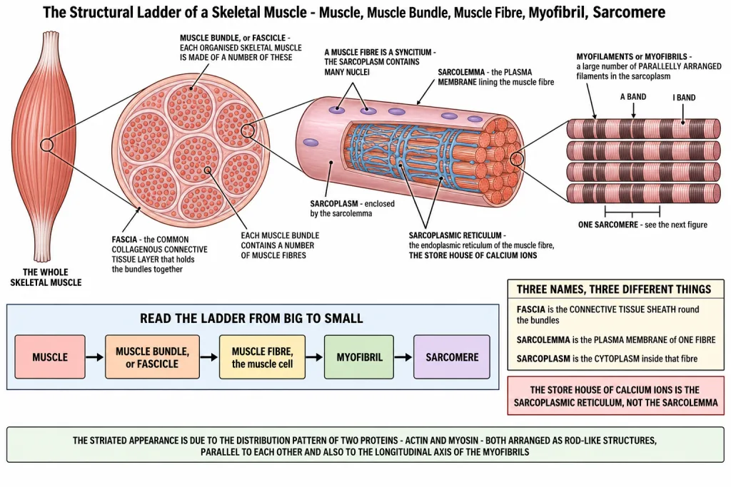
So the whole chapter's structural ladder is just this, read from big to small: muscle -> muscle bundle (fascicle) -> muscle fibre (muscle cell) -> myofibril -> sarcomere. Learn it in that order and every naming question in this section answers itself.
Now look inside one muscle fibre.
- Each muscle fibre is lined by the plasma membrane called the SARCOLEMMA, enclosing the SARCOPLASM.
- A muscle fibre is a SYNCITIUM, as the sarcoplasm contains MANY NUCLEI. One fibre, many nuclei - that is what the word syncitium means here.
- The endoplasmic reticulum of the muscle fibre, the SARCOPLASMIC RETICULUM, is the STORE HOUSE OF CALCIUM IONS.
That last line deserves a line of its own: in a muscle fibre, is stored in the SARCOPLASMIC RETICULUM. It is a fill in the blank in the chapter-end exercises, and it is the single most repeated one-word answer from this part of the chapter.
[NEET Important] Three membrane and compartment names, three different things. Fascia is the connective tissue sheath around the bundles, sarcolemma is the plasma membrane of one fibre, and sarcoplasm is the cytoplasm inside it. The favourite distractor is offering sarcolemma as the calcium store - the store house of calcium ions is the SARCOPLASMIC RETICULUM, not the membrane.
Myofibrils, and the Two Bands That Stripe Them
A characteristic feature of the muscle fibre is a large number of parallelly arranged filaments in the sarcoplasm called MYOFILAMENTS or MYOFIBRILS.
Each myofibril has alternate dark and light bands on it. That is where the word striated comes from, and the chapter is precise about the cause: the striated appearance is due to the distribution pattern of two proteins - ACTIN and MYOSIN.
Which protein sits in which band is asked directly, and the two names get swapped constantly. Say the pairing out loud:
| Band | Other name | Protein it contains | Appearance |
|---|---|---|---|
| 'I' band | ISOTROPIC band | ACTIN | LIGHT |
| 'A' band | ANISOTROPIC band | MYOSIN | DARK |
The memory hook is the first letter: 'A' band goes with Anisotropic and with the letter A of dArk; 'I' band goes with Isotropic and with the lIght band.
Both proteins are arranged as rod-like structures, parallel to each other and also to the longitudinal axis of the myofibrils. They are not tangled or coiled around the fibre - they run straight along it.
Actin filaments are THINNER as compared to the myosin filaments, hence they are commonly called THIN and THICK filaments respectively.
| Filament | Protein | Band |
|---|---|---|
| THIN filament | ACTIN | 'I' band |
| THICK filament | MYOSIN | 'A' band |
[NEET Important] Actin = thin = 'I' band = isotropic = light. Myosin = thick = 'A' band = anisotropic = dark. Every question in this part of the chapter is one row of that chain with one item removed. The commonest trap is calling the 'A' band isotropic - it is anisotropic.
The Sarcomere - Its Two 'Z' Lines, Its 'M' Line and Its 'H' Zone
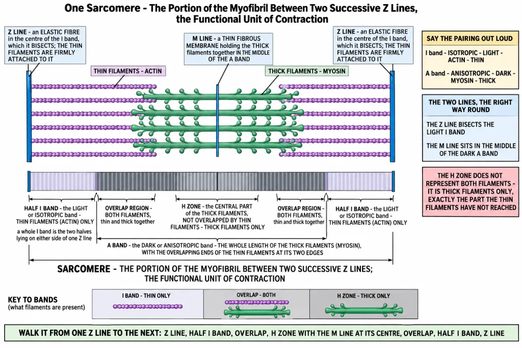
In the centre of each 'I' band is an elastic fibre called the 'Z' LINE, which bisects it. The THIN filaments are firmly attached to the 'Z' line.
The thick filaments in the 'A' band are also held together in the middle of that band by a thin fibrous membrane called the 'M' LINE.
The 'A' and 'I' bands are arranged alternately throughout the length of the myofibrils - dark, light, dark, light, all the way along.
The portion of the myofibril between two successive 'Z' lines is considered the functional unit of contraction and is called a SARCOMERE.
Now the 'H' zone, which is the one region students get wrong. In a resting state, the edges of the thin filaments on either side of the thick filaments PARTIALLY OVERLAP the free ends of the thick filaments, leaving the central part of the thick filaments free. That central part of the thick filament, not overlapped by thin filaments, is called the 'H' ZONE.
So the 'H' zone contains THICK FILAMENTS ONLY. It is not a region of overlap - it is exactly the part of the thick filament that the thin filaments have not reached.
| Region | What lies there |
|---|---|
| 'Z' line | the elastic fibre bisecting the 'I' band; the thin filaments are firmly attached to it |
| 'I' band (light, isotropic) | thin filaments (actin) only |
| 'A' band (dark, anisotropic) | the whole length of the thick filaments (myosin), with the overlapping ends of the thin filaments at its two edges |
| 'H' zone | the central part of the thick filaments, not overlapped by thin filaments - thick filaments only |
| 'M' line | the thin fibrous membrane holding the thick filaments together in the middle of the 'A' band |
| Sarcomere | everything between two successive 'Z' lines - the functional unit of contraction |
[NEET Important] Two definitions must be word-perfect. A sarcomere is the portion of the myofibril between two successive 'Z' lines, and it is the functional unit of contraction. The 'H' zone is the central part of the thick filament that is NOT overlapped by thin filaments. The standard false statement is that the 'H' zone represents both thick and thin filaments - it does not; it is thick filaments only. Note also that the 'Z' line bisects the 'I' band while the 'M' line lies in the middle of the 'A' band - swapping those two is the other easy mark to lose.
Quick Recap
- Each organised skeletal muscle is made of a number of MUSCLE BUNDLES or FASCICLES, held together by a common collagenous connective tissue layer called FASCIA.
- Each muscle bundle contains a number of MUSCLE FIBRES.
- Each muscle fibre is lined by the plasma membrane called the SARCOLEMMA, enclosing the SARCOPLASM.
- A muscle fibre is a SYNCITIUM, as the sarcoplasm contains many nuclei.
- The sarcoplasmic reticulum - the endoplasmic reticulum of the muscle fibre - is the STORE HOUSE OF CALCIUM IONS.
- A large number of parallelly arranged filaments in the sarcoplasm are called MYOFILAMENTS or MYOFIBRILS.
- Each myofibril has alternate dark and light bands, and the striated appearance is due to the distribution pattern of ACTIN and MYOSIN.
- The light bands contain ACTIN and are called the 'I' band or ISOTROPIC band.
- The dark band, the 'A' or ANISOTROPIC band, contains MYOSIN.
- Both proteins are arranged as rod-like structures, parallel to each other and to the longitudinal axis of the myofibrils.
- Actin filaments are THINNER than myosin filaments, hence they are called THIN and THICK filaments respectively.
- In the centre of each 'I' band is an elastic fibre called the 'Z' LINE, which bisects it, and the thin filaments are firmly attached to the 'Z' line.
- The thick filaments in the 'A' band are held together in the middle of that band by a thin fibrous membrane called the 'M' LINE.
- The 'A' and 'I' bands are arranged alternately throughout the length of the myofibrils.
- The portion of the myofibril between two successive 'Z' lines is the functional unit of contraction and is called a SARCOMERE.
- In a resting state the edges of the thin filaments partially overlap the free ends of the thick filaments, leaving the central part of the thick filaments free.
- That central part of the thick filament, not overlapped by thin filaments, is the 'H' ZONE - thick filaments only.
Solved Examples
Question 1
Q. What is a fascicle, and what holds the fascicles of a muscle together?
Answer. A fascicle is a MUSCLE BUNDLE - each organised skeletal muscle is made of a number of muscle bundles or fascicles, and each muscle bundle contains a number of muscle fibres. They are held together by a common collagenous connective tissue layer called FASCIA.
Question 2
Q. Name the plasma membrane of a muscle fibre and the material it encloses.
Answer. The plasma membrane of a muscle fibre is the SARCOLEMMA, and it encloses the SARCOPLASM.
Question 3
Q. Why is a muscle fibre called a syncitium?
Answer. Because the sarcoplasm of one muscle fibre contains MANY NUCLEI. A single fibre with many nuclei in it is a syncitium.
Question 4
Q. What is the sarcoplasmic reticulum, and what is its role?
Answer. It is the endoplasmic reticulum of the muscle fibre. It is the STORE HOUSE OF CALCIUM IONS - in a muscle fibre is stored in the sarcoplasmic reticulum.
Question 5
Q. What are myofibrils, and why does a muscle fibre look striped?
Answer. Myofibrils, also called myofilaments, are the large number of parallelly arranged filaments in the sarcoplasm - they are a characteristic feature of the muscle fibre. Each myofibril has alternate dark and light bands on it, and the striated appearance is due to the distribution pattern of two proteins, ACTIN and MYOSIN.
Question 6
Q. Which band contains actin and which contains myosin, and what is the other name of each band?
Answer.
| Band | Other name | Protein | Appearance |
|---|---|---|---|
| 'I' band | ISOTROPIC band | ACTIN | light |
| 'A' band | ANISOTROPIC band | MYOSIN | dark |
The light bands contain actin and are called the 'I' band or isotropic band, whereas the dark band, called the 'A' or anisotropic band, contains myosin.
Question 7
Q. Why are actin and myosin called thin and thick filaments?
Answer. Because actin filaments are THINNER as compared to the myosin filaments, so they are commonly called thin and thick filaments respectively. Both are arranged as rod-like structures, parallel to each other and also to the longitudinal axis of the myofibrils.
Question 8
Q. What is the 'Z' line, and what is attached to it?
Answer. In the centre of each 'I' band is an elastic fibre called the 'Z' line, which bisects it. The THIN filaments are firmly attached to the 'Z' line.
Question 9
Q. What is the 'M' line?
Answer. A thin fibrous membrane that holds the thick filaments of the 'A' band together in the middle of that band. Note the contrast with the previous answer: the 'Z' line sits in the middle of the 'I' band, the 'M' line in the middle of the 'A' band.
Question 10
Q. Define a sarcomere.
Answer. The portion of the myofibril between two successive 'Z' lines. It is considered the functional unit of contraction.
Question 11
Q. Describe how the thin and thick filaments lie with respect to each other in a resting muscle, and say what the 'H' zone is.
Answer. In a resting state, the edges of the thin filaments on either side of the thick filaments PARTIALLY OVERLAP the free ends of the thick filaments, which leaves the central part of the thick filaments free. That central part of the thick filament, not overlapped by thin filaments, is called the 'H' zone.
Question 12
Q. Draw the diagram of a sarcomere of skeletal muscle showing different regions. This is one of the chapter-end exercises.
Answer. Draw it as a straight strip and put the parts in this order, left to right.
Step 1 - the boundaries. Draw two vertical lines, one at each end of the strip. These are the two 'Z' lines, and everything between them is one SARCOMERE - the functional unit of contraction.
Step 2 - the thin filaments. From each 'Z' line, draw several fine horizontal lines running inwards, towards the middle. These are the THIN filaments (ACTIN), and they are firmly attached to the 'Z' line.
Step 3 - the thick filaments. In the middle of the strip, draw a bundle of thicker horizontal rods that does not reach either 'Z' line. These are the THICK filaments (MYOSIN).
Step 4 - the overlap. Make the inner ends of the thin filaments run alongside the outer ends of the thick filaments, so that at each end of the thick bundle the two sets partially overlap. Leave the central part of the thick filaments unreached by any thin filament.
Step 5 - label the regions.
- The 'A' band - mark it across the whole length of the thick filaments, including the overlapped ends. It is the dark, anisotropic band.
- The 'H' zone - mark it in the middle of the 'A' band, over the central part of the thick filaments that is not overlapped by thin filaments.
- The 'M' line - a short vertical line at the exact centre of the 'A' band, the thin fibrous membrane holding the thick filaments together.
- The 'I' band - mark it at each end of the sarcomere, around each 'Z' line, over the thin filaments alone. It is the light, isotropic band. A full sarcomere carries one whole 'A' band in the middle and one half 'I' band at either end.
Step 6 - the label list of what each region contains.
| Region | Contents |
|---|---|
| 'Z' line | elastic fibre bisecting the 'I' band; the thin filaments are attached to it |
| 'I' band (light, isotropic) | thin filaments (actin) only |
| 'A' band (dark, anisotropic) | the full length of the thick filaments (myosin), with the thin filament ends overlapping at its two edges |
| 'H' zone | the central part of the thick filaments, not overlapped by thin filaments - thick filaments only |
| 'M' line | thin fibrous membrane in the middle of the 'A' band, holding the thick filaments together |
| Sarcomere | the whole portion between two successive 'Z' lines; the functional unit of contraction |
Question 13
Q. State whether true or false, and if false change the statement so that it is true: "H-zone of striated muscle fibre represents both thick and thin filaments." This is one of the chapter-end exercises.
Answer. FALSE.
The corrected statement: the 'H' zone of a striated muscle fibre represents the central part of the thick filaments that is NOT overlapped by the thin filaments, and so it contains THICK FILAMENTS ONLY.
Why. In a resting state the edges of the thin filaments on either side partially overlap the free ends of the thick filaments, leaving the central part of the thick filaments free, and that free central part is the 'H' zone. The region where both thick and thin filaments are present is the overlap zone at the two edges of the 'A' band, not the 'H' zone.
Question 14
Q. Fill in the blank and restate the sentence: "In a muscle fibre Ca++ is stored in __." This is one of the chapter-end exercises.
Answer. In a muscle fibre is stored in the SARCOPLASMIC RETICULUM.
The sarcoplasmic reticulum is the endoplasmic reticulum of the muscle fibre, and the chapter calls it the store house of calcium ions.
Question 15
Q. Arrange these from largest to smallest: sarcomere, muscle fibre, fascicle, myofibril, muscle.
Answer. Muscle -> fascicle (muscle bundle) -> muscle fibre -> myofibril -> sarcomere. A muscle is made of fascicles, each fascicle contains muscle fibres, each fibre contains parallelly arranged myofibrils, and the portion of a myofibril between two successive 'Z' lines is a sarcomere.