
Female Reproductive System — Overview
The female reproductive system is located in the pelvic region and is structured for gamete production, sperm reception, fertilisation, embryo development, and childbirth.
Components
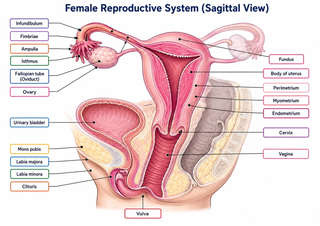
The female system consists of:
- A pair of ovaries (the gonads) — produce ova and female sex hormones.
- A pair of fallopian tubes (oviducts) — carry the egg from ovary to uterus; site of fertilisation.
- Uterus — single, hollow, muscular organ where the embryo develops.
- Vagina — birth canal and copulation organ.
- External genitalia (vulva) — labia majora, labia minora, clitoris, hymen.
- Mammary glands (breasts) — accessory glands for lactation.
\
Key features
- Unlike males, the female urethra and reproductive tract are completely separate (urethra carries only urine).
- All female reproductive organs are internal except the external genitalia.
- The female system shows cyclic activity (menstrual cycle) governed by hormones — covered in Section 9.
The Ovaries — Female Gonads
The ovaries are paired, almond-shaped female gonads located on either side of the uterus in the pelvic cavity.
Anatomy
- Size: ~2-4 cm in length × 1.5-3 cm wide × 1 cm thick (in adult women).
- Position: lateral to the uterus, in the ovarian fossa of the pelvic wall.
- Attachment: held in place by the ovarian ligament and broad ligament of the uterus.
Functions
- Ovum production (oogenesis) — release ~one ovum per menstrual cycle (~400 in lifetime).
- Hormone secretion — produce oestrogen (mainly from follicles) and progesterone (mainly from corpus luteum).
Internal structure (covered in Section 6)
- Outer cortex — contains follicles at various stages.
- Inner medulla — contains blood vessels, nerves, connective tissue.
Note on follicle development
Inside each ovary, primordial follicles (with arrested primary oocytes) gradually develop into:
primordial → primary → secondary → tertiary → Graafian follicle → ovulation
Detailed coverage in Sections 6 and 8.
The Fallopian Tubes (Oviducts)
The fallopian tubes (also called oviducts) are paired, narrow muscular tubes connecting each ovary to the uterus. Length: ~10-12 cm.
Four parts of the fallopian tube (NCERT names three: infundibulum, ampulla, isthmus; the intramural part is extra)
From ovary to uterus:
- Infundibulum — funnel-shaped opening near the ovary, with finger-like projections called fimbriae that catch the released egg.
- Ampulla — the widest, longest part; fertilisation occurs at the ampullary-isthmic junction (where the ampulla meets the isthmus — NCERT).
- Isthmus — narrow part close to the uterus.
- Uterine part (intramural) — passes through the wall of the uterus to open into it.
Functions
- Egg pickup: Fimbriae of infundibulum catch the released ovum from ovary.
- Egg transport: Cilia + smooth muscle contractions move the egg toward uterus.
- Fertilisation site: The ampulla — where sperm meets egg.
- Early embryo transport: Carries the cleaving embryo to the uterus.
Tubal pregnancy
If a fertilised embryo gets stuck in the fallopian tube and implants there (instead of in the uterus), it's called an ectopic / tubal pregnancy — life-threatening and requires immediate medical intervention.
[NEET Important] "Where does fertilisation occur?" Answer: Ampulla of the fallopian tube. Memorise — common NEET MCQ.
The Uterus — Where Life Develops
The uterus (Greek: hystera) is a single, hollow, pear-shaped, muscular organ in the centre of the pelvis. It is the site of embryo implantation and fetal development.
Anatomy
- Size in non-pregnant state: ~7-8 cm long × 5 cm wide × 3 cm thick (about the size of a small pear).
- Pregnant state: can expand 500-1000× to accommodate the fetus.
- Three regions:
- Fundus — top, dome-shaped portion above the entry of fallopian tubes.
- Body — main central portion.
- Cervix — narrow lower portion that opens into the vagina.
Three wall layers (memorise!)
The uterine wall has three layers (from outside to inside):
- Perimetrium — outermost serous covering.
- Myometrium — middle, thick, muscular layer; provides the powerful contractions during labour.
- Endometrium — innermost, glandular, vascular layer; thickens during the menstrual cycle to prepare for implantation; sheds during menstruation.
Cervix and birth canal
- The cervix is the narrow neck of the uterus that protrudes into the vagina.
- It contains a small cervical canal that connects the uterine cavity to the vagina.
- During childbirth, the cervix dilates (opens up to ~10 cm) to allow passage of the baby.
- Cervical mucus changes throughout the menstrual cycle, becoming more permeable around ovulation to allow sperm passage.
The birth canal
- The vagina + cervical canal together form the birth canal.
- During labour, the baby passes through this canal from the uterus to the outside.
[NEET Important] Memorise the 3 layers of uterine wall: Perimetrium (outer) → Myometrium (middle, muscular) → Endometrium (inner). The endometrium is the layer that menstruates and supports implantation.
Vagina and External Genitalia
Vagina
The vagina is a muscular, elastic tube ~7-10 cm long that connects the cervix (uterus) to the outside (at the vaginal orifice).
Functions:
- Receives the penis during sexual intercourse.
- Birth canal — passage through which the baby is delivered.
- Outflow for menstrual blood.
The vagina is acidic (pH 3.5-4.5) — maintained by lactobacilli that produce lactic acid. This acidity protects against bacterial infection but is hostile to sperms (hence the need for alkaline semen).
External Genitalia (Vulva)
The vulva is the collective term for the external female genitalia. It includes:
- Mons pubis — fatty tissue mound over the pubic bone, covered with hair after puberty.
- Labia majora — outer "large lips," fleshy folds outside the vaginal opening.
- Labia minora — inner "small lips," thinner folds within the labia majora.
- Clitoris — small erectile tissue at the front (anterior) part of the vulva, homologous to the penis (developmentally similar). The most sensitive part of the female genitalia; has many nerve endings.
- Hymen — a thin membrane that partially covers the vaginal orifice in young girls. May be torn during first intercourse, vigorous physical activity, or other reasons. Note: the presence or absence of hymen is NOT a reliable indicator of sexual activity (a common social misconception).
Two openings in the vulva
- Urethral opening (anterior) — for urine.
- Vaginal opening (posterior) — for vagina.
Note that in females (unlike males), the urethra and vagina are separate — urethra is just for urine, vagina just for reproduction.
[NEET Important] Hymen is NOT a reliable indicator of virginity (NCERT explicitly states this). The hymen can be torn by exercise, tampons, or other non-sexual causes.
Small Memory Capsule
Section 5 in 60 seconds.
Female reproductive system components
- Ovaries (pair) — gonads; produce ova + oestrogen/progesterone
- Fallopian tubes (pair) — fertilisation site
- Uterus — embryo development site
- Vagina — birth canal + copulation organ
- Vulva — external genitalia
- Mammary glands — accessory (lactation)
Fallopian tube (4 parts)
Infundibulum (with fimbriae) → Ampulla (fertilisation!) → Isthmus → Uterine part
Uterus
- 3 regions: Fundus (top) → Body → Cervix (bottom)
- 3 wall layers: Perimetrium → Myometrium (muscular) → Endometrium (sheds in menstruation)
- Pear-shaped in non-pregnant state
Vagina
- pH ~3.5-4.5 (acidic, hostile to sperm)
- Functions: copulation + birth canal + menstrual outflow
Vulva (5 parts)
- Mons pubis
- Labia majora (outer lips)
- Labia minora (inner lips)
- Clitoris (sensitive erectile tissue)
- Hymen (thin membrane; NOT a virginity indicator)
Two openings
- Urethra → urine only
- Vagina → reproduction (separate from urethra in females)
Where things happen
- Ovum production: Ovary
- Fertilisation: Ampulla of fallopian tube
- Implantation: Uterus (endometrium)
- Childbirth: Through cervix → vagina
One-line takeaway
"Female reproductive system has internal organs (ovaries, oviducts, uterus, vagina) + external vulva; ova are made in ovaries, fertilisation occurs in ampulla of fallopian tube, embryo develops in the endometrium of the uterus, baby is born through the vagina."
Solved Examples
Example 1: Six female reproductive components
List the six main components of the female reproductive system.
Solution:
- Ovaries (pair) — female gonads; produce ova + sex hormones.
- Fallopian tubes (oviducts) (pair) — connect ovary to uterus; fertilisation site.
- Uterus — embryo implantation and development.
- Vagina — birth canal, copulation organ.
- External genitalia (vulva) — mons pubis, labia majora/minora, clitoris, hymen.
- Mammary glands (breasts) — accessory glands for lactation.
Answer: Ovaries + fallopian tubes + uterus + vagina + vulva + mammary glands.
Example 2: Four parts of fallopian tube
Name the four parts of the fallopian tube in order from ovary to uterus.
Solution:
From ovary side to uterus side:
- Infundibulum — funnel-shaped end near ovary, with fimbriae (finger-like projections) that catch the released egg.
- Ampulla — the longest, widest portion. Fertilisation site!
- Isthmus — narrow part close to uterus.
- Uterine part (intramural) — short part embedded in the uterine wall, opens into uterine cavity.
Answer: Infundibulum → Ampulla → Isthmus → Uterine part.
[NEET Important] Memorise this 4-part order. Common NEET MCQ.
Example 3: Three layers of uterus
Name the three layers of the uterine wall from outside to inside. State the function of each.
Solution:
| Layer | Position | Function |
|---|---|---|
| Perimetrium | Outermost | Serous covering; protects uterus |
| Myometrium | Middle | Thick muscular layer; provides labour contractions |
| Endometrium | Innermost | Glandular, vascular; thickens during cycle, sheds in menstruation, supports implantation |
Answer: Perimetrium → Myometrium → Endometrium (outside to inside).
[Board Important] A standard 3-mark CBSE question.
Example 4: Three regions of uterus
Name the three regions of the uterus.
Solution:
- Fundus — top, dome-shaped portion above the entry of fallopian tubes.
- Body — central, main portion.
- Cervix — narrow lower portion opening into the vagina via the cervical canal.
Answer: Fundus + Body + Cervix.
Function note: The cervix dilates to ~10 cm during labour to allow baby's passage.
Example 5: Vulva components
Name the components of the vulva (external female genitalia).
Solution:
The vulva comprises:
- Mons pubis — fatty mound over pubic bone; hair-covered after puberty.
- Labia majora — outer "large lips" — fleshy folds.
- Labia minora — inner "small lips" — thinner folds inside labia majora.
- Clitoris — small erectile tissue at the anterior; homologous to penis; very sensitive.
- Hymen — thin membrane partially covering vaginal opening (in young girls).
Plus the vaginal opening and the urethral opening (which is separate in females).
Answer: Mons pubis + Labia majora + Labia minora + Clitoris + Hymen.
[NEET Important] Standard NCERT-canonical question.
Example 6: Hymen — virginity indicator?
Is the hymen a reliable indicator of virginity?
Solution:
No. NCERT explicitly states that the hymen is NOT a reliable indicator of virginity or sexual activity.
Why?
- The hymen can be torn by non-sexual causes: vigorous exercise, sports (cycling, gymnastics), tampon use, masturbation, surgical procedures.
- In some women, the hymen is naturally absent or very thin.
- In others, the hymen is flexible and may not tear during first intercourse.
The hymen's presence/absence/state therefore tells you nothing reliable about whether someone has had intercourse.
Cultural significance: This biological fact is important to challenge harmful social practices that treat the hymen as a "purity" test.
Answer: No — the hymen is NOT a reliable indicator of virginity. NCERT explicitly states this.
[Board Important] A direct NCERT-canonical question.