
Inside an Ovary
We've placed the ovaries in the female pelvis (Section 5). Now let's look inside one to understand where ova are produced and where the menstrual cycle plays out.
Ovary structure
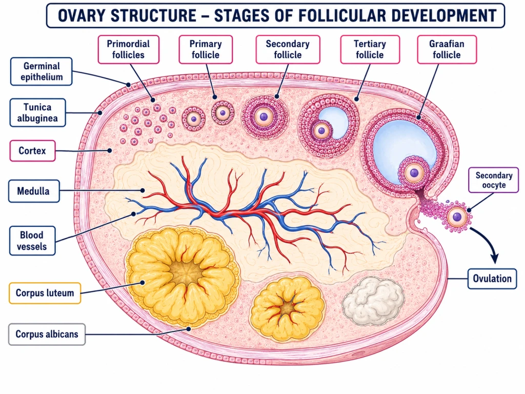
A cross-section of an ovary shows two distinct regions:
- Outer cortex — contains the follicles at various stages of development.
- Inner medulla — contains blood vessels, nerves, and connective tissue.
Plus an outer protective covering:
- Tunica albuginea — fibrous capsule under the surface epithelium.
\
Follicles — The Egg Factories
Inside the cortex, follicles at various stages are visible. A follicle is essentially the oocyte (egg cell) plus its surrounding support cells.
Stages of follicular development
Each follicle goes through these stages during a menstrual cycle:
- Primordial follicle — earliest stage; contains a primary oocyte surrounded by a single flat layer of granulosa cells.
- Primary follicle — granulosa cells become cuboidal; oocyte enlarges.
- Secondary follicle — multiple layers of granulosa cells form; theca cells develop outside.
- Tertiary follicle — fluid-filled cavity (antrum) starts forming.
- Graafian follicle (mature) — fully developed; large antrum; oocyte ready for release.
Ovulation
Around day 14 of the menstrual cycle:
- The Graafian follicle ruptures.
- The secondary oocyte is released into the fallopian tube — this is ovulation.
After ovulation: corpus luteum
The ruptured follicle transforms into the corpus luteum ("yellow body"):
- Endocrine structure that secretes progesterone (and some oestrogen).
- Maintains the endometrium for potential implantation.
- If pregnancy occurs: corpus luteum persists for ~3 months (then placenta takes over).
- If no pregnancy: corpus luteum degenerates into corpus albicans ("white body") around day 28.
Quick stages summary
[NEET Important] Memorise this 8-stage sequence. It's a NEET-classic 'arrange in order' MCQ.
Mammary Glands — Anatomy
The mammary glands (breasts) are accessory reproductive organs that produce milk for the newborn after childbirth — the process of lactation.
Anatomy
\
The major structures
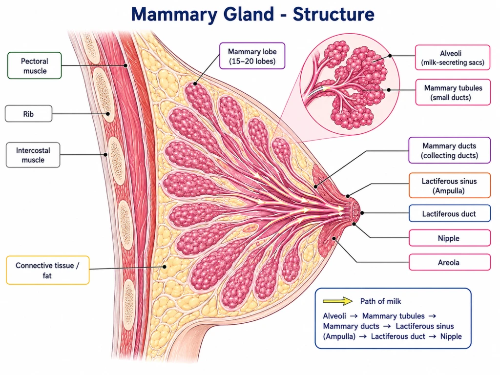
Each mammary gland has:
- Glandular tissue — divided into 15-20 mammary lobes.
- Each lobe contains clusters of alveoli (singular: alveolus) — milk-secreting cells.
- The alveoli open into mammary tubules.
- Tubules of each lobe join to form a mammary duct.
- Several mammary ducts join to form a wider mammary ampulla, connected to the lactiferous duct. Milk is stored in the cavities (lumens) of the alveoli (NCERT).
- Each ampulla narrows into a lactiferous duct that opens at the nipple.
The path of milk (memorise!)
External features
- Nipple — the projecting tip; lactiferous ducts open here.
- Areola — the circular pigmented area around the nipple; contains small bumps (Montgomery's tubercles) that secrete protective oils.
[NEET Important] Memorise the milk pathway: Alveoli → Tubules → Ducts → Ampulla → Lactiferous duct → Nipple.
Mammary Gland Development & Function
Development
- Mammary glands are present in both sexes from birth.
- At puberty in females: under oestrogen and progesterone, mammary glands enlarge significantly:
- Fat deposition increases.
- Ducts proliferate.
- Alveoli develop (but milk production not yet active).
- During pregnancy: under high oestrogen, progesterone, prolactin, and human placental lactogen (hPL):
- Alveoli mature fully.
- Glands prepare for milk production.
- After childbirth: prolactin triggers milk synthesis; oxytocin triggers milk ejection.
Lactation
Lactation = the process of milk secretion and release. Detailed coverage in Section 13.
Two key hormones:
- Prolactin (anterior pituitary) — drives milk synthesis in alveoli.
- Oxytocin (posterior pituitary) — triggers milk ejection (let-down reflex) when baby suckles.
Milk types
- Colostrum — yellowish, thick milk produced for first 2-3 days postpartum. Rich in antibodies (IgA), proteins, vitamins. Provides passive immunity to newborn.
- Mature milk — produced from day 4 onwards. Optimal nutritional balance.
[NEET Important] Colostrum is the FIRST milk, antibody-rich, very important for newborn immunity. Common NEET fact.
Small Memory Capsule
Section 6 in 60 seconds.
Ovary structure
- Cortex (outer) — contains follicles at various stages.
- Medulla (inner) — blood vessels, nerves.
- Tunica albuginea — fibrous capsule.
Follicle stages (memorise the 8!)
Primordial → Primary → Secondary → Tertiary → Graafian → Ovulation → Corpus luteum → Corpus albicans
Key facts
- Graafian follicle — fully mature, ready for ovulation (~day 14).
- Ovulation — release of secondary oocyte from Graafian follicle.
- Corpus luteum — yellow body; secretes progesterone; maintains endometrium.
- Corpus albicans — white body; degenerated corpus luteum; forms by day 28 if no pregnancy.
Mammary gland structure
- 15-20 lobes per breast
- Each lobe → alveoli (milk-secreting cells)
- Alveoli → Tubules → Ducts → Ampulla (lactiferous sinus) → Lactiferous duct → Nipple
External features
- Nipple — milk emerges
- Areola — pigmented ring around nipple (Montgomery's tubercles)
Lactation hormones
- Prolactin (ant. pituitary) → milk synthesis in alveoli
- Oxytocin (post. pituitary) → milk ejection (let-down)
Milk types
- Colostrum (first 2-3 days) — antibody-rich, immune protection
- Mature milk (day 4+) — optimal nutrition
One-line takeaway
"Ovary cortex contains follicles maturing through 8 stages (primordial → corpus albicans); mammary glands have 15-20 lobes of alveoli that secrete milk into ducts → ampulla → nipple, with prolactin driving synthesis and oxytocin driving ejection."
Solved Examples
Example 1: Two regions of ovary
Name the two regions of the ovary and state what is found in each.
Solution:
Cortex (outer) — contains follicles at various stages of development (primordial → Graafian) and the corpus luteum after ovulation.
Medulla (inner) — contains blood vessels, lymph vessels, nerves, and connective tissue. No follicles here.
Plus: Tunica albuginea — fibrous capsule just under the surface (germinal) epithelium of the ovary.
Answer: Cortex (with follicles) + Medulla (vascular/connective tissue).
Example 2: Eight stages of follicular development
List the eight stages of follicular development in correct order.
Solution:
- Primordial follicle — earliest; primary oocyte + single flat layer of granulosa cells.
- Primary follicle — granulosa cells become cuboidal.
- Secondary follicle — multiple granulosa cell layers + theca cells outside.
- Tertiary follicle — antrum (fluid-filled cavity) forms.
- Graafian follicle — fully mature; large antrum; ready to ovulate.
- Ovulation — Graafian follicle ruptures, releases secondary oocyte.
- Corpus luteum — ruptured follicle becomes yellow body; secretes progesterone.
- Corpus albicans — corpus luteum degenerates to white scar (if no pregnancy).
Answer: Memorise this 8-stage sequence. NEET-classic.
Example 3: What is the corpus luteum?
What is the corpus luteum? When does it form? What does it secrete?
Solution:
The corpus luteum ("yellow body") is an endocrine structure that forms from the ruptured Graafian follicle AFTER ovulation (day 14 onwards).
Function: Secretes:
- Progesterone (main hormone) — maintains the endometrium for potential implantation.
- Some oestrogen.
Duration:
- If pregnancy occurs: corpus luteum persists for ~3 months (until placenta takes over hormone production).
- If no pregnancy: corpus luteum degenerates into corpus albicans by day 28 of cycle.
Answer: Corpus luteum = post-ovulation yellow body; secretes progesterone to maintain endometrium.
Example 4: Path of milk
Trace the path of milk from the alveoli to the nipple.
Solution:
- Alveoli — milk-secreting cells (in clusters within each mammary lobe).
- Mammary tubules — small ducts collecting milk from alveoli.
- Mammary ducts — larger ducts formed by junction of tubules.
- Mammary ampulla (lactiferous sinus) — wider area where milk pools before release.
- Lactiferous duct — narrowing of ampulla; opens at nipple.
- Nipple — exit point.
Answer: Alveoli → Tubules → Ducts → Ampulla (lactiferous sinus) → Lactiferous duct → Nipple.
[NEET Important] Memorise this 6-step path.
Example 5: Number of mammary lobes
How many mammary lobes does each breast have?
Solution:
Each mammary gland (breast) has 15-20 mammary lobes of glandular tissue.
Each lobe consists of:
- Multiple alveoli (milk-secreting cells)
- A network of mammary tubules and ducts converging toward the nipple
Lobes are separated by connective tissue and fat.
Answer: 15-20 mammary lobes per breast.
Example 6: What is colostrum?
What is colostrum? Why is it important for the newborn?
Solution:
Colostrum is the first milk produced by the mammary glands during the first 2-3 days after childbirth. Characteristics:
- Yellowish, thick consistency.
- Lower in fat and lactose than mature milk.
- Very rich in:
- Antibodies (IgA) — provide passive immunity to the newborn.
- Proteins, including immune-active proteins.
- Vitamins A, E, K.
Importance:
- Passive immunity — antibodies in colostrum protect the newborn from infections during the first few days when the baby's own immune system is immature.
- Nutrient-rich — easily digested.
- Laxative effect — helps the newborn pass meconium (first stool).
WHO recommends exclusive breastfeeding (with colostrum first) for the first 6 months of life.
Answer: Colostrum = first milk (days 1-3 postpartum); rich in antibodies for newborn immunity.
[NEET Important] Frequent NEET MCQ.