
Inside the Seminiferous Tubule
We've established that seminiferous tubules are where sperm production happens. Now let's go inside one.
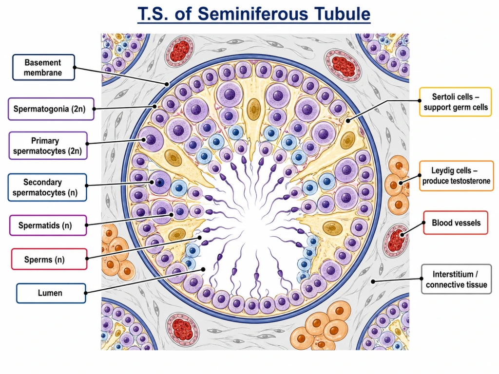
If you look at a transverse section of a single seminiferous tubule under a microscope, you see two main types of cells:
- Male germ cells — at various stages of spermatogenesis
- Sertoli cells — large supporting "nurse" cells
Plus, just OUTSIDE the tubule (in the connective tissue between tubules):
- Leydig cells (also called interstitial cells) — produce testosterone
\
NCERT direct quote: "The seminiferous tubules are lined on its inside by two types of cells called male germ cells (spermatogonia) and Sertoli cells."
Male Germ Cells — From Spermatogonium to Sperm
The male germ cells line the inside of the seminiferous tubule. They are the cells that will eventually become sperms.
The lineage
The germ cells exist at multiple stages, all of which can be seen in a single T.S. of an active tubule:
- Spermatogonium (, diploid) — outermost, against the basement membrane.
- Primary spermatocyte () — formed by mitosis of spermatogonium; this is the cell that undergoes meiosis.
- Secondary spermatocyte (, haploid) — formed by Meiosis I.
- Spermatid () — formed by Meiosis II.
- Spermatozoon (sperm) () — formed by spermiogenesis; the final mature gamete.
The cells are arranged from periphery (least mature) to centre (most mature) — like layers of an onion.
Mitotic divisions
The undifferentiated spermatogonia divide mitotically to:
- Maintain the stem cell pool (so spermatogenesis can continue indefinitely).
- Produce primary spermatocytes that will undergo meiosis.
This continuous self-renewal is why men can produce sperm into old age — the spermatogonia don't get used up.
[NEET Important] A common NEET MCQ: "Which cell undergoes meiosis in the testis?" Answer: Primary spermatocyte () → undergoes meiosis I to give Secondary spermatocyte ().
Sertoli Cells — The Nurses
Sertoli cells (named after Italian histologist Enrico Sertoli, 1865) are large, irregularly shaped cells that span the entire thickness of the seminiferous tubule — from the basement membrane to the lumen.
What Sertoli cells do
NCERT describes them as "cells that provide nutrition to the germ cells." Their key roles:
- Nourish developing germ cells — provide nutrients (amino acids, lipids, growth factors).
- Form the blood-testis barrier — tight junctions between adjacent Sertoli cells create a barrier that protects developing germ cells from the body's immune system. This is critical because haploid germ cells have surface antigens that would otherwise be attacked.
- Coordinate spermatogenesis — release signals that synchronise germ cell divisions.
- Phagocytose excess cytoplasm — when spermatids transform into sperms (spermiogenesis), excess cytoplasm is shed; Sertoli cells eat it up.
- Secrete inhibin — a hormone that negatively regulates FSH secretion (a feedback loop).
Hormonal control of Sertoli cells
- FSH (from pituitary) → activates Sertoli cells → spermatogenic support.
- Sertoli cells → secrete inhibin → inhibits FSH (negative feedback).
Quick summary
| Sertoli cell function | Mechanism |
|---|---|
| Nourishment | Provides nutrients to germ cells |
| Protection | Blood-testis barrier (tight junctions) |
| Coordination | Synchronises spermatogenic cycles |
| Cleanup | Phagocytoses excess cytoplasm |
| Hormone secretion | Releases inhibin (suppresses FSH) |
[NEET Important] Memorise: "Sertoli cells are stimulated by FSH and secrete inhibin to inhibit FSH." Has appeared in NEET 2019, 2022.
Leydig Cells — The Hormone Producers
Leydig cells (named after German anatomist Franz Leydig, 1850), also called interstitial cells, are located in the connective tissue between adjacent seminiferous tubules — NOT inside the tubules.
What Leydig cells do
Function: Synthesise and secrete testosterone — the male sex hormone.
That's it. One job. But that one job is critical:
- Testosterone drives spermatogenesis (acts on Sertoli cells).
- Testosterone produces secondary sexual characteristics at puberty (deep voice, facial hair, muscle mass, libido).
- Testosterone maintains male reproductive organs.
- Testosterone influences behaviour (aggression, libido).
Hormonal control of Leydig cells
- LH (from pituitary) → stimulates Leydig cells → secrete testosterone.
- Testosterone → negative feedback on hypothalamus + pituitary (slows LH).
Critical Sertoli vs Leydig table (NEET classic)
| Feature | Sertoli cells | Leydig cells |
|---|---|---|
| Location | INSIDE seminiferous tubule | OUTSIDE tubule, in interstitium |
| Function | Nourish germ cells; secrete inhibin | Secrete testosterone |
| Stimulated by | FSH | LH |
| Hormone produced | Inhibin | Testosterone |
| Synonym | "Nurse cells" | Interstitial cells |
[NEET Important] This comparison is tested EVERY year in NEET. Memorise it cold.
Hormonal Control of the Testis
Putting it all together — the testis is under tight hormonal control by the brain.
The HPG axis in males
- Hypothalamus secretes GnRH (Gonadotropin-Releasing Hormone).
- GnRH triggers anterior pituitary to release:
- FSH (Follicle-Stimulating Hormone)
- LH (Luteinizing Hormone, called ICSH — Interstitial Cell Stimulating Hormone — in males)
- FSH → acts on Sertoli cells → spermatogenesis support.
- LH → acts on Leydig cells → testosterone production.
- Testosterone + Inhibin → feedback to hypothalamus + pituitary (negative feedback).
Note on LH naming
In males, LH is sometimes called ICSH (Interstitial Cell Stimulating Hormone) because its target is the interstitial (Leydig) cells. ICSH and LH are the same hormone — just named differently in male vs female contexts.
A clean summary diagram
[NEET Important] This 4-hormone system (GnRH, FSH, LH, testosterone) plus inhibin is the master control of male reproduction. Master it before NEET.
Small Memory Capsule
Section 3 in 60 seconds.
Inside the seminiferous tubule
| Cell type | Location | Function |
|---|---|---|
| Spermatogonia () | At the basement membrane | Stem cells; divide mitotically; become primary spermatocytes |
| Primary spermatocyte () | Above basement membrane | Undergoes MEIOSIS I |
| Secondary spermatocyte () | Middle | Undergoes MEIOSIS II |
| Spermatids () | Near lumen | Mature into sperms (spermiogenesis) |
| Sperms () | In the lumen | Released, travel down the tubule |
| Sertoli cells | Throughout, span basement membrane to lumen | Nurse + protect germ cells; secrete inhibin |
Outside the seminiferous tubule (interstitium)
| Cell type | Function |
|---|---|
| Leydig (interstitial) cells | Secrete testosterone |
Sertoli vs Leydig — the NEET classic
| Sertoli | Leydig | |
|---|---|---|
| Location | INSIDE tubule | OUTSIDE tubule |
| Stimulated by | FSH | LH (ICSH) |
| Secrete | Inhibin | Testosterone |
| Function | Nourish germ cells | Hormone production |
HPG axis (males)
- Hypothalamus → GnRH
- Pituitary → FSH (→ Sertoli) + LH/ICSH (→ Leydig)
- Sertoli → inhibin (negative feedback on FSH)
- Leydig → testosterone (negative feedback on LH)
One-line takeaway
"Inside seminiferous tubules: germ cells (becoming sperm) supported by Sertoli cells (FSH-driven, secrete inhibin); outside tubules: Leydig cells (LH-driven, secrete testosterone). Both controlled by hypothalamus → pituitary axis."
Solved Examples
Example 1: Two cell types inside seminiferous tubule
Name the two main types of cells found inside the seminiferous tubule.
Solution:
- Male germ cells (spermatogonia, primary/secondary spermatocytes, spermatids, sperms) — at various stages of spermatogenesis.
- Sertoli cells — large supporting "nurse" cells that span from basement membrane to lumen.
NCERT direct: "The seminiferous tubules are lined on its inside by two types of cells called male germ cells and Sertoli cells."
Answer: Male germ cells + Sertoli cells.
Example 2: Sertoli cells — what do they do?
State the main functions of Sertoli cells.
Solution:
- Nourish developing germ cells — provide nutrients (amino acids, lipids, growth factors).
- Form the blood-testis barrier — tight junctions between adjacent Sertoli cells protect haploid germ cells from immune attack.
- Coordinate spermatogenesis — synchronise germ cell divisions.
- Phagocytose excess cytoplasm — clean up after spermiogenesis.
- Secrete inhibin — feedback hormone that inhibits FSH from pituitary.
Answer: Nourishment + immune protection (blood-testis barrier) + spermatogenesis coordination + cleanup + inhibin secretion.
[Board Important] Standard 3-mark CBSE question.
Example 3: Where are Leydig cells?
Where exactly are Leydig (interstitial) cells located? What do they secrete?
Solution:
Location: Leydig cells are located in the connective tissue (interstitium) BETWEEN adjacent seminiferous tubules — NOT inside the tubules.
This is critical: Leydig cells are OUTSIDE the tubules; the inside of the tubules has only germ cells + Sertoli cells.
Secretion: Testosterone — the primary male sex hormone.
Testosterone is responsible for:
- Driving spermatogenesis (acts on Sertoli cells)
- Producing secondary sexual characteristics (deep voice, facial hair, muscle mass)
- Maintaining male reproductive organs
- Libido
Answer: Located in connective tissue between seminiferous tubules; secrete testosterone.
[NEET Important] The "inside vs outside tubule" distinction is a NEET classic trap.
Example 4: Sertoli vs Leydig comparison
Construct a comparison table for Sertoli cells and Leydig cells covering: location, function, stimulating hormone, hormone secreted.
Solution:
| Feature | Sertoli cells | Leydig cells |
|---|---|---|
| Location | INSIDE seminiferous tubule | OUTSIDE tubule (interstitium) |
| Stimulating hormone | FSH | LH (ICSH) |
| Hormone secreted | Inhibin (feedback on FSH) | Testosterone |
| Main function | Nourish + protect germ cells | Produce testosterone |
| Synonym | "Nurse cells" | Interstitial cells |
| Cell shape | Large, pyramidal, spans full tubule thickness | Round, in clusters in interstitium |
Answer: Sertoli = inside, FSH-driven, secrete inhibin; Leydig = outside, LH-driven, secrete testosterone.
[NEET Important] This comparison appears in NEET every other year.
Example 5: Which cell undergoes meiosis?
In the testis, which cell type undergoes meiosis? What are the products?
Solution:
The PRIMARY SPERMATOCYTE () is the cell that undergoes meiosis in the male reproductive system.
Process:
- Spermatogonium () divides mitotically → primary spermatocyte ().
- Primary spermatocyte () → Meiosis I → 2 secondary spermatocytes ( each).
- Secondary spermatocyte () → Meiosis II → 2 spermatids ( each).
- So 1 primary spermatocyte → 4 spermatids → 4 sperms.
Answer: Primary spermatocyte undergoes meiosis. Result: 4 haploid spermatids per primary spermatocyte.
[NEET Important] A perennial NEET MCQ — primary spermatocyte (NOT spermatogonium, NOT secondary spermatocyte) is THE meiotic cell.
Example 6: Hormonal pathway
Describe the HPG axis in males — i.e., the hormonal control of the testis.
Solution:
HPG = Hypothalamic-Pituitary-Gonadal axis:
- Hypothalamus secretes GnRH (Gonadotropin-Releasing Hormone).
- GnRH triggers the anterior pituitary to release:
- FSH (Follicle-Stimulating Hormone)
- LH (Luteinizing Hormone, also called ICSH in males)
- FSH → acts on Sertoli cells → supports spermatogenesis. Sertoli cells in turn secrete inhibin.
- LH (ICSH) → acts on Leydig cells → secrete testosterone.
- Negative feedback:
- Testosterone → inhibits hypothalamus and pituitary (reduces LH).
- Inhibin → inhibits pituitary FSH release.
Answer: GnRH (hypothalamus) → FSH/LH (pituitary) → FSH→Sertoli, LH→Leydig → spermatogenesis + testosterone, with feedback loops via inhibin and testosterone.
[NEET Important] Master this 4-hormone system.